ASD CLOSURE
An atrial septal defect (ASD) is a hole in the wall between the two upper chambers of your heart (atria). The condition is present at birth (congenital).
Small defects may never cause a problem and may be found incidentally. It's also possible that small atrial septal defects may close on their own during infancy or early childhood.
Large and long-standing atrial septal defects can damage your heart and lungs. An adult who has had an undetected atrial septal defect for decades may have a shortened life span from heart failure or high blood pressure that affects the arteries in the lungs (pulmonary hypertension). Surgery may be necessary to repair atrial septal defects to prevent complications.
Symptoms
Many babies born with atrial septal defects don't have associated signs or symptoms. In adults, signs or symptoms may begin around age 30, but in some cases signs and symptoms may not occur until decades later.
Atrial septal defect signs and symptoms may include:
Shortness of breath, especially when exercising
Fatigue
Swelling of legs, feet or abdomen
Heart palpitations or skipped beats
Stroke
Heart murmur, a whooshing sound that can be heard through a stethoscope
When to see a doctor
Contact your doctor if you or your child has any of these signs or symptoms:
Shortness of breath
Tiring easily, especially after activity
Swelling of legs, feet or abdomen
Heart palpitations or skipped beats
These could be signs or symptoms of heart failure or another complication of congenital heart disease.
How the heart works with an atrial septal defect
An atrial septal defect (ASD) allows freshly oxygenated blood to flow from the left upper chamber of the heart (left atrium) into the right upper chamber of the heart (right atrium). There, it mixes with deoxygenated blood and is pumped to the lungs, even though it's already refreshed with oxygen.
If the atrial septal defect is large, this extra blood volume can overfill the lungs and overwork the right side of the heart. If not treated, the right side of the heart eventually enlarges and weakens. If this process continues, the blood pressure in your lungs may increase as well, leading to pulmonary hypertension.
Atrial septal defects can be several types, including:
Secundum. This is the most common type of ASD, and occurs in the middle of the wall between the atria (atrial septum).
Primum. This defect occurs in the lower part of the atrial septum, and may occur with other congenital heart problems.
Sinus venosus. This rare defect usually occurs in the upper part of the atrial septum.
Coronary sinus. In this rare defect, part of the wall between the coronary sinus — which is part of the vein system of the heart — and the left atrium is missing.
Complications
A small atrial septal defect may never cause any problems. Small atrial septal defects often close during infancy.
Larger defects can cause serious problems, including:
Right-sided heart failure
Heart rhythm abnormalities (arrhythmias)
Increased risk of a stroke
Shortened life span
ASD CLOSURE
(MIKE MULLEN/AMIT BHAN/AJAY JAIN)
TOE guided
under General Anaesthetic
1 8Fr Femoral Sheath
1 6Fr MPA1 diagnostic catheter
1 12Fr COOK Mullins 63cm sheath
1 6Fr JR4 diagnostic catheter (optional)
1 Amplatz Super Stiff J-tip wire 0.035” x 260cm (Dr A Jain/ Dr A Bhan)
1 ASD plug (ask for size)
1 Delivery system (appropriate for ASD size choice)
2 30mL luerlock syringe (for prepping the device)
1 3-way tap with extension (BD Connecta)
1 500mL Saline bag
1 Spike
1 Backstop
1 Ethilon 1-0 Suture for closure (Z-suture)
1 10mL Heparin (10,000iu)
1 10ml Bupivacaine 0.5%
1 10ml Lidocaine 1%
1 Ultrasound probe cover
For sizing balloon:
30mm PTS-X balloon
Contrast mixture: 25ml contrast/75ml saline
3 way tap
60ml luerlock syringe
30ml luerlock syringe
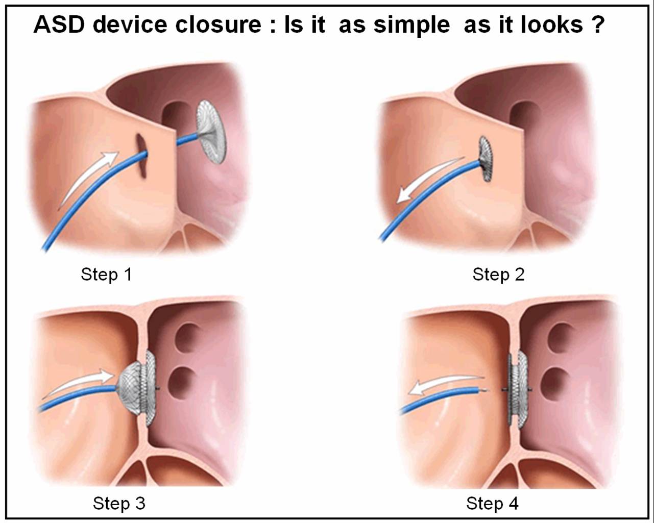
Procedure:
Right femoral venous puncture, then insert 8Fr femoral sheath
Introduce MPA diagnostic catheter with guidewire and cannulate ASD
Remove 0.035 guidewire and replace with Amplatz Super Stiff J-tip wire 0.035” x 260cm
Remove MPA diagnostic catheter and replace with delivery system with ASD plug
Deploy plug under fluoroscopy and TOE guidance
Close access


