Ventricular septal defect (VSD) closure: a Cath Lab 101 guide
Reviewed by Rodnie Oro, cath lab charge nurse · Last reviewed: October 2026 · Education only, not medical advice
VSD in one minute
What it is: a ventricular septal defect (VSD) is a hole in the wall (the septum) between the heart's two lower pumping chambers, the ventricles. Most are present from birth (congenital). Blood leaks through the hole from the left side to the right, so the heart and lungs work harder.
Who needs treatment: many small VSDs cause no problems or close on their own. Larger holes, or holes that strain the heart, may be closed by open-heart surgery or, for suitable holes, with a small occluder (a fabric-filled, double-disc metal mesh plug) placed through blood vessels in the groin.
Main risks: groin bruising or bleeding is the most common. Serious problems, such as a heart rhythm problem needing a pacemaker, the device moving, a leak through a heart valve or a small leftover leak, are uncommon. Your heart team will explain your own risk.
Usually: general anaesthetic with echo guidance, a few hours in the lab, home after one or two nights.
On this page: What is a VSD? · Who is it for? · When is it done? · Step by step · Recovery · Risks · Kit list · First time in the lab? · FAQ
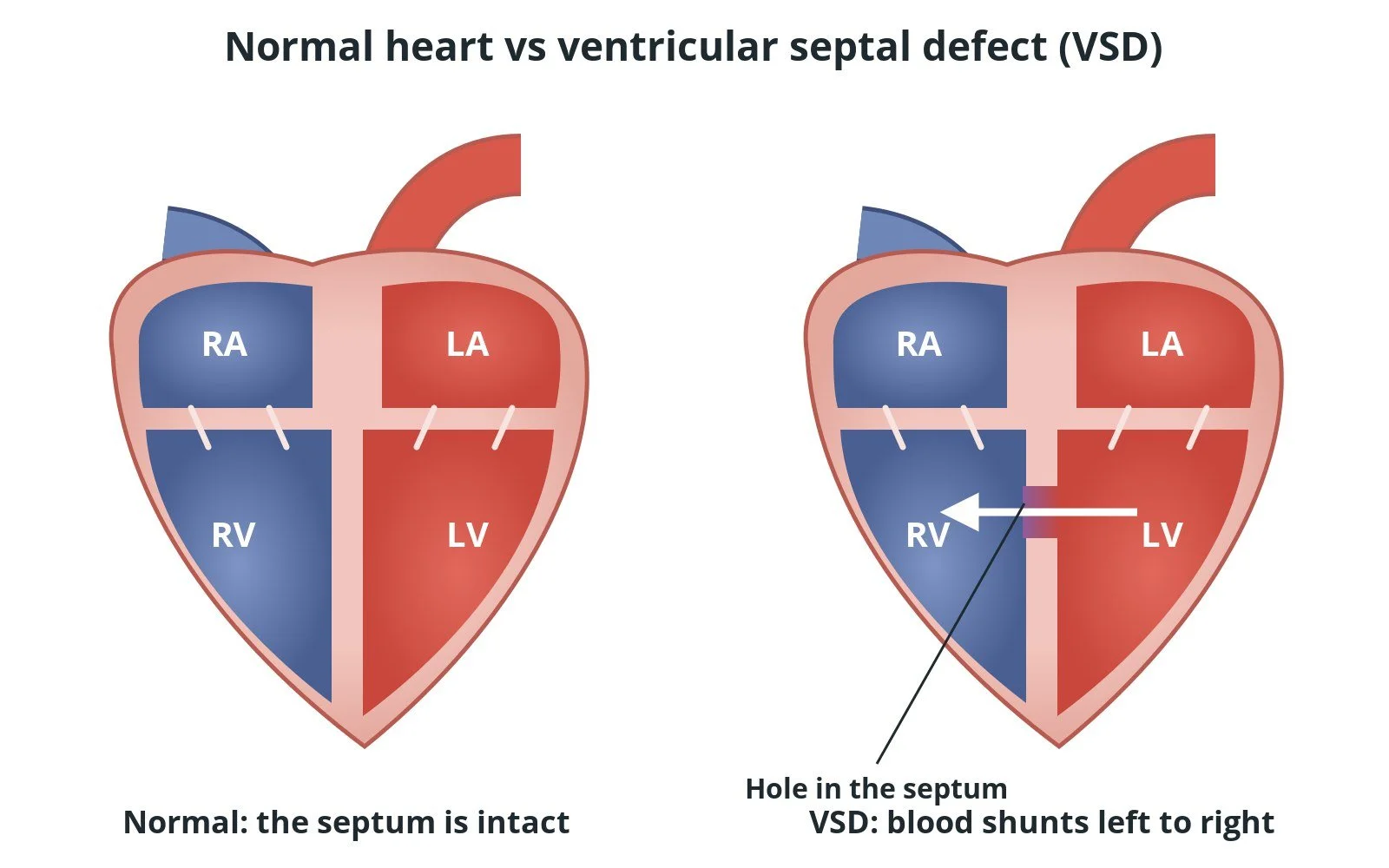
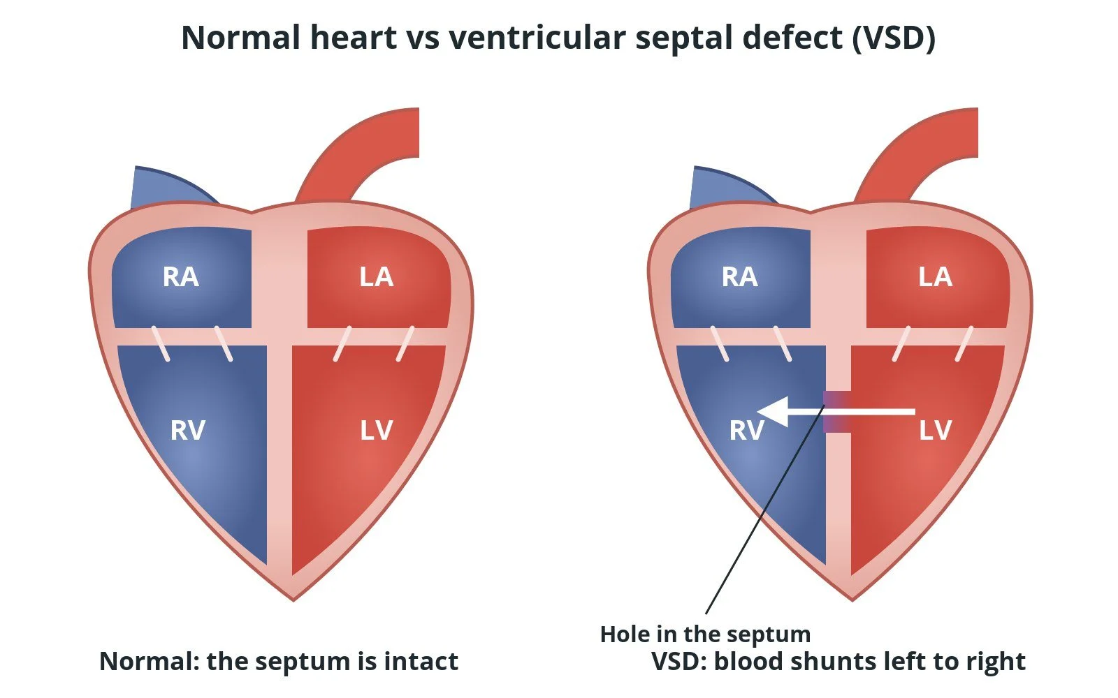
Left: a normal heart, with the wall (septum) between the lower chambers intact. Right: a VSD. Blood flows through the hole from the left ventricle (LV) to the right ventricle (RV). RA and LA are the right and left atria.
What is a VSD?
The heart has four chambers. The right side pumps blood to the lungs to pick up oxygen. The oxygen-rich blood returns to the left side, which pumps it out to the body. A muscular wall, the ventricular septum, keeps the two lower chambers apart.
With a VSD there is a hole in that wall. Pressure is higher on the left, so oxygen-rich blood is pushed through the hole into the right ventricle and back to the lungs (a left-to-right shunt). A small hole makes a loud murmur but little extra work. A large hole sends a lot of extra blood to the lungs. Over time this can enlarge the heart, cause heart failure and raise the pressure in the lungs' blood vessels (pulmonary hypertension).
VSDs are named by where they sit. Perimembranous defects, high in the septum near the heart valves, and muscular defects, in the thick lower part, are the most common. Inlet and outlet defects are rarer. Which type it is matters, because it decides whether a device can be used.
This guide covers congenital VSD. A VSD can very rarely tear open after a heart attack (post-infarct VSD). That is an emergency with its own treatment, and it isn't covered here. New to the acronyms? See the Cath Lab Abbreviations A–Z.
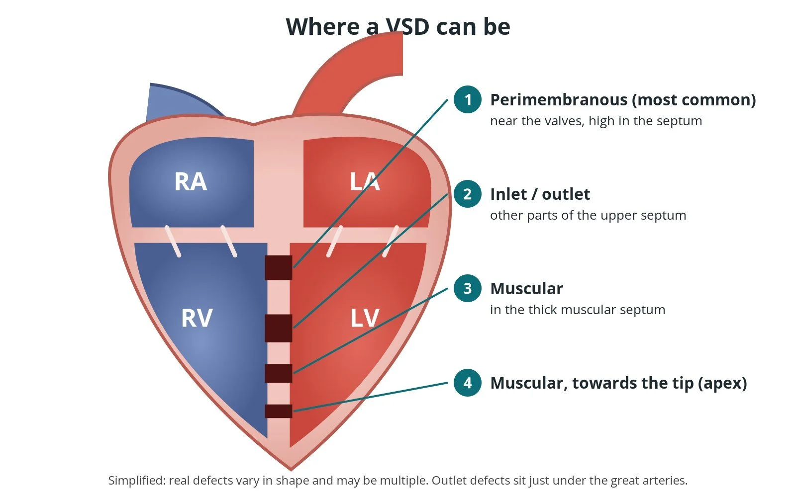
Where a VSD can be: (1) perimembranous, high in the septum near the valves, the most common; (2) inlet or outlet, other parts of the upper septum; (3) and (4) muscular, in the thick lower septum, sometimes several holes. Simplified.
Who is it for?
The decision is made by a specialist congenital heart team, including an interventional cardiologist and a congenital heart surgeon. Closure is usually considered when:
the hole is straining the heart, for example the left ventricle is enlarged from the extra blood, or there are symptoms
the lung pressures are still safe for closure. If they have become very high (Eisenmenger syndrome), closing the hole can be harmful
there is a related problem, such as a leaking aortic valve or a previous heart infection (endocarditis) linked to the VSD
Device closure suits muscular VSDs well, and some perimembranous VSDs in experienced centres. NICE says closure of perimembranous VSD by catheter is safe and effective enough for use with careful patient selection by a multidisciplinary team, and only by trained cardiologists with cardiac surgery available. Holes close to the valves, or with other heart problems that need repair, usually go to surgery instead.
When is it done?
Device closure is a planned procedure. Before it, the team usually:
images the hole with echocardiography (heart ultrasound), and sometimes MRI or CT, to measure it and check how close it is to the valves
checks the lung pressures, sometimes with a cardiac catheter study
records an ECG as a baseline for the heart's rhythm and conduction
discusses it at a congenital heart team meeting
Step by step
1. Preparation
The team checks bloods, allergies (especially to contrast dye), heart rhythm and kidney function. VSD closure is usually done under a general anaesthetic, with a transoesophageal echo (TOE) probe to guide the procedure. Children are treated only in specialist paediatric units.
2. Access and crossing the hole
After cleaning the skin, the operator places sheaths in the femoral artery and femoral vein at the top of the leg, usually with ultrasound. Heparin is given as per centre protocol. Pressures and pictures are taken to confirm the size and position of the hole.
The hole is usually easiest to cross from the left side. A shaped catheter goes up the artery into the left ventricle, and a soft wire is steered through the VSD into the right side of the heart. A snare (a small wire lasso) from the vein catches the wire and pulls it out of the groin vein. This makes an arteriovenous (AV) loop: one wire running from artery to vein through the hole, which gives a stable rail for the delivery sheath.
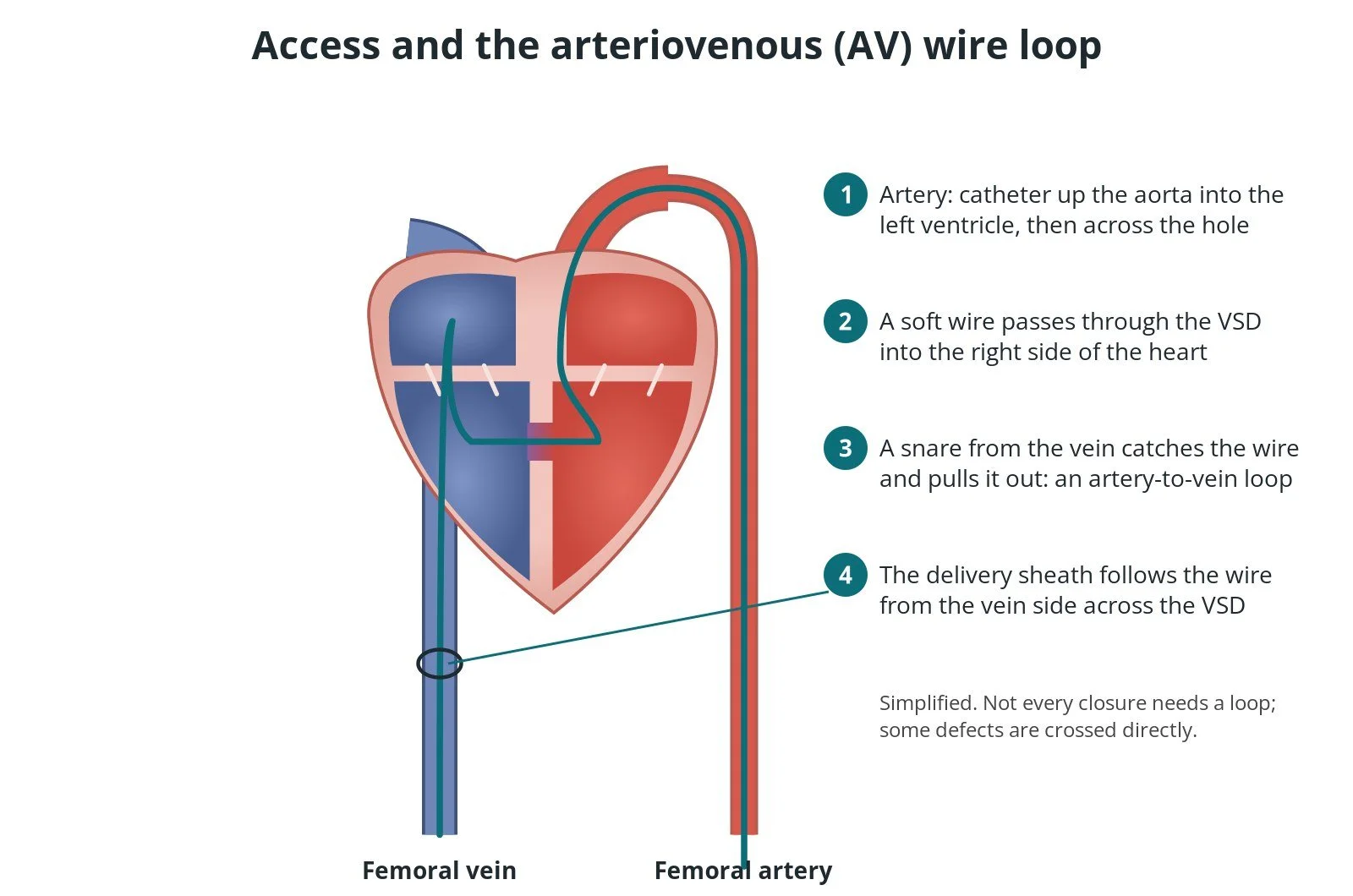
The arteriovenous (AV) loop: (1) a catheter goes up the artery into the left ventricle; (2) a wire crosses the hole; (3) a snare from the vein catches it and pulls it out; (4) the delivery sheath follows the wire from the vein side.
3. Delivery sheath
The delivery sheath is pushed over the loop from the vein side, through the right ventricle and across the hole into the left ventricle. The arterial catheter helps guide its tip. Sheath and wire sizes are per device and operator.
4. Placing the device
The operator chooses a device size from the echo and angiography measurements. The occluder is pushed down the sheath. The left-sided disc opens first in the left ventricle and is pulled back against the septum. The sheath is then withdrawn so the waist fills the hole and the right-sided disc opens on the right side.
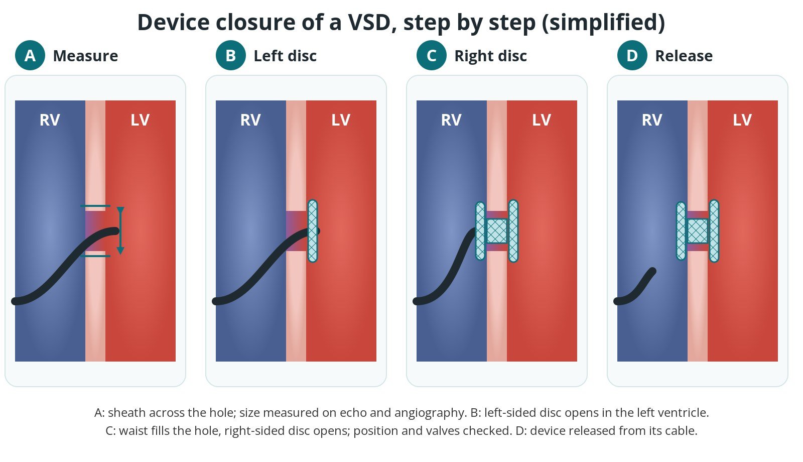
The wall between the right ventricle (RV) and left ventricle (LV), zoomed in. A: the sheath crosses the hole, which is measured. B: the left-sided disc opens in the LV. C: the waist fills the hole and the right-sided disc opens. D: after checks, the device is released from its cable.
5. Checking and release
Before release, echo and contrast confirm that the device is stable, the hole is sealed, and the aortic and tricuspid valves are not being pushed on or made leaky. The heart rhythm is watched closely. Only then is the device unscrewed from its cable. The sheaths are removed and the groin is closed with a closure device, a stitch or pressure, as per centre protocol.
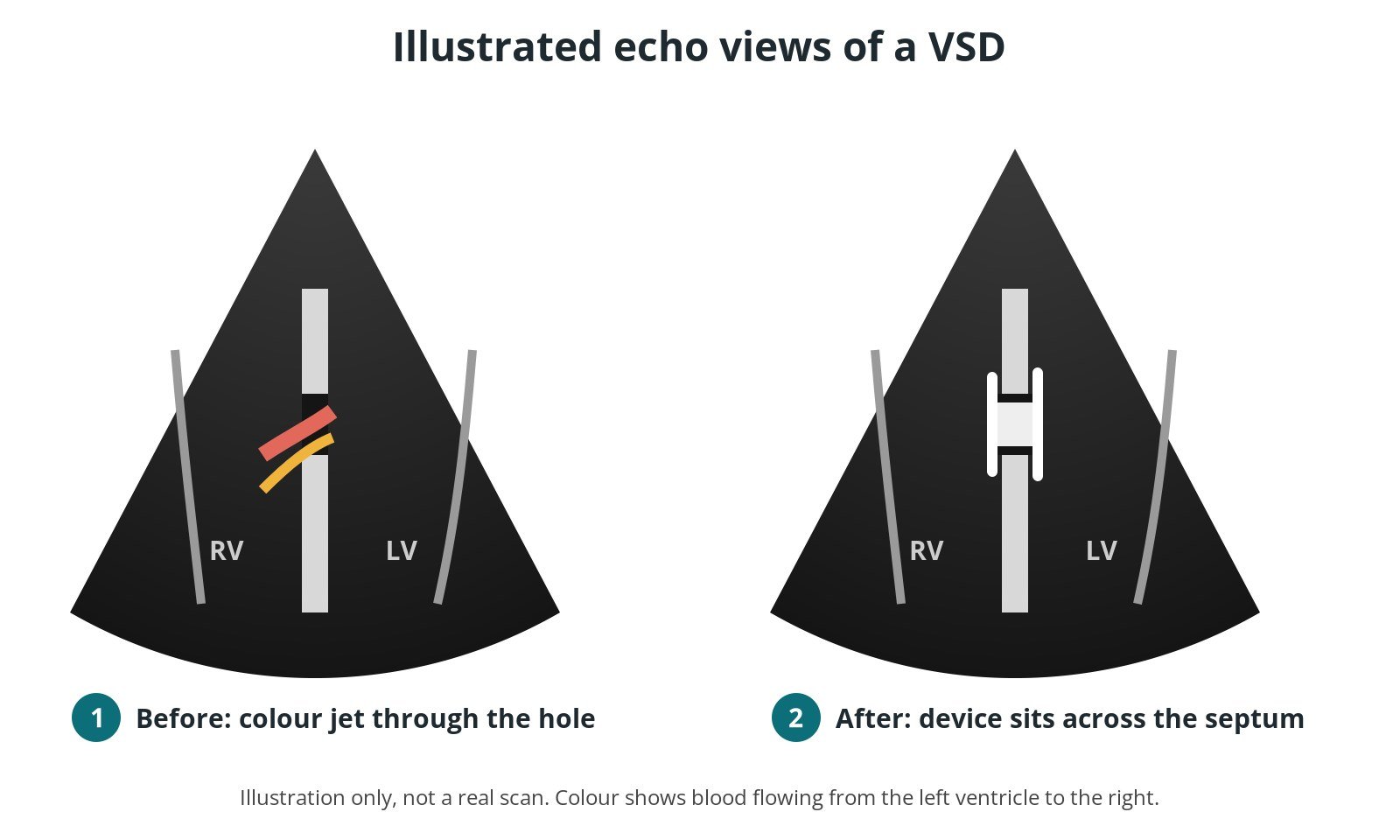
Illustration of the echo pictures: (1) before, a colour jet shows blood flowing through the hole; (2) after, the device sits across the septum and the jet has gone.
Recovery and aftercare
Usually one or two nights in hospital. An echo and ECG are done before going home to check the device and the heart rhythm.
Groin care. Lie flat for a few hours. Avoid heavy lifting and strenuous exercise for about a week, or as your team advises.
Medicines. Antiplatelet medicine (often aspirin) is usually given for several months while the device heals in. Don't stop or change it without speaking to your heart team.
Follow-up. Clinic visits with echo and ECG checks continue over time, because heart rhythm problems can rarely appear later.
Teeth and infection. Look after your teeth and gums, and tell any doctor or dentist you have a heart device.
Call 999 for chest pain, collapse, fainting or severe breathlessness. Call your team for groin swelling or bleeding, fever, a very slow or irregular pulse, or dark urine.
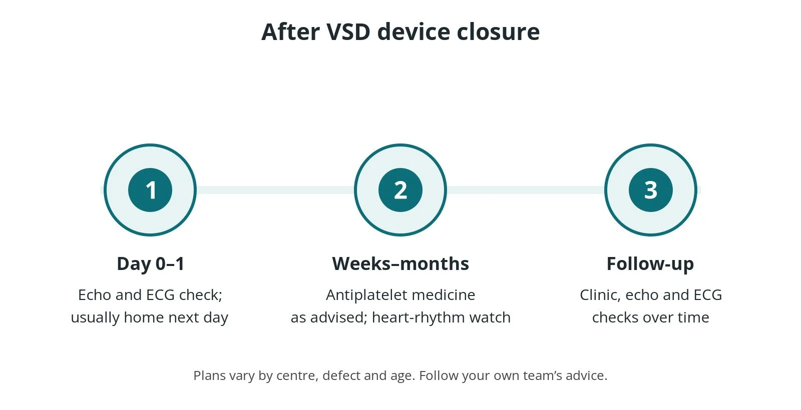
After VSD device closure: (1) an echo and ECG check, usually home after one or two nights; (2) antiplatelet medicine for several months; (3) clinic, echo and ECG follow-up over time.
Risks and complications
Most procedures go smoothly. Possible problems include:
Bleeding or bruising at the groin, the most common problem
Heart block: the device can press on the heart's wiring, which runs close to perimembranous defects. A pacemaker may be needed. Rarely this happens weeks or months later
Other heart rhythm problems during or after the procedure
Valve leak: new or worse leakage of the aortic or tricuspid valve
Residual shunt: a small leftover leak around the device
Device embolisation: the device moves out of place and has to be retrieved, sometimes surgically
Haemolysis: red blood cells damaged by a leak through the device (dark urine is a warning sign)
Damage to the blood vessels used for access, infection, or a reaction to contrast dye
Your heart team will explain how these apply to you.
Cath lab kit list (printable)
Drugs, doses and dilutions: always as per centre protocol. Equipment, sizes and catheter shapes also vary by unit, operator and device. Device names below are generic. Always check the sheath and wire required in the chosen device's instructions for use.
Example: Barts Heart Centre kit list — always follow your own centre protocol
Typical setup for a femoral VSD device closure with an arteriovenous loop, shared as one real-world example. It is not a protocol for your unit.
Sterile table
angiography pack
large chlorhexidine skin prep applicator
ultrasound probe cover (for ultrasound-guided access)
1 L saline for flushing and preparing the device
Access and closure
femoral access needle
6 Fr femoral sheath × 2 (one venous, one arterial)
suture-based vascular closure device
VSD delivery sheath, size per device and operator
Catheters, wires and snare
6 Fr Judkins Right 4 (JR4) diagnostic catheter (crossing the VSD from the left ventricle)
6 Fr pigtail diagnostic catheter (left ventriculogram)
260 cm hydrophilic J-tip guidewire (wire for the loop per device and operator)
25 mm single-loop snare
Device
VSD occluder, type and size chosen in the lab from echo and angiography measurements
Medications (scrub nurse): heparin, contrast and any local anaesthetic as per centre protocol.
Imaging: TOE under general anaesthetic, fluoroscopy and contrast, as per centre protocol.
Procedural steps (Barts example)
Ultrasound-guided femoral venous and arterial access, 6 Fr sheaths
Heparin as per centre protocol; left ventriculogram with the pigtail to show the defect
Cross the VSD from the left ventricle with the JR4 catheter and J-tip wire
Snare the wire in the right heart or pulmonary artery from the venous side and bring it out of the femoral vein (AV loop)
Advance the delivery sheath from the vein over the loop into the left ventricle
Prepare the occluder; open the left disc, pull back to the septum, then open the waist and right disc
Check position, valves, residual shunt and ECG with TOE and contrast before release
Remove sheaths; close per centre protocol
First time in the cath lab?
💡 First-time tip
Watch the ECG the whole time. Heart block or slow rhythms can appear as the wire, sheath or device crosses the septum. Know where the pacing kit is. Keep the AV loop tension-free. Pulling on both ends of the loop can damage the valves or the hole. Follow the operator's lead. Have the snare ready early. Open and flush it before the wire crosses. Flush and de-air everything. Air in a large sheath on the left side of the heart can travel to the brain. Have the next device size available. And always check the device and sheath against the instructions for use.
FAQ
Is VSD closure open-heart surgery?
No. Device closure is a keyhole catheter procedure through blood vessels in the groin. Some VSDs still need open-heart surgery.
Will a small VSD close by itself?
Many small muscular VSDs in babies close on their own in the first years of life. Your child's cardiologist will check with echo.
Can adults have a VSD closed?
Yes. Some adults have a VSD that strains the heart or causes other problems, and it can be closed by surgery or a device if lung pressures are safe.
Why is heart rhythm such a concern?
The heart's electrical wiring runs close to the upper part of the septum, so a device in a perimembranous VSD can press on it. This is why ECGs continue after the procedure.
Can I have an MRI scan with a VSD device?
Most modern septal occluders are MRI-conditional, so scans can usually be done under set conditions. Tell the scanning team you have one and bring your device card.
How long until I can get back to normal?
Most people return to normal daily activities within about a week. Your team will advise on sport and work.
Related Cath Lab 101 guides
Sources
All checked on 10 October 2026. Each page's own date is shown where it gives one.
NICE. Transcatheter endovascular closure of perimembranous ventricular septal defect (HTG213, which replaced IPG172). nice.org.uk/guidance/htg213 (published 24 Mar 2010)
Baumgartner H, et al. 2020 ESC Guidelines for the management of adult congenital heart disease. Eur Heart J 2021;42(6):563–645. PubMed 32860028
Stout KK, et al. 2018 AHA/ACC Guideline for the management of adults with congenital heart disease. J Am Coll Cardiol 2019;73(12):e81–e192. PubMed 30121239
NHS. Congenital heart disease. nhs.uk/conditions/congenital-heart-disease (page last reviewed 11 Dec 2025)
British Heart Foundation. Ventricular septal defect (VSD). bhf.org.uk
Disclaimer
Education only. This page is for general education for healthcare students, cath lab staff and curious readers. It isn't medical advice and doesn't replace advice from your own doctor, heart team or local unit protocols. Equipment and medicines are described generically. No product or manufacturer is recommended. If you think someone is having a heart emergency, call 999.
Disclosure: I work in the medical device industry. This page is independent education and isn't sponsored or reviewed by any company.
All diagrams on this page are original illustrations created for rodnieoro.com.