Heart attack (acute MI) and primary PCI: a Cath Lab 101 guide
Reviewed by Rodnie Oro, cath lab charge nurse · Last reviewed: October 2026 · Education only, not medical advice
Heart attack and primary PCI in one minute
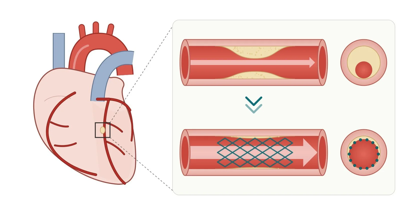
What it is: a heart attack (acute myocardial infarction, MI) happens when a coronary artery is suddenly blocked, usually by a clot on a burst fatty plaque, and part of the heart muscle loses its blood supply. In a STEMI, the artery is usually fully blocked, and the fastest treatment is primary PCI: opening the artery with a balloon and a stent through the wrist or groin.
Who it is for: people with a heart attack confirmed on the ECG and symptoms, brought straight to a heart attack centre. People with an NSTEMI usually have an angiogram within a few days.
Main risks: bleeding at the wrist or groin, irregular heart rhythms, kidney strain from the dye and, less often, stroke or damage to the artery. The heart attack itself carries its own risks. Your team will explain.
Usually: awake under local anaesthetic, about 30 to 90 minutes, then a few days in hospital.
On this page: What is a heart attack? · Who is it for? · When is it done? · Step by step · Recovery · Risks · Kit list · First time in the lab? · FAQ
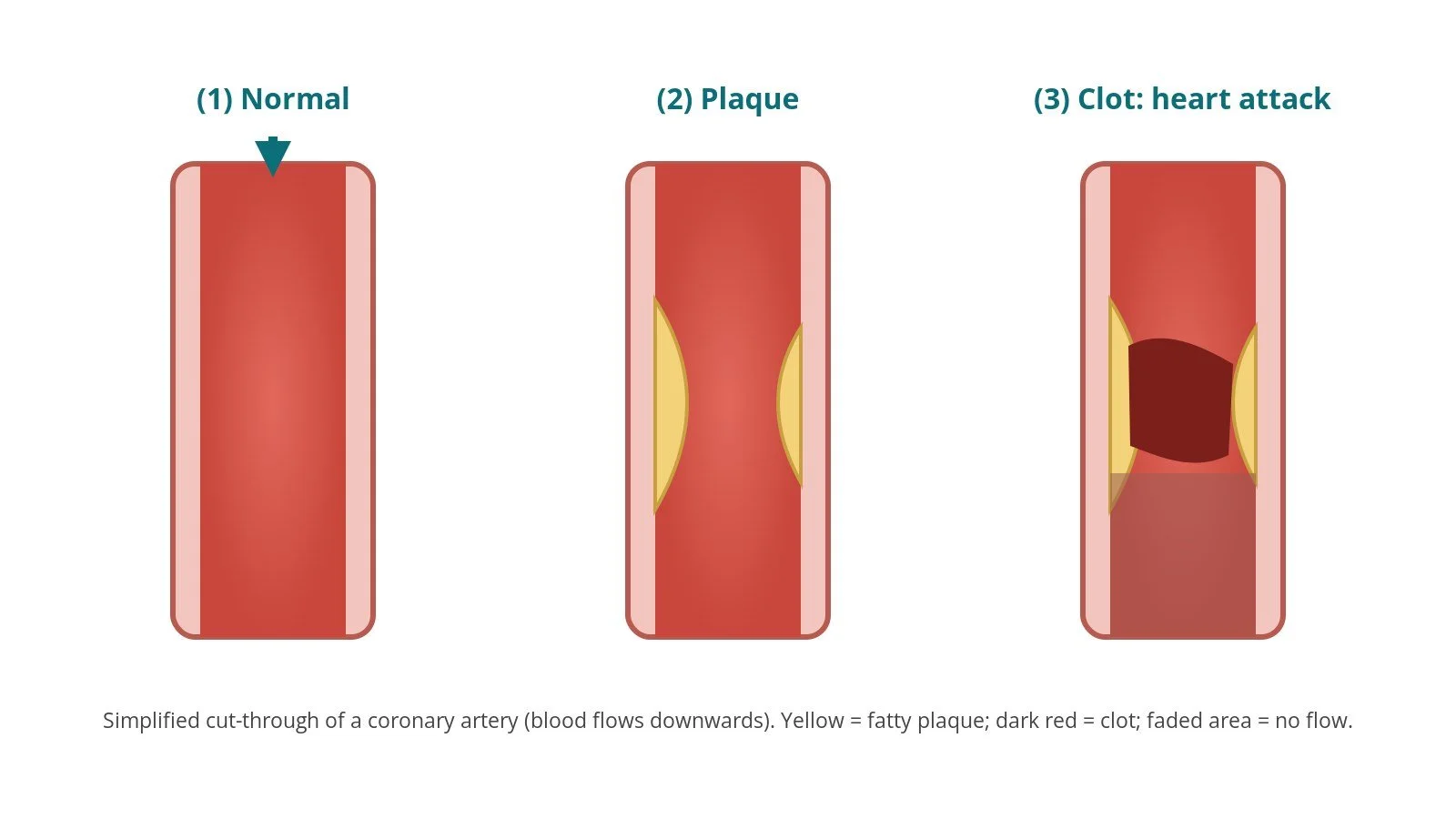
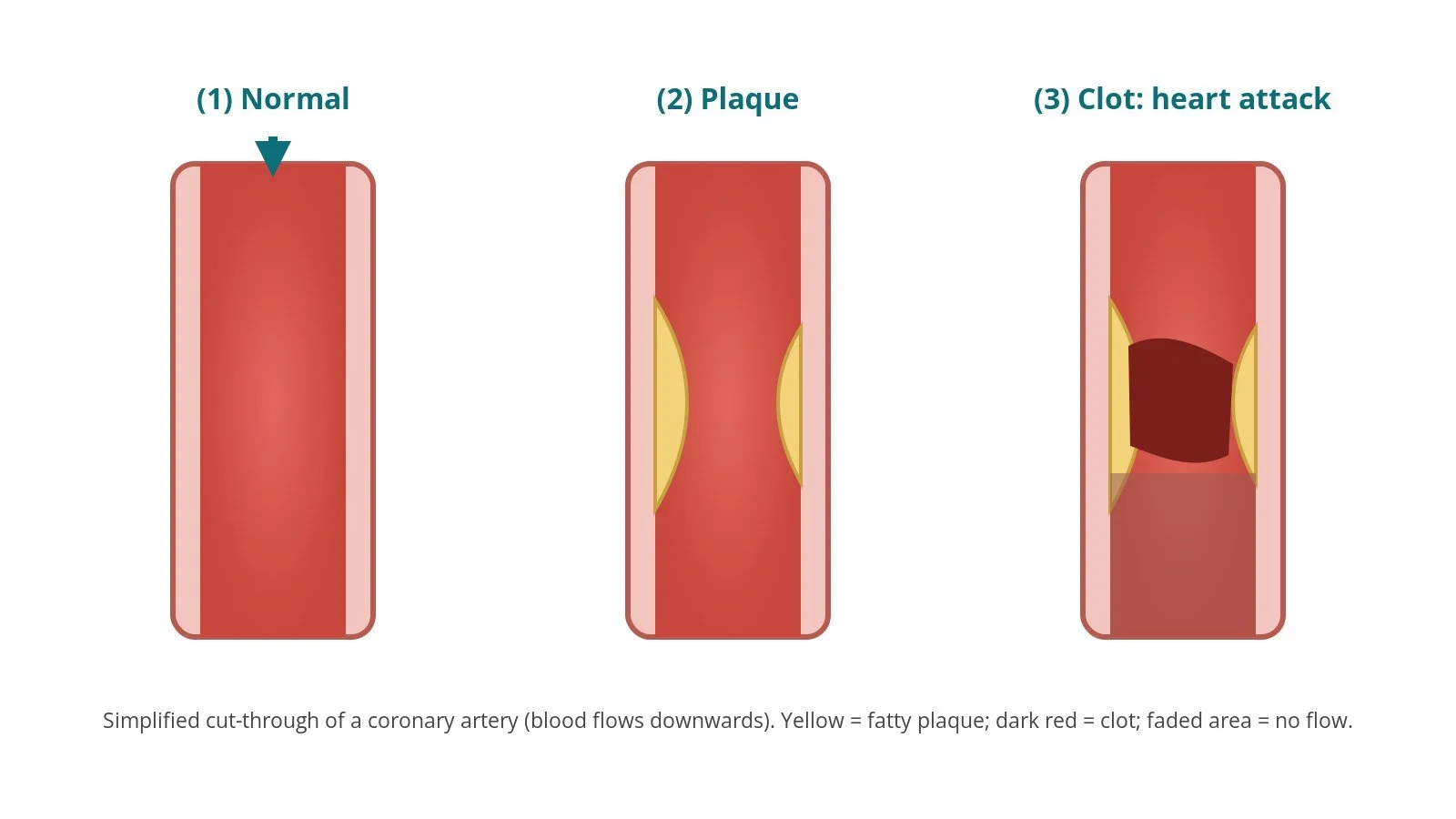
(1) Normal artery. (2) Plaque narrows the artery. (3) A plaque bursts and a clot blocks the artery: a heart attack.
What is a heart attack?
The coronary arteries supply the heart muscle with blood (see the coronary angiogram guide for the anatomy). Over years, fatty plaque can build up in their walls. If a plaque cracks or bursts, a clot forms on it and can suddenly block the artery. The heart muscle beyond the blockage starts to die within minutes to hours, so time is muscle.
Typical symptoms are chest pain or pressure, often spreading to the arms, neck, jaw or back, with breathlessness, sweating or feeling sick. Some people, particularly women, older people and people with diabetes, have less typical symptoms. If you think someone is having a heart attack, call 999.
Heart attacks are grouped by the ECG:
STEMI (ST-elevation MI): the ECG shows a pattern ("ST elevation") that usually means the artery is fully blocked. The aim is to open it as fast as possible, ideally with primary PCI.
NSTEMI (non-ST-elevation MI): a blood test (troponin) shows heart muscle damage without that ECG pattern. Treatment starts with medicines, and most people have an angiogram, and PCI if needed, within days.
Less common causes include a tear in the artery wall (SCAD), artery spasm, and heart attacks with no blocked artery (MINOCA).
New to the acronyms? See the Cath Lab Abbreviations A–Z.
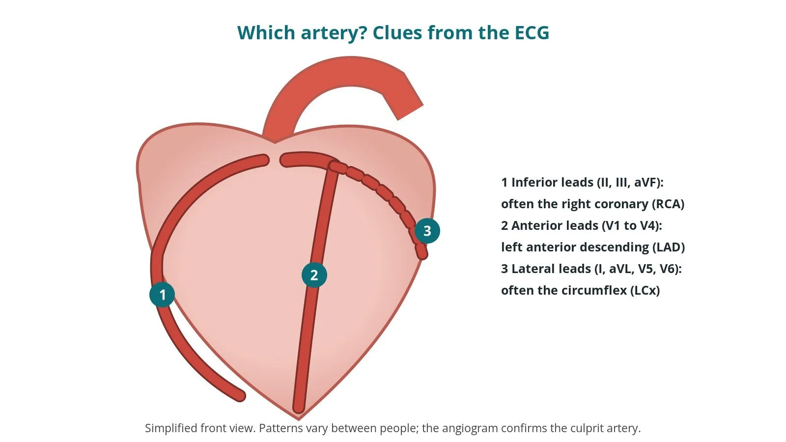
The ECG pattern suggests the blocked artery: (1) inferior leads, often the right coronary; (2) anterior leads, the LAD; (3) lateral leads, often the circumflex.
Who is it for?
Primary PCI is the standard treatment for STEMI in the UK. NICE (NG185) recommends primary PCI for STEMI when it can be delivered within 120 minutes of the time when fibrinolysis (clot-busting drugs) could have been given, and fibrinolysis when it cannot. The ESC 2023 acute coronary syndrome guidelines give similar advice. Ambulance crews send the ECG ahead and take the person straight to a heart attack centre, bypassing the emergency department where possible.
For NSTEMI, NICE and ESC recommend assessing the risk and offering angiography, with PCI or bypass surgery if needed, usually within 72 hours, sooner if the person is unstable.
When is it done?
For STEMI, immediately, 24 hours a day. The heart attack call works like this:
the ambulance team records an ECG, gives aspirin and pain relief and alerts the centre
the cath lab team (often a heart attack centre nurse, a scrub nurse, a circulating nurse, a radiographer, a physiologist and the interventional cardiologist) prepares the lab
on arrival the team confirms the history and ECG, checks allergies, blood thinners and consent, and moves the person straight onto the table
the ECG pattern suggests which artery is blocked, which helps the team choose the first catheters (see the diagram above)
Step by step
1. Getting ready
You lie on the X-ray table, awake. ECG stickers, a blood pressure cuff and an IV line are attached. Pain relief and sedation may be given, as per centre protocol. The wrist (or groin) is cleaned and covered with sterile drapes.
2. Access
Radial (wrist) access is preferred in most UK centres; femoral (groin) is used when needed. A sheath is inserted using the Seldinger technique; sheath sizes are per device and operator.
Medicines to prevent artery spasm and clots (including heparin) are given, as per centre protocol. Antiplatelet medicines are given as per centre protocol.
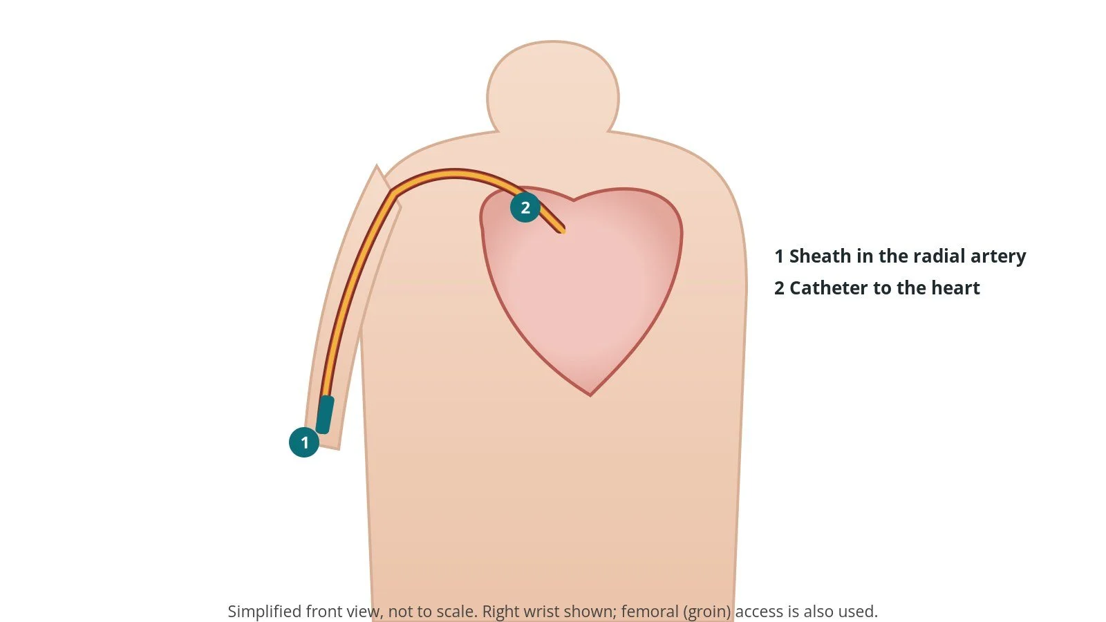
Access: (1) sheath in the radial artery at the wrist; (2) catheter to the heart.
3. Angiogram and opening the artery
A diagnostic catheter first takes pictures of the artery that is thought not to be blocked; then a guide catheter is placed in the blocked artery. Many centres start with the artery suggested by the ECG.
A. Wire. A thin coronary guidewire is steered through the clot and parked well beyond it. Often this alone starts to restore some flow.
B. Balloon. A semi-compliant balloon is inflated in the blockage to open it (pre-dilatation). Some operators also remove clot with an aspiration catheter when there is a lot of it, per operator.
C. Stent. A drug-eluting stent, sized to the artery, is placed across the narrowing and expanded with its balloon.
D. Post-dilate. A non-compliant (high-pressure) balloon may be used to fully expand the stent. Final pictures check the artery is open with good flow.
Other narrowings in other arteries may be treated at the same time or at a later date, as decided by the team.
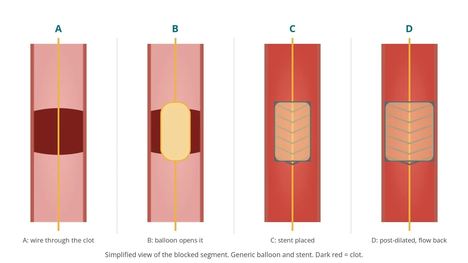
A: wire through the clot. B: balloon opens the blockage. C: stent placed. D: stent fully expanded; flow restored.
4. Finishing
The sheath is removed. A compression band goes on the wrist, or the groin is closed with pressure or a closure device, per operator. You go to the coronary care unit (CCU) for monitoring.
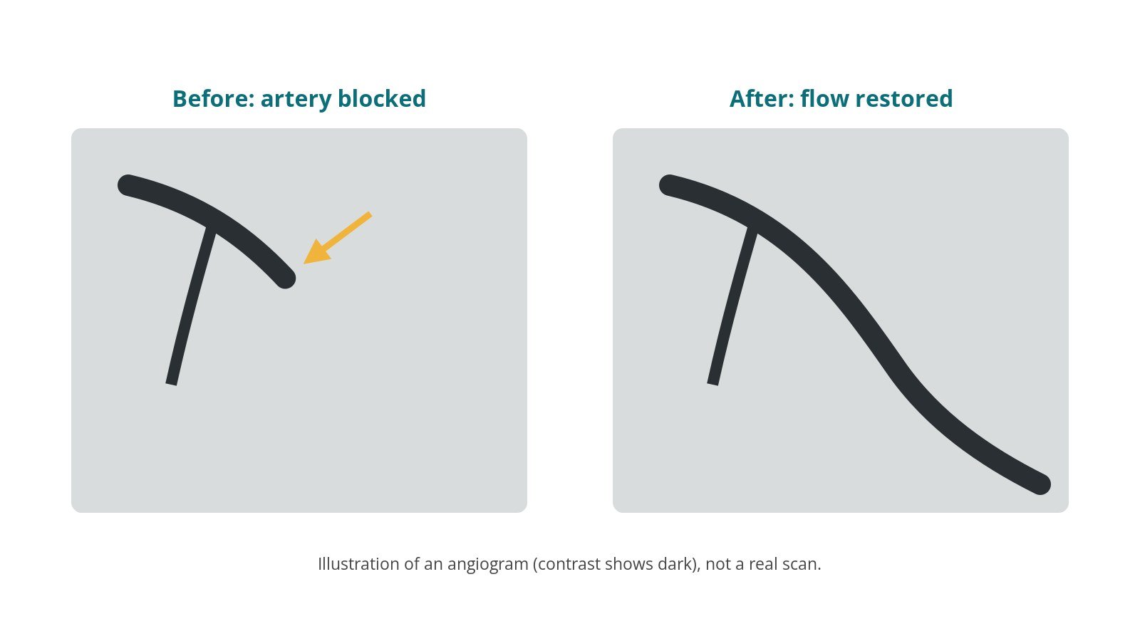
Illustration, not a real scan. Before: the artery stops abruptly. After: flow restored beyond the stent.
Recovery and aftercare
CCU and ward: heart rhythm monitoring, wrist or groin checks, blood tests and an echo to check the heart's pumping strength.
Going home: often after 2 to 3 days if there are no complications.
Medicines: usually dual antiplatelet therapy (aspirin plus a second antiplatelet), typically for 12 months, as advised by your team, plus a statin and other heart medicines. Do not stop antiplatelets without speaking to your cardiology team, as this can cause the stent to block.
Cardiac rehabilitation: NICE (CG172) recommends offering a cardiac rehabilitation programme to everyone after a heart attack. It covers exercise, diet, stopping smoking, stress and returning to work.
Driving: for a car licence (Group 1), DVLA guidance says you must not drive for 1 week after a heart attack if you had successful PCI, no other urgent treatment is planned and your heart's pumping function (ejection fraction) is at least 40%; otherwise 4 weeks. You need not tell the DVLA for a car licence. Bus and lorry drivers must tell the DVLA.
Call 999 if you have chest pain that does not settle within a few minutes of rest (or your GTN spray, if you have one), or bleeding from the wrist or groin that does not stop with firm pressure.
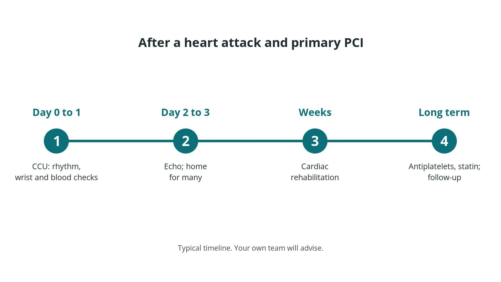
After primary PCI: (1) CCU monitoring; (2) echo, home after 2 to 3 days for many; (3) cardiac rehabilitation; (4) medicines and follow-up.
Risks and complications
Your team will discuss your own risks. Possible problems include:
bleeding or bruising at the wrist or groin
irregular heart rhythms during or after the procedure
the "no-reflow" problem, where flow is slow despite an open artery
damage to the artery, stroke or the need for emergency bypass surgery (uncommon)
kidney strain from the contrast dye
a clot forming in the stent (stent thrombosis), especially if antiplatelets are stopped early
Cath lab kit list (printable)
Barts example kit list
An example from one UK centre, written generically. Every lab has its own protocol, and sizes and doses vary.
Guide and diagnostic catheters (prepared by the heart attack centre nurse)
anterior or lateral ST elevation (often the left coronary): extra-backup left guide catheter, plus a right coronary-shaped diagnostic catheter, sizes per operator
inferior or posterior ST elevation (often the right coronary): right coronary-shaped guide catheter, plus a left coronary-shaped diagnostic catheter, sizes per operator
Sedation and pain relief (circulating nurse)
sedative and opioid, doses and increments as per centre protocol
IV drug line
saline bag, giving set, long extension line, 3-way tap, labelled syringes, blunt drawing-up needle
Supplies (scrub nurse)
angiography pack, skin antiseptic, sterile saline and spike, transparent dressing for a radial sheath
radial or femoral sheath, size per device and operator
inflation device, short haemostasis valve extension, contrast–saline balloon mix as per centre protocol, Luer-lock syringe
coronary guidewire (operator preference)
semi-compliant balloons (pre-dilatation) and non-compliant balloons (post-dilatation), sized to the vessel
drug-eluting stents, sized to the vessel
aspiration catheter if there is a large clot, per operator
Medicines (all as per centre protocol)
local anaesthetic, heparin, nitrate and calcium-channel blocker for radial spasm
Haemostasis
radial compression band or femoral closure device, per operator
Procedural steps (Barts example)
Local anaesthetic; sheath inserted (Seldinger); anti-spasm cocktail through a radial sheath
Diagnostic pictures of the non-culprit artery; guide catheter into the culprit artery
Coronary wire across the blockage, parked distally
Pre-dilatation with a semi-compliant balloon (aspiration if a large clot)
Stent deployed; post-dilatation with a non-compliant balloon if needed
Final pictures; sheath removed; band or closure device; transfer to CCU
First time in the cath lab?
💡 First-time tip
Be ready before the doors open. Know your role in a heart attack call, and have the catheters for both arteries ready. Watch the monitor. Opening a blocked artery often triggers rhythm changes ("reperfusion arrhythmias"). Know where the defibrillator and pacing kit are. Label every syringe. Heparin, sedatives and spasm drugs look the same. Talk to the patient. They are awake, frightened and often in pain. A calm explanation helps. Record the times. Door-to-balloon time and other key times are audited nationally.
FAQ
How quickly does the artery need opening?
As quickly as possible. Heart attack networks aim to open the artery within about 2 hours of the first call for help, ideally much sooner.
Am I awake?
Usually yes, with local anaesthetic and some sedation if needed.
Will I need more stents later?
Sometimes. Other narrowings may be treated at the same time or at a later date.
When can I go back to work?
It depends on your job and recovery. Many people return after a few weeks; cardiac rehabilitation helps you plan this.
ST changes and the culprit artery
The ECG pattern suggests which artery is blocked, which helps the team choose the first catheters.
Each group of leads looks at one wall of the heart, and each wall is usually supplied by one artery.
| Pattern | Leads | Usual artery |
|---|---|---|
| Inferior | ST elevation in II, III, aVF | Usually RCA (right coronary), sometimes circumflex |
| Anterior and septal | ST elevation in V1–V4 (septal V1–V2) | LAD (left anterior descending) |
| Lateral | ST elevation in I, aVL, V5–V6 | Circumflex or an LAD diagonal |
| High lateral | ST elevation in I, aVL | First diagonal or circumflex |
| Posterior | ST depression in V1–V3, often with tall R waves and upright T waves; ST elevation in V7–V9 | Circumflex or RCA |
| Right ventricle | ST elevation in V4R, with an inferior STEMI | Proximal RCA |
| Reciprocal changes | ST depression in leads on the opposite side of the heart | Not a separate artery: reciprocal changes support the diagnosis |
| ST elevation in aVR with widespread ST depression | ST elevation in aVR (and often V1), ST depression in six or more leads | Left main or severe multi-vessel disease |
| Wellens syndrome | Biphasic T waves (type A) or deep symmetrical T wave inversion (type B) in V2–V3, once the chest pain has settled | Critical narrowing of the proximal LAD |
| de Winter pattern | 1–3 mm upsloping ST depression at the J point in V1–V6, running into tall, symmetrical T waves; often slight ST elevation in aVR | Usually a blocked proximal LAD |
ST changes by territory
Inferior
Leads: ST elevation in II, III, aVF
Artery: Usually RCA (right coronary), sometimes circumflex
- ST elevation bigger in III than II, with ST depression in I and aVL, points to the RCA.
- Inferior STEMIs often bring slow rhythms and low blood pressure.
Anterior and septal
Leads: ST elevation in V1–V4 (septal V1–V2)
Artery: LAD (left anterior descending)
- Convex (“tombstone”) ST elevation and new Q waves may follow.
Lateral
Leads: ST elevation in I, aVL, V5–V6
Artery: Circumflex or an LAD diagonal
- The diagonal is a branch of the LAD. It is not drawn separately on the schematic.
High lateral
Leads: ST elevation in I, aVL
Artery: First diagonal or circumflex
- The first diagonal is a branch of the LAD. It is not drawn separately on the schematic.
Posterior
Leads: ST depression in V1–V3, often with tall R waves and upright T waves; ST elevation in V7–V9
Artery: Circumflex or RCA
- A “mirror image” of ST elevation. Often comes with an inferior or lateral STEMI.
- If you see ST depression in V1–V3 in a patient with chest pain, suggest posterior leads. It is easily missed.
Right ventricle
Leads: ST elevation in V4R, with an inferior STEMI
Artery: Proximal RCA
- ST elevation in V1, and in III more than II, are clues.
- Record V4R when an inferior STEMI is suspected. Watch the blood pressure closely.
Reciprocal changes
Leads: ST depression in leads on the opposite side of the heart
Artery: Not a separate artery: reciprocal changes support the diagnosis
- For example, an inferior STEMI often shows ST depression in I and aVL.
ST elevation in aVR with widespread ST depression
Leads: ST elevation in aVR (and often V1), ST depression in six or more leads
Artery: Left main or severe multi-vessel disease
- Especially if the patient is unstable. Needs urgent senior review.
Patterns without classic ST elevation
Some blocked or critically narrowed arteries do not show classic ST elevation. These patterns need urgent senior review.
Wellens syndrome
Leads: Biphasic T waves (type A) or deep symmetrical T wave inversion (type B) in V2–V3, once the chest pain has settled
Artery: Critical narrowing of the proximal LAD
- Little or no ST elevation, no Q waves, R waves preserved.
- Urgent coronary angiography. Exercise stress testing is avoided, because it can trigger a large anterior heart attack.
de Winter pattern
Leads: 1–3 mm upsloping ST depression at the J point in V1–V6, running into tall, symmetrical T waves; often slight ST elevation in aVR
Artery: Usually a blocked proximal LAD
- Recognised as a STEMI equivalent. Treat as an anterior STEMI: same preparation, same urgency.
The ECG points to the artery. The coronary angiogram confirms it, because anatomy varies. Strips are simplified illustrations, not real patient ECGs. Educational use only.
More: STEMI territories (ECG basics) · Ischaemia and infarction patterns (ECG advanced)
Related Cath Lab 101 guides
Sources
All checked on 10 October 2026. Each page's own date is shown where it gives one.
NICE. Acute coronary syndromes (NG185). nice.org.uk/guidance/ng185
NICE. Myocardial infarction: cardiac rehabilitation and prevention of further cardiovascular disease (CG172). nice.org.uk/guidance/cg172
Byrne RA, et al. 2023 ESC Guidelines for the management of acute coronary syndromes. Eur Heart J 2023;44(38):3720–3826. PubMed 37622654
NHS. Heart attack. nhs.uk/conditions/heart-attack
British Heart Foundation. Heart attack. bhf.org.uk
British Heart Foundation. Coronary angioplasty and stents (PCI). bhf.org.uk
DVLA / GOV.UK. Cardiovascular disorders: assessing fitness to drive (acute coronary syndromes). gov.uk
Disclaimer
Education only. This page is for general education for healthcare students, cath lab staff and curious readers. It isn't medical advice and doesn't replace advice from your own doctor, heart team or local unit protocols. Equipment and medicines are described generically. No product or manufacturer is recommended. If you think someone is having a heart emergency, call 999.
Disclosure: I work for a company that makes a device used in this procedure. This page is independent education, isn't sponsored or reviewed by any company, and doesn't recommend any product.
All diagrams on this page are original illustrations created for rodnieoro.com.