Alcohol septal ablation for HOCM: a Cath Lab 101 guide
Reviewed by Rodnie Oro, cath lab charge nurse · Last reviewed: October 2026 · Education only, not medical advice
Alcohol septal ablation in one minute
What it is: alcohol septal ablation is a keyhole (catheter) treatment for obstructive hypertrophic cardiomyopathy (HOCM). A tiny balloon is placed in a small artery that feeds the thickened part of the heart wall, and a small amount of pure alcohol is injected. This makes a controlled scar, so the bulge thins and blood can leave the heart more easily.
Who it is for: people whose thick heart muscle blocks blood flow out of the left ventricle and who still have significant symptoms, such as breathlessness, chest pain or fainting, despite the best tolerated medicines. The choice between alcohol septal ablation and heart surgery (septal myectomy) is made by a specialist heart team.
Main risks: the most important is heart block, a slow heartbeat that may need a permanent pacemaker. A temporary pacing wire is placed during the procedure for this reason. Your heart team will explain your own risk.
Usually: awake with local anaesthetic, sedation and pain relief, about 1 to 2 hours, then a few days on a heart monitor in hospital.
On this page: What is alcohol septal ablation? · Who is it for? · When is it done? · Step by step · Recovery · Risks · Kit list · First time in the lab? · FAQ
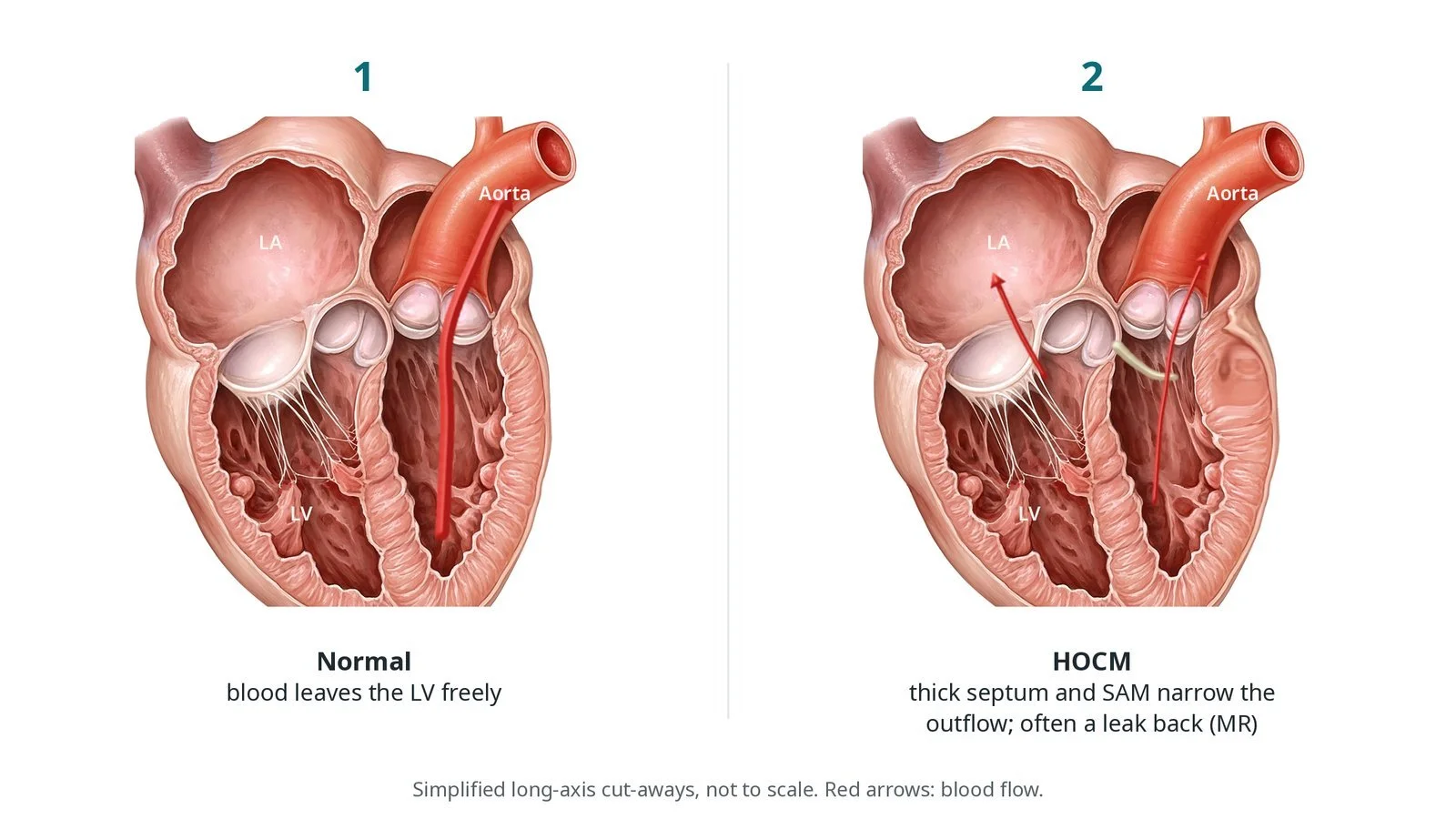
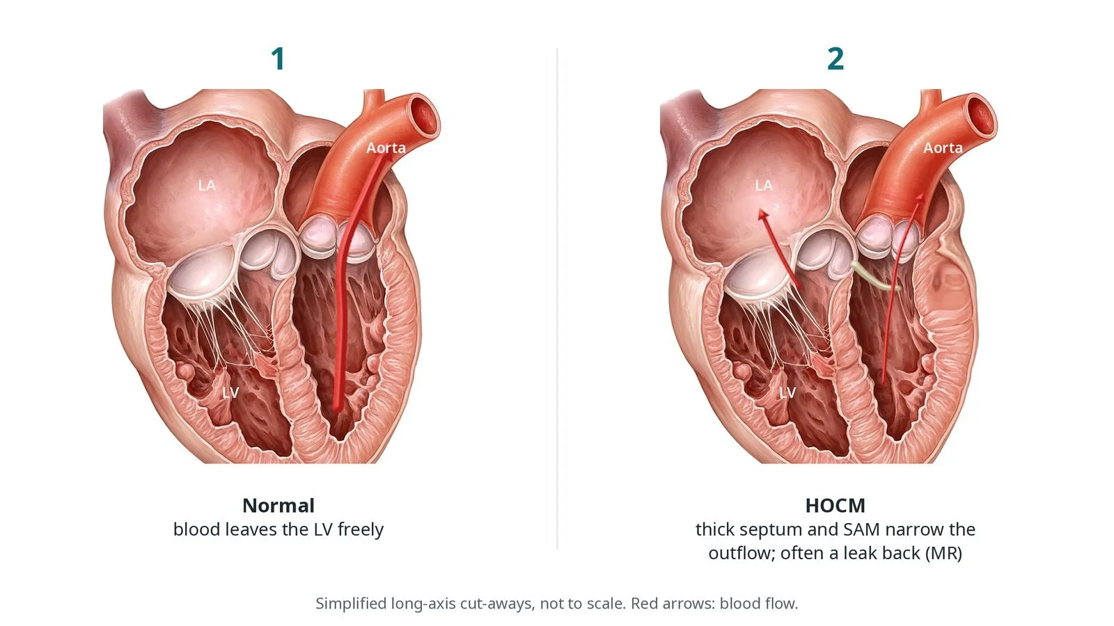
Normal heart vs HOCM. (1) Normal: blood leaves the left ventricle (LV) freely. (2) HOCM: a thick basal septum and the mitral leaflet pulled towards it (SAM) narrow the outflow tract, often with a leak back into the left atrium (MR).
What is alcohol septal ablation?
In hypertrophic cardiomyopathy (HCM) the heart muscle is thicker than it should be, without another cause such as high blood pressure or a narrowed aortic valve. It is often inherited. In many people the thickening is greatest in the septum, the wall between the two pumping chambers, just below the aortic valve.
When the heart squeezes, a thick upper (basal) septum can narrow the left ventricular outflow tract (LVOT), the channel blood passes through on its way to the aorta. The fast flow can also pull the front leaflet of the mitral valve towards the septum. This is called systolic anterior motion (SAM). It narrows the channel further and often causes a leak back through the mitral valve. HCM with this blockage is called obstructive HCM or HOCM. The blockage is measured on echo as a pressure gradient across the outflow tract.
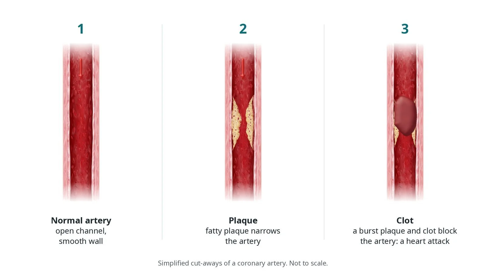
Alcohol septal ablation treats the blockage from the inside, through an artery. The septum gets its blood from small septal branches of the left anterior descending (LAD) coronary artery. The operator finds the branch that feeds the bulge, seals it with a small balloon and injects alcohol into it. The muscle it supplies dies in a small, controlled heart attack. Over the following weeks to months that area scars and thins, the outflow tract widens and the gradient falls.
The surgical alternative is septal myectomy, where a surgeon removes part of the septum during open-heart surgery. Both are called septal reduction therapy.
New to the acronyms? See the Cath Lab Abbreviations A–Z.
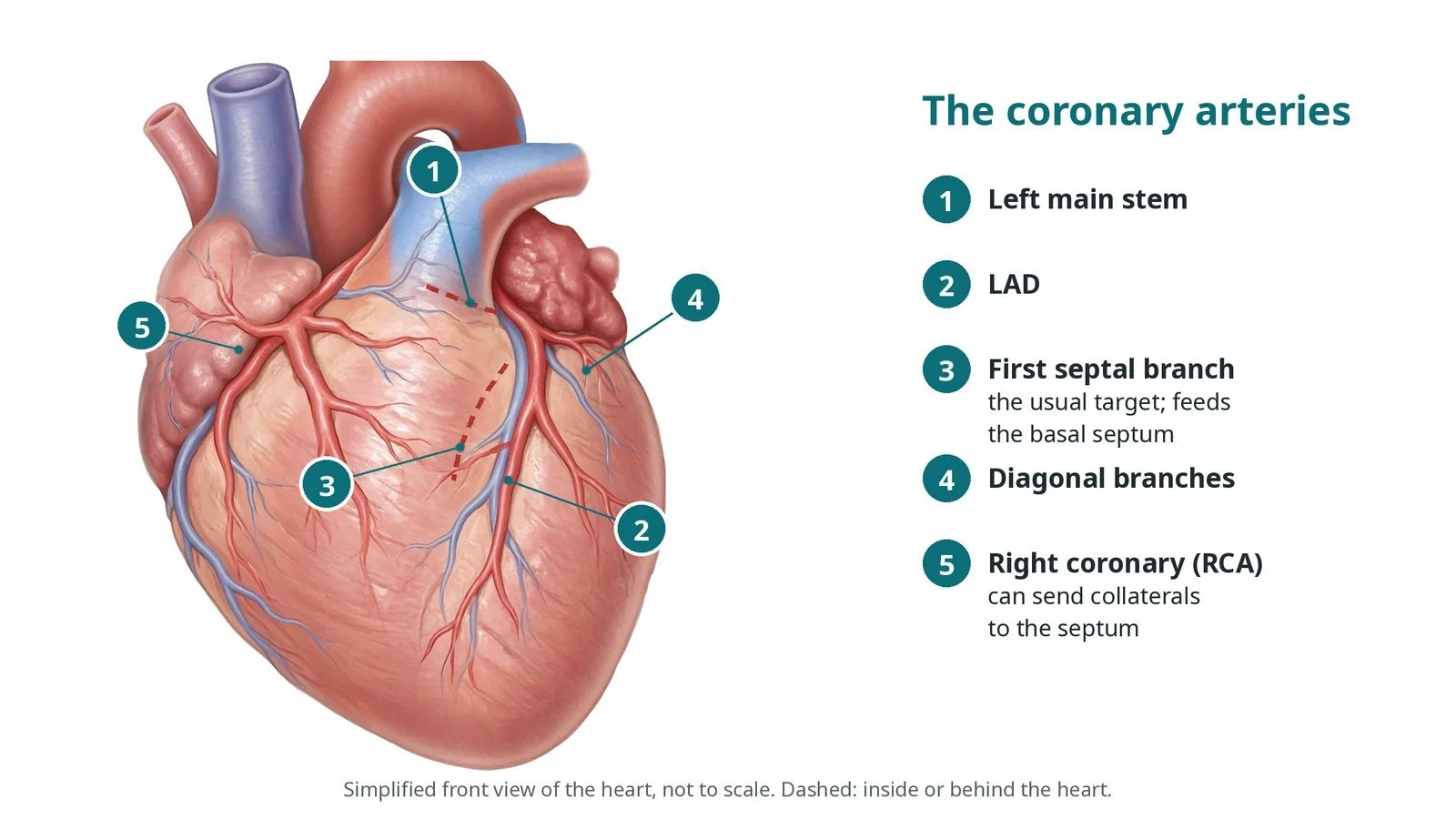
The coronary arteries: (1) left main stem; (2) LAD; (3) the first septal perforator, the usual target, which feeds the basal septum; (4) diagonal branches; (5) the RCA, which can send collaterals to the septum.
Who is it for?
The decision is made by a specialist cardiomyopathy and heart team after echo, heart MRI and coronary angiography. European (ESC 2023) and American (AHA/ACC 2024) guidelines broadly agree. Septal reduction is considered when:
the outflow gradient is 50 mmHg or more, at rest or when provoked (for example on exercise or standing)
symptoms stay significant (breathlessness, chest pain, dizziness or fainting on effort) despite the best tolerated medicines
the procedure is done in an experienced centre
Medicines come first. They include beta-blockers, some calcium channel blockers, disopyramide and, more recently, a class called cardiac myosin inhibitors.
Alcohol septal ablation is more likely to be chosen for people who are older or at higher risk from surgery, or who prefer to avoid it, when the anatomy suits. Surgery is usually preferred when:
another heart operation is needed at the same time, such as for the mitral valve or coronary arteries
the septum is very thick, or no suitable septal branch can be found
the person is young, depending on the team's assessment
NICE says the procedure should only be done in specialist units by clinicians with adequate training (HTG18, formerly IPG40).
When is it done?
Alcohol septal ablation is a planned procedure. Before it, the team usually:
confirms the gradient on echo, including provoked measurements, and checks the mitral valve and SAM
reviews the ECG for existing conduction problems, such as right bundle branch block, which raise the chance of needing a pacemaker
does a coronary angiogram, often the same day, to see the septal branches
checks bloods, kidney function and medicines, including any blood thinners, as per centre protocol
plans a bed on a coronary care or high-dependency unit for monitoring afterwards
Step by step
1. Preparation and temporary pacing
The patient lies on the cath lab table with ECG, blood pressure and oxygen monitoring. An echo machine and a sonographer or echo-trained doctor are ready. A temporary pacing wire is placed through a vein, usually in the neck or groin, with its tip in the right ventricle. It protects against heart block during and after the procedure. Sheath sizes are per device and operator.
2. Arterial access and angiography
The operator enters an artery at the wrist or groin. The right coronary artery may be imaged first, to check for vessels that connect to the septum. A guide catheter is then placed at the start of the left coronary artery, and angiograms show the LAD and its septal branches. Anticoagulation is given as per centre protocol.
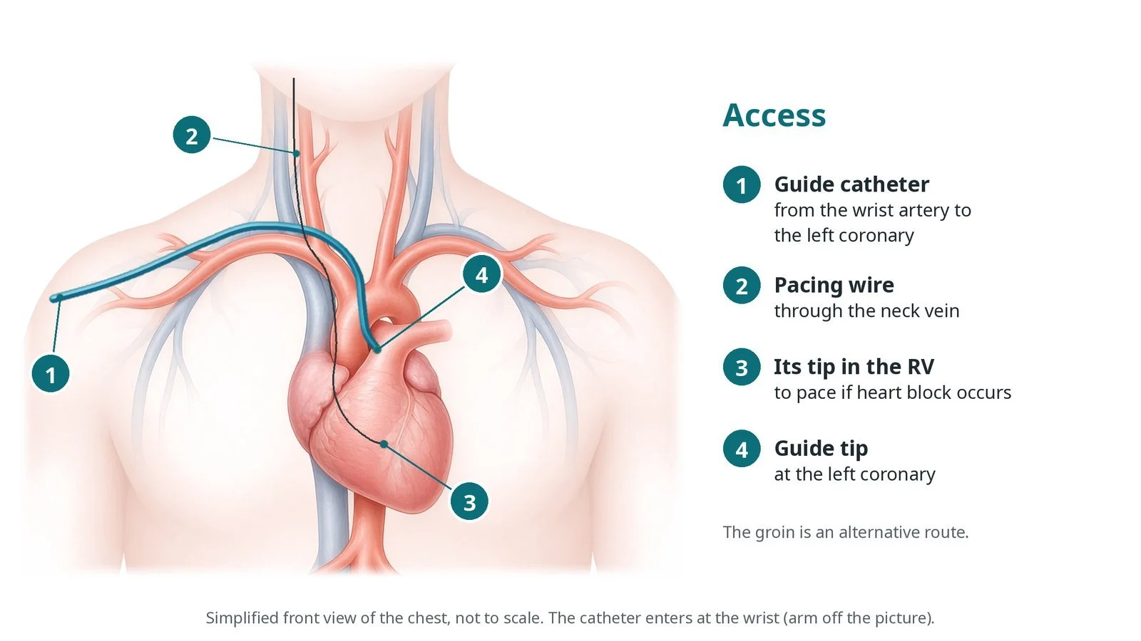
Access: (1) a guide catheter from the wrist artery to the left coronary; (2) a temporary pacing wire through the neck vein; (3) its tip in the right ventricle (RV), to pace the heart if heart block occurs; (4) the guide tip at the left coronary. The groin is an alternative.
3. Wiring and sealing the septal branch
A fine coronary guidewire is passed down the LAD and into the chosen septal branch, usually the first major septal perforator. A short, small over-the-wire balloon is advanced over it into the branch. The balloon is inflated to seal the branch, and contrast is injected through the balloon's central channel. This confirms the balloon is sealing properly and that nothing flows back into the LAD.
4. Echo contrast: the target check
This is the key safety step. With the balloon still inflated, a small amount of echo (ultrasound) contrast is injected through the balloon. On echo, the muscle that the branch feeds lights up. The team checks that it is the basal septum at the point where SAM touches it, and not the papillary muscles, the right ventricle or other parts of the heart. If the area is wrong, a different branch is chosen, or the procedure is stopped.
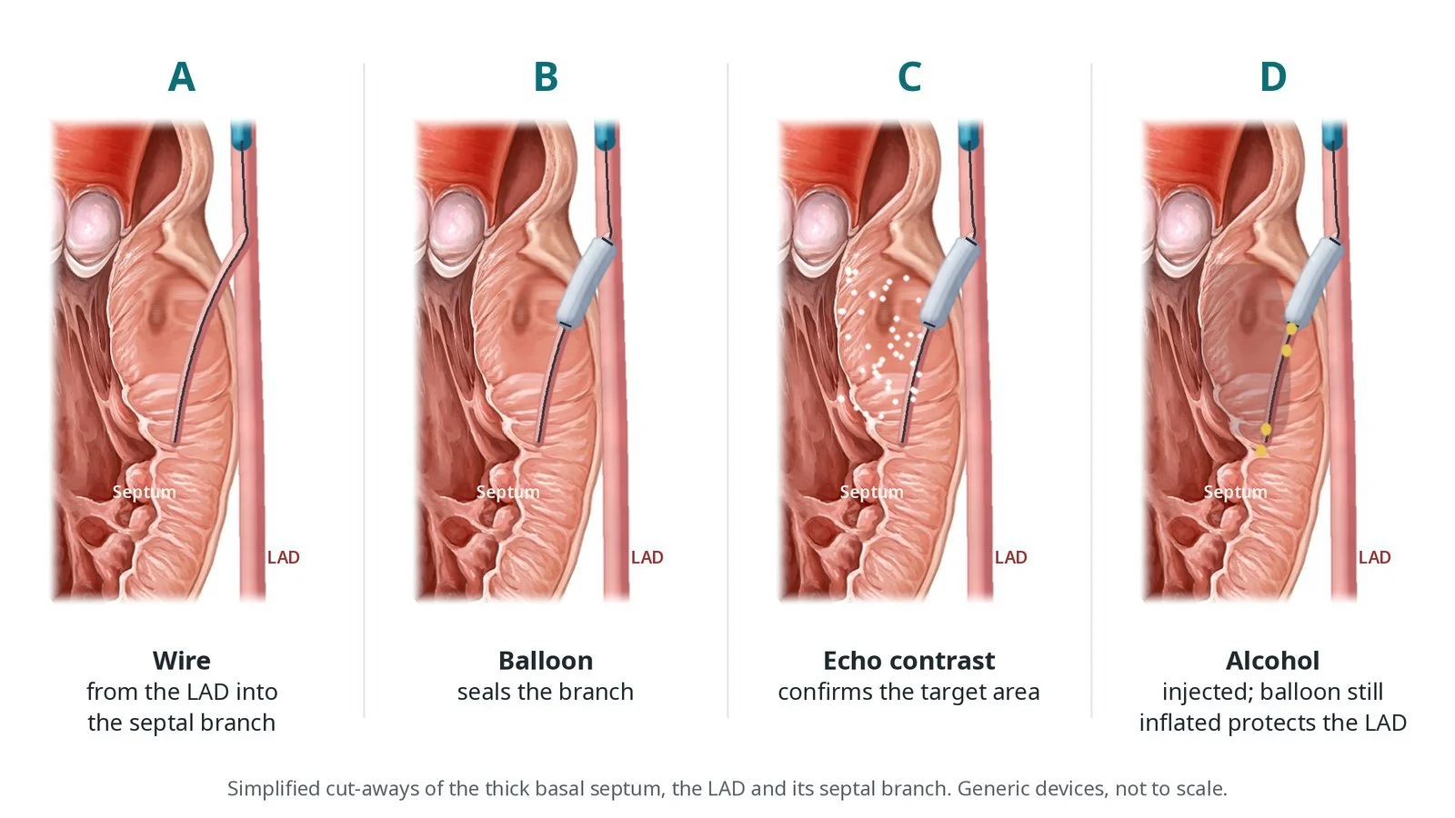
A: a wire goes from the LAD into the septal branch. B: a small over-the-wire balloon seals the branch. C: echo contrast confirms the branch feeds the bulge. D: alcohol is injected with the balloon still inflated, protecting the LAD.
5. Alcohol injection
Pain relief, an anti-sickness medicine and sedation are given first, as per centre protocol, because the alcohol causes chest pain. Dehydrated (absolute) alcohol is then injected slowly through the balloon. The volume is decided by the operator for the size of the branch and the area that lit up, as per centre protocol. The ECG is watched closely for heart block. The balloon stays inflated for several minutes afterwards, so alcohol cannot leak back into the LAD.
6. Checking the result and finishing
The balloon is deflated and removed, and an angiogram confirms that the septal branch is closed and the LAD is intact. Echo shows less movement in the targeted septum and remeasures the gradient. It often falls straight away, but the full effect builds over the following months. The arterial sheath is removed, and the wrist is closed with a compression band or the groin with a closure method per operator. The temporary pacing wire stays in.
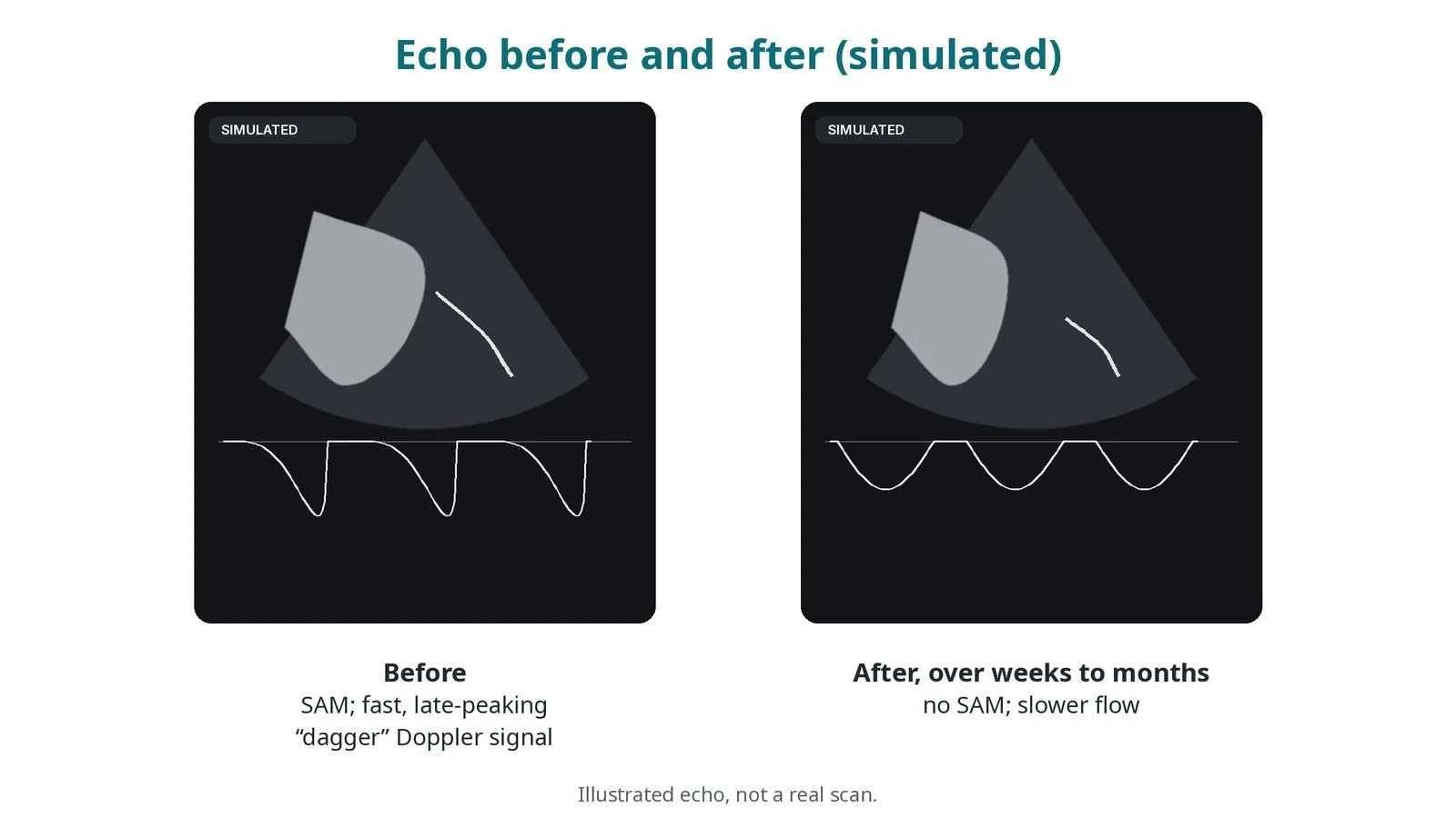
Illustrated echo, not a real scan. Before: SAM and a fast, late-peaking “dagger” Doppler signal (high gradient). After, over weeks to months: no SAM and slower flow (much lower gradient).
Recovery and aftercare
First days. Care is on a coronary care or high-dependency unit with continuous ECG monitoring. Blood tests show a rise in troponin, which is expected, because the treatment works by creating a small, controlled heart attack.
Temporary pacing wire. This usually stays in for a day or two and is removed if the heart rhythm is stable. Heart block can appear late, so monitoring continues afterwards.
Going home. Most people stay in hospital for several days. If heart block persists, a permanent pacemaker is fitted before discharge. See the Pacemaker guide.
Access site. Look after the wrist or groin puncture as for an angiogram, and avoid heavy use of that arm or leg for a few days.
Medicines. Your team will tell you which HCM medicines to continue. Some can be reduced later if symptoms improve.
Follow-up. A clinic review and an echo, often within a few months, to measure the gradient. Improvement usually continues for months as the septum thins. Some people need a second procedure or surgery if the gradient stays high.
Driving (UK). Ask your team about driving. If a pacemaker is fitted, the DVLA pacemaker rules apply. Check the current GOV.UK guidance.
Contact your team or NHS 111 if the access site swells, bleeds or becomes painful, or if you feel unwell. Call 999 for chest pain, fainting, a very slow heartbeat or severe breathlessness.
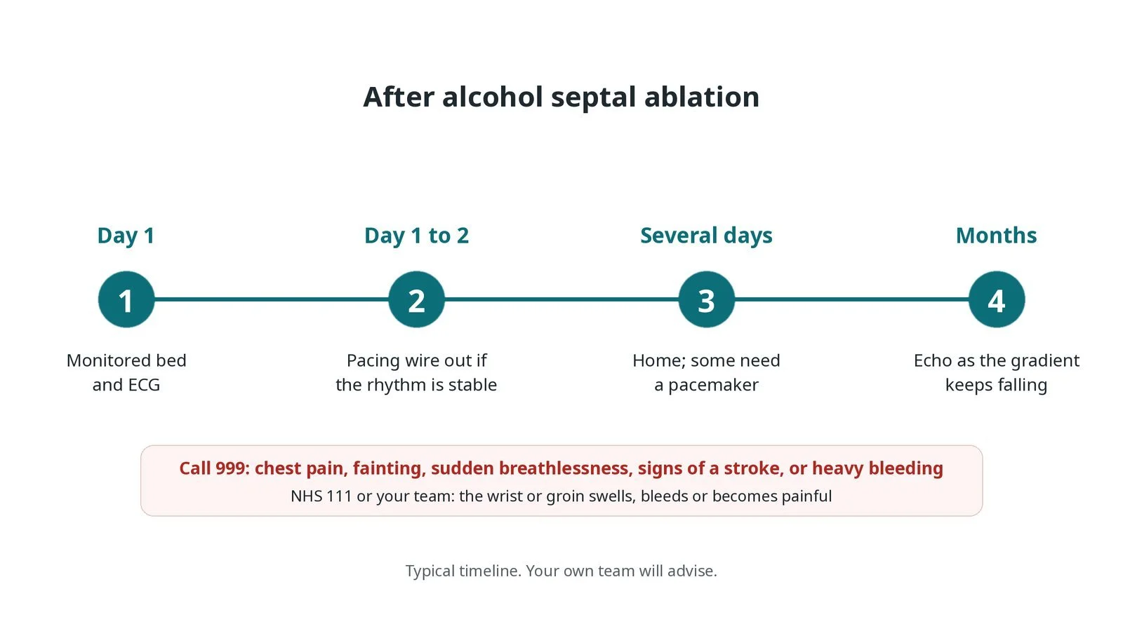
After alcohol septal ablation: (1) monitored bed and ECG on the day; (2) temporary pacing wire out after a day or so if the rhythm is stable; (3) home after several days, some with a pacemaker; (4) follow-up echo as the gradient keeps falling over months.
Risks and complications
In experienced centres alcohol septal ablation is usually safe and relieves symptoms for most suitable patients. Possible problems include:
Heart block needing a permanent pacemaker. This is the most common important complication: around 1 in 10 people in many published series, and more often when there is existing conduction disease. Right bundle branch block on the ECG afterwards is common.
Alcohol reaching the wrong area, such as the LAD, causing a larger heart attack. The sealed balloon and the echo contrast check are there to prevent this.
Dangerous heart rhythms during or soon after the procedure
Coronary artery injury from the wire or balloon
Fluid around the heart (tamponade), for example from the temporary pacing wire (uncommon)
Bleeding or bruising at the access site
Not enough gradient reduction, needing a repeat procedure or surgery
Death is rare in experienced centres
Your heart team will explain how these apply to you.
Cath lab kit list (printable)
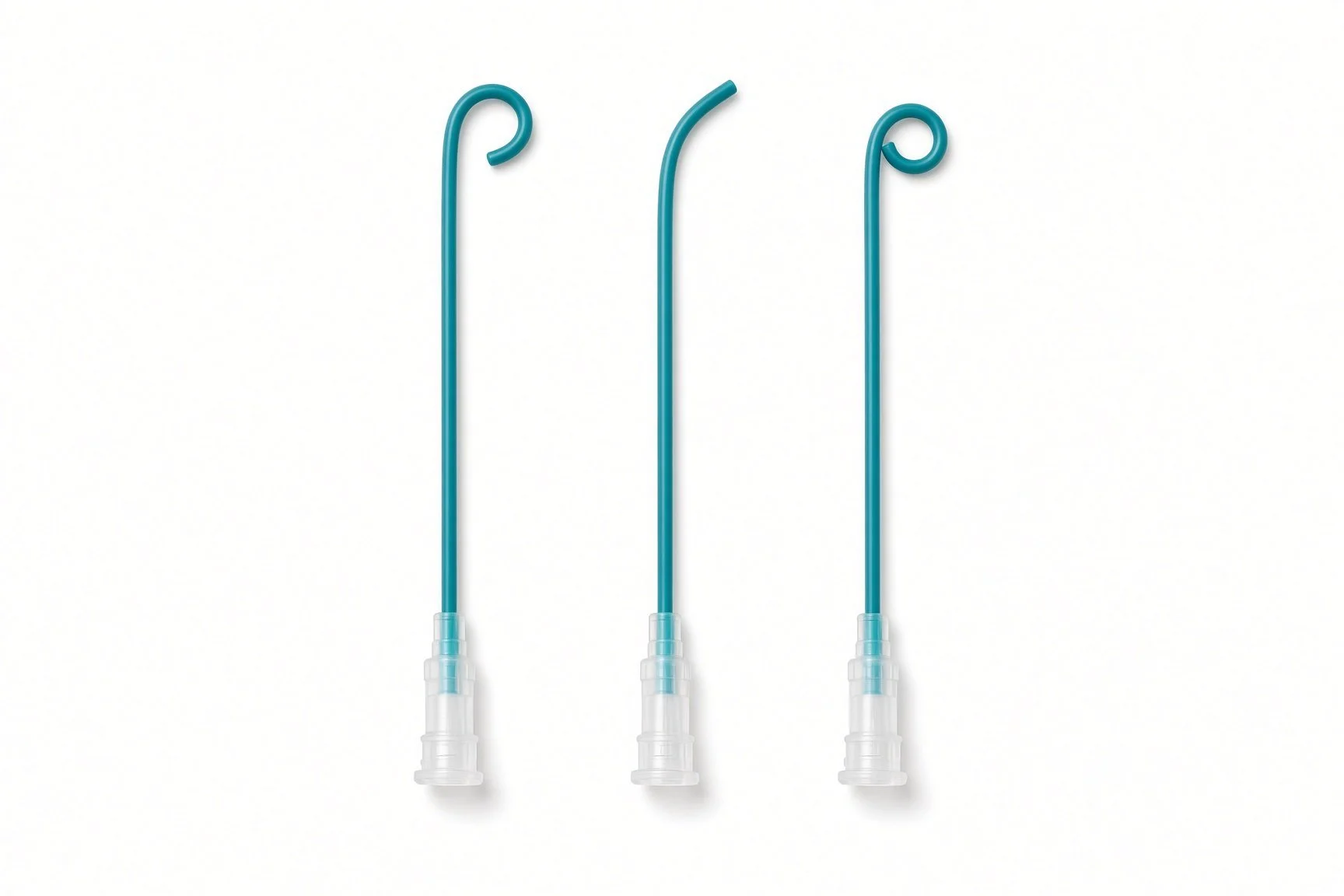
5 Fr right Judkins (JR4) diagnostic catheter, for the right coronary artery (from the guide kit list; no published kit-page picture)
Drugs, doses and dilutions: always as per centre protocol. Equipment, sizes and choices also vary by unit and operator. Device and product names below are generic. Always check the sheath, wire and balloon required in each product's instructions for use.
Example: Barts Heart Centre kit list — always follow your own centre protocol
Typical setup for alcohol septal ablation under local anaesthetic with mild to moderate sedation, shared as one real-world example. It is not a protocol for your unit.
Access and pacing
angiography pack
radial artery sheath (6 Fr in this example), per device and operator
venous sheath for the temporary pacing wire (neck or groin), per device and operator
temporary pacing wire (4 Fr in this example) and pacing box
non-absorbable suture to secure the pacing sheath, and a transparent film dressing
Catheters, wires and balloons
5 Fr right Judkins (JR4) diagnostic catheter, for the right coronary artery
6 Fr extra-backup left guide catheter (curve per operator)
PCI kit
0.014" floppy workhorse coronary guidewire (straight tip in this example), per operator
selection of small, short over-the-wire (OTW) balloons, sized to the septal branch per operator
contrast for angiography, and balloon inflation mix as per centre protocol
Echo and imaging
echo machine, with device interrogation if needed
echo (ultrasound) contrast agent, prepared as per centre protocol
portable ultrasound for vascular access, with a sterile probe cover
Alcohol
dehydrated (absolute) alcohol, stored, checked and drawn up as per centre protocol
small Luer-lock syringe for delivery through the balloon (volume decided by the operator, as per centre protocol)
Closure
radial compression device, or groin closure per operator
Medications (scrub nurse): sedation, analgesia, anti-emetic and anticoagulation as per centre protocol. No stent is placed, so antiplatelet loading is not routinely needed. Follow your centre protocol.
Procedural steps (Barts example)
Venous access (neck in this example) and temporary pacing wire to the right ventricle
Radial artery access
Diagnostic catheter: right coronary angiogram
Guide catheter to the left main stem; LAD angiogram to choose and size the septal branch
Wire the septal branch; advance the OTW balloon
Inflate the balloon; contrast through the central lumen to confirm the seal
Echo contrast through the central lumen; confirm the target area on echo
If the target is right: analgesia, anti-emetic and sedation as per centre protocol
Inject dehydrated alcohol slowly through the balloon (volume as per operator and centre protocol); balloon stays inflated
Deflate and remove; check angiogram of the septal branch and LAD
Repeat echo to reassess septal movement and the gradient
Remove the arterial sheath, apply a radial compression device; temporary pacing wire stays in; transfer to a monitored bed
First time in the cath lab?
💡 First-time tip
Watch the ECG closely, especially during the alcohol injection. Heart block can come on suddenly. Check the temporary pacing wire is capturing and that the pacing box is set and within reach before the alcohol goes in. Know the echo step. The contrast check decides whether the procedure goes ahead. Make sure the echo machine, contrast and operator are ready before the balloon goes up. Treat the alcohol like a high-risk drug. Check it with a second person, label the syringe clearly and keep it separate from flush and contrast syringes. Have pain relief and anti-sickness medicine drawn up as per centre protocol before the injection. The chest pain can be strong. Don't pull the pacing wire. It goes to the monitored bed with the patient. Secure it well and hand it over clearly.
FAQ
What does alcohol septal ablation mean?
In the cath lab, alcohol septal ablation is a catheter treatment for obstructive hypertrophic cardiomyopathy.
Is it the same as a cardiac ablation for an irregular heartbeat?
No. Catheter ablation for rhythm problems uses heat or cold to treat electrical pathways. Alcohol septal ablation uses alcohol in a coronary artery branch to shrink a thick part of the heart wall.
Am I awake for it?
Usually yes. Local anaesthetic is used at the access sites, with sedation and strong pain relief during the alcohol injection.
Will I feel better straight away?
Many people notice improvement soon, but the septum keeps thinning for months, so the full benefit can take time to show.
Why might I need a pacemaker afterwards?
The septum carries part of the heart's electrical wiring. Scarring there can cause heart block. Usually this is temporary, but around 1 in 10 people need a permanent pacemaker.
Alcohol septal ablation or myectomy: which is better?
Both reduce the blockage and relieve symptoms in most suitable people, and they have not been compared head to head in a large randomised trial. Surgery tends to give a larger, more reliable fall in gradient and avoids a pacemaker more often. Alcohol septal ablation avoids open-heart surgery and is a shorter recovery. The right choice depends on your anatomy, age, other heart problems and preference, and is made with a specialist team.
Related Cath Lab 101 guides
Sources
All checked on 10 October 2026. Each page's own date is shown where it gives one.
NICE. Non-surgical reduction of the myocardial septum (HTG18, formerly interventional procedures guidance IPG40). nice.org.uk/guidance/htg18 (published 25 Feb 2004; moved to HealthTech guidance with the recommendations unchanged)
Arbelo E, et al. 2023 ESC Guidelines for the management of cardiomyopathies. Eur Heart J 2023;44(37):3503–3626. PubMed 37622657
Ommen SR, et al. 2024 AHA/ACC/AMSSM/HRS/PACES/SCMR Guideline for the Management of Hypertrophic Cardiomyopathy. Circulation 2024;149(23):e1239–e1311. PubMed 38718139
NHS. Cardiomyopathy. nhs.uk/conditions/cardiomyopathy (page last reviewed 25 May 2023)
British Heart Foundation. Hypertrophic cardiomyopathy. bhf.org.uk (published 21 Jun 2024)
Cardiomyopathy UK. Asymmetrical septal hypertrophy with obstruction. cardiomyopathy.org (no date shown)
DVLA. Pacemakers and driving. gov.uk/pacemakers-and-driving
Disclaimer
Education only. This page is for general education for healthcare students, cath lab staff and curious readers. It isn't medical advice and doesn't replace advice from your own doctor, heart team or local unit protocols. Equipment and medicines are described generically. No product or manufacturer is recommended. If you think someone is having a heart emergency, call 999.
Disclosure: I work in the medical device industry. This page is independent education and isn't sponsored or reviewed by any company.
All diagrams on this page are original illustrations created for rodnieoro.com.