PCI (coronary angioplasty and stents): a Cath Lab 101 guide
Reviewed by Rodnie Oro, cath lab charge nurse · Last reviewed: October 2026 · Education only, not medical advice
PCI in one minute
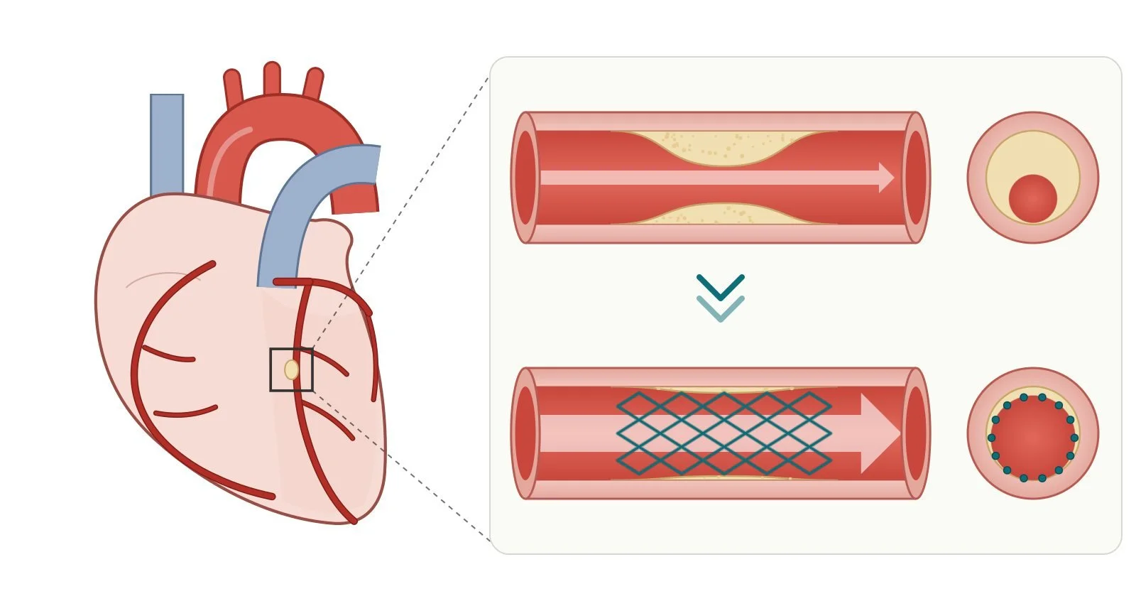
What it is: PCI (percutaneous coronary intervention) is a keyhole procedure that opens a narrowed or blocked heart artery. A thin tube goes in through the wrist (sometimes the groin), a tiny balloon widens the artery, and usually a stent (a small metal mesh tube) is left in to hold it open.
Who it's for: people with angina that medicines don't control, and people having a heart attack, when it's an emergency.
Main risks: bruising or bleeding where the tube went in is common. Serious problems, such as damage to an artery, heart attack, stroke, kidney injury or a reaction to the X-ray dye, are uncommon. Your cardiology team will explain your own risk.
Usually: awake, local anaesthetic, 30 minutes to 2 hours, home the same or next day if planned.
On this page: What is PCI? · Who is it for? · When is it done? · Step by step · Recovery · Risks · Kit list · First time in the lab? · FAQ
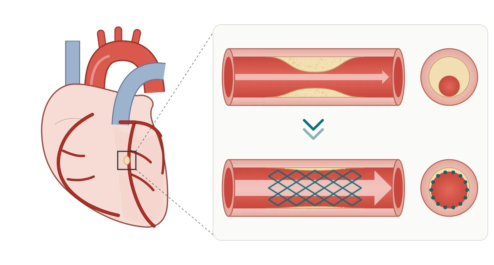
Top: plaque narrows a coronary artery and squeezes blood flow. Bottom: after PCI, a stent holds the artery open and flow is restored.
What is PCI?
Your heart muscle gets its blood from the coronary arteries. Over time, fatty deposits (plaque) can build up inside them and narrow them. This is called atherosclerosis, and it's the cause of coronary heart disease.
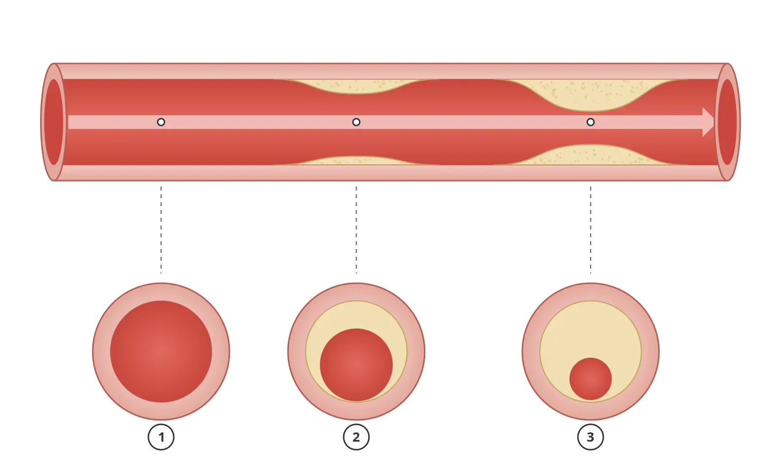
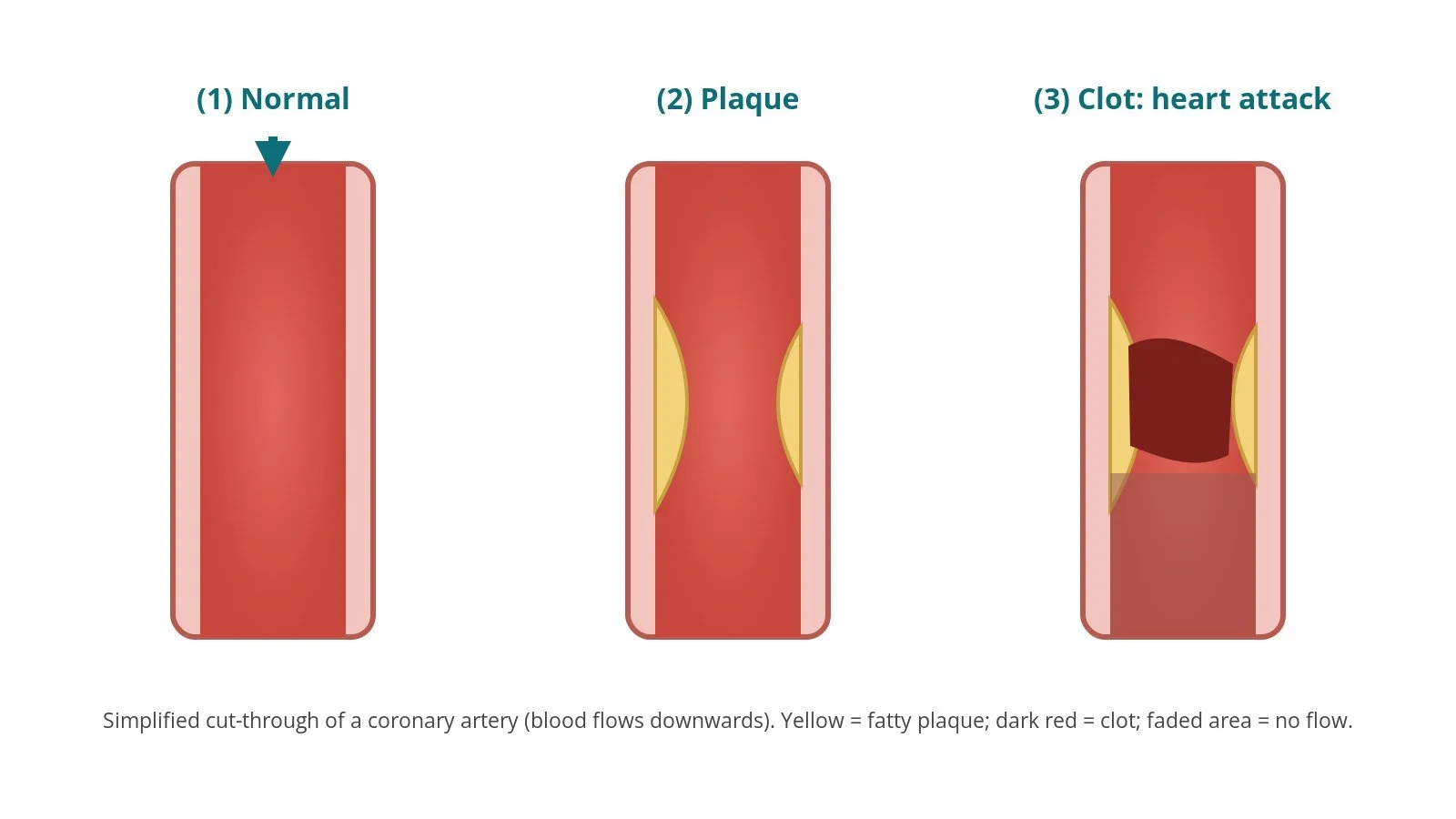
As plaque builds up, the channel for blood gets smaller: (1) healthy artery, (2) moderate narrowing, (3) severe narrowing that can cause angina.
PCI treats a narrowing from the inside. A cardiologist passes a fine guidewire across the narrowing. They then run a balloon along the wire and inflate it to squash the plaque back against the artery wall.
Arteries tend to spring back after ballooning, so a stent is usually placed. It arrives crimped onto a balloon, expands as the balloon inflates, and stays in for good. Most stents used today are drug-eluting: they slowly release a medicine that lowers the chance of the artery narrowing again.
You may also hear PCI called coronary angioplasty, angioplasty and stenting, or PTCA (percutaneous transluminal coronary angioplasty). New to the acronyms? See the Cath Lab Abbreviations A–Z.
Who is it for?
PCI is used for two main groups:
Planned (elective) PCI: people with angina (chest pain on effort) that hasn't settled with medicines and lifestyle changes.
Emergency PCI: people having a heart attack, to reopen a blocked artery as fast as possible and limit damage to the heart muscle. In a STEMI this is called primary PCI. More on this in Acute Myocardial Infarction (AMI).
PCI isn't right for everyone. With several narrowed arteries or complex anatomy, the heart team may recommend bypass surgery (CABG) or medicines alone.
PCI relieves angina and reopens arteries in a heart attack, but it doesn't cure the underlying disease. Medicines and lifestyle still matter afterwards.
When is it done?
PCI almost always follows a coronary angiogram: X-ray pictures of the arteries taken with contrast dye. If the angiogram shows a narrowing that's suitable and worth treating, PCI can happen:
straight away, on the same table, as one continuous procedure (common for heart attacks)
at a later planned date, after the heart team has discussed the options
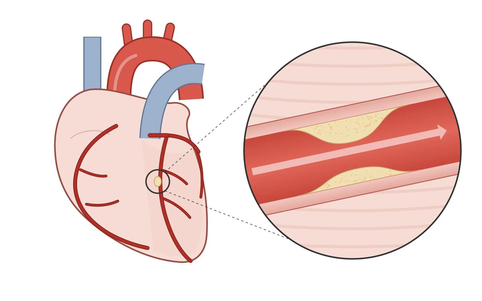
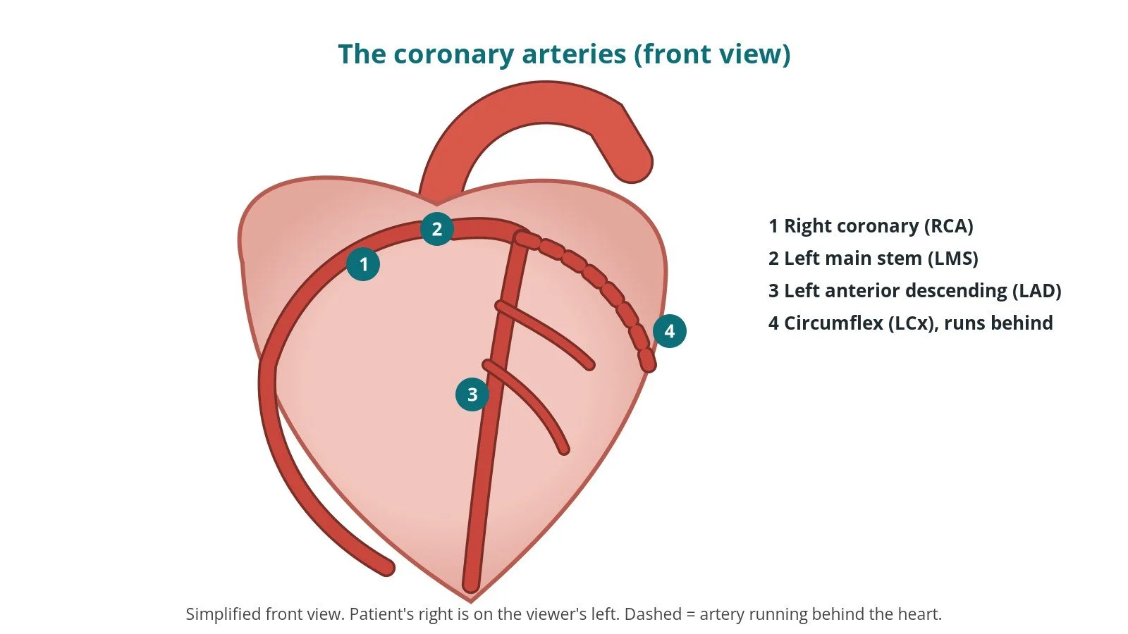
The coronary arteries run over the surface of the heart. The circle magnifies a narrowing caused by plaque, the target for PCI.
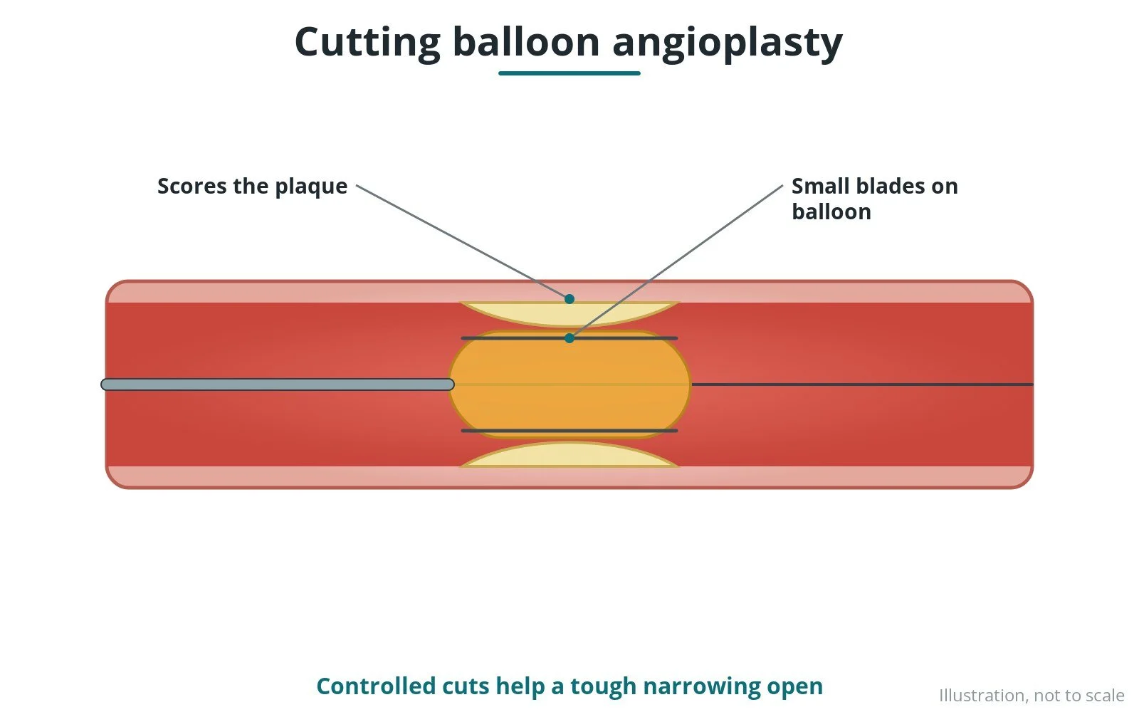
Some arteries need extra tools: heavily calcified narrowings (see intravascular lithotripsy, IVL) or long-standing complete blockages (see Chronic Total Occlusion (CTO PCI)).
Step by step
1. Preparation
The team checks bloods, allergies (especially to contrast dye) and medicines. Blood thinners and diabetes medicines may need adjusting, so follow your hospital's instructions. On the table: ECG stickers, blood pressure monitoring and a cannula. Most people are awake, with a mild sedative if needed.
2. Access: wrist or groin
After local anaesthetic, the operator places a short tube called a sheath into an artery, most often the radial artery in the wrist, and sometimes the femoral artery in the groin. In the UK the wrist is the most common route. Medicines may be given through the sheath to stop the artery going into spasm.
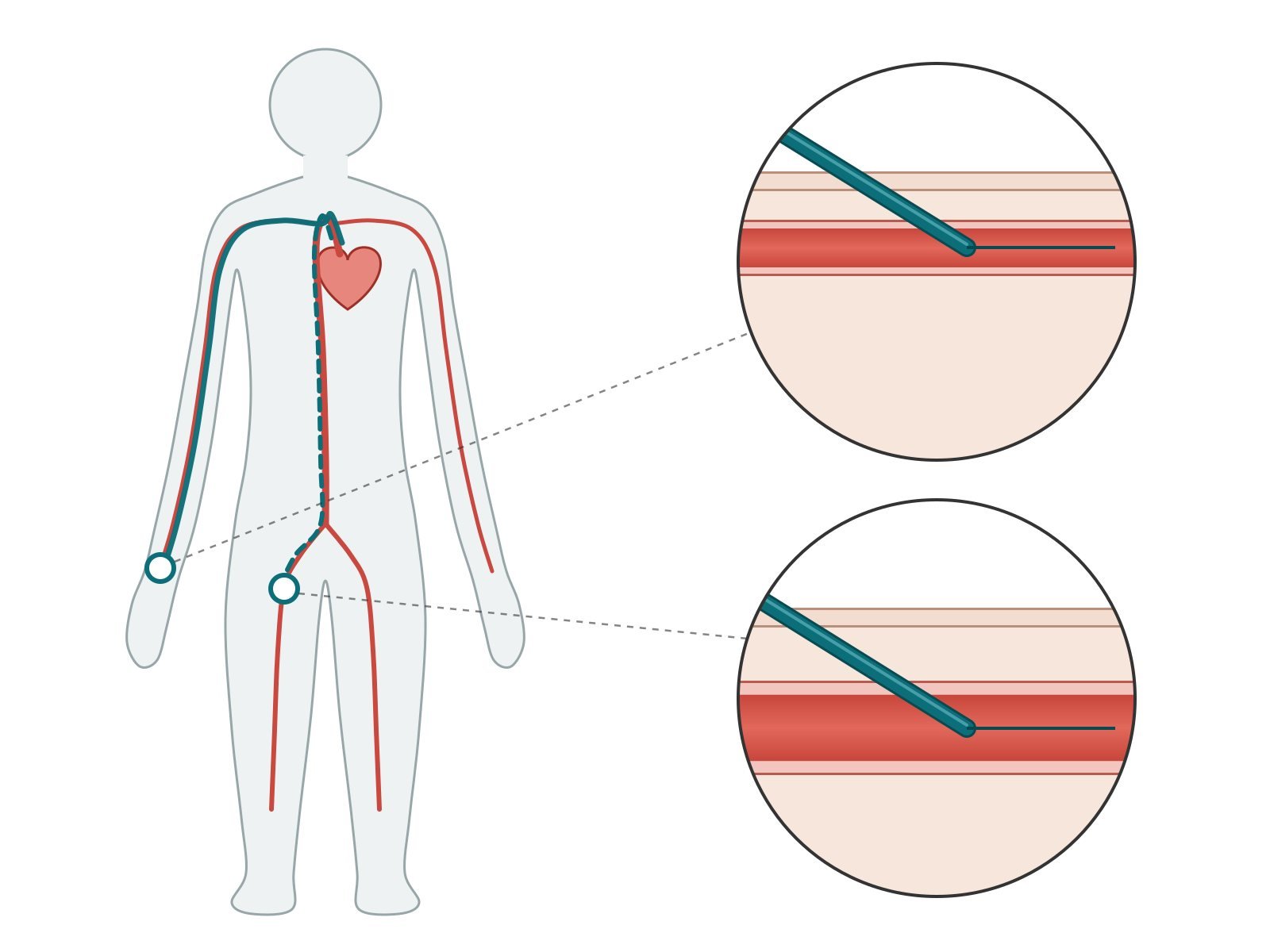
Wrist (radial) is the usual route (solid line). Groin (femoral) is used when needed (dashed line). Close-ups: the sheath enters the radial artery just under the skin at the wrist (top) and the larger, deeper femoral artery in the groin (bottom).
3. Wiring the artery
A guide catheter is steered up to the opening of the left or right coronary artery. Contrast pictures confirm the target. Blood thinners are given to prevent clots. A very fine guidewire is then steered across the narrowing and parked beyond it. From here on, the wire is the "rail" every other device runs on.
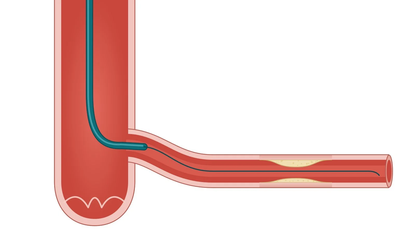
The guide catheter (thick teal tube) sits at the opening of the coronary artery. The guidewire (thin teal line) crosses the narrowing and becomes the rail for the balloon and stent.
4. Balloon and stent
A. Wire across: the guidewire sits across the narrowing.
B. Balloon: a balloon is passed over the wire and inflated in the narrowing (pre-dilation), usually for a few seconds at a time.
C. Stent: the stent on its delivery balloon is positioned and expanded against the artery wall.
D. Result: the balloon is withdrawn, leaving the stent holding the artery open. A high-pressure balloon may be used to fully expand it (post-dilation).
Patients may feel brief chest tightness while the balloon is up. This is expected and should be reported, so the team can reassure you or act on it.
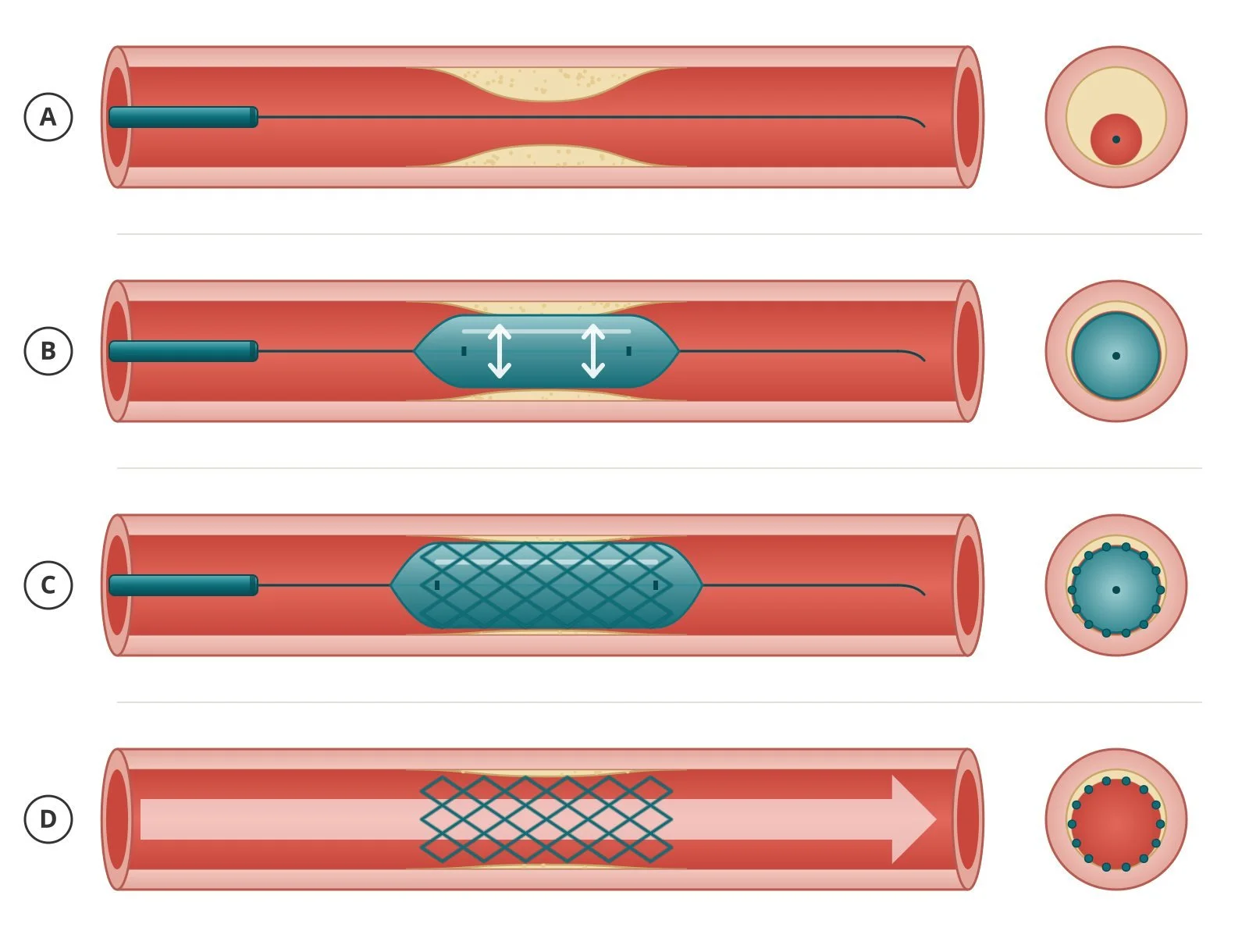
A: wire across the narrowing. B: balloon inflated to widen it. C: stent expanded on its balloon. D: balloon removed, stent left in place, blood flow restored. The circles show the artery in cross-section at each step.
5. Final checks and closure
Final contrast pictures check the flow and the result, sometimes with intracoronary imaging or pressure measurements. The wire and catheter come out. A wrist sheath is removed and a compression band is applied. Groin access may be closed with a closure device or by firm pressure.
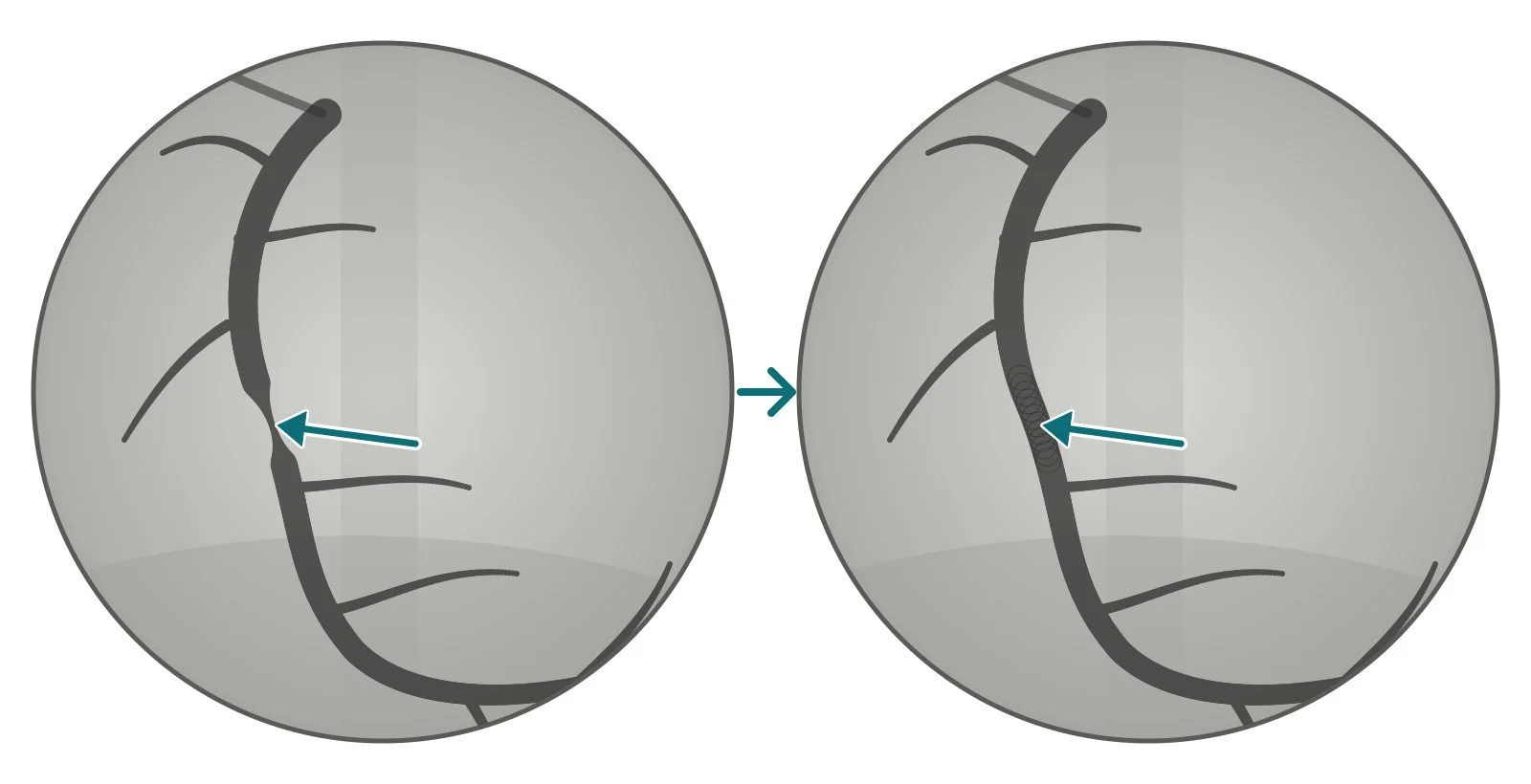
Illustration of what the final pictures show: before PCI, the arrow points to a tight narrowing; after PCI, the same artery is smooth and open.
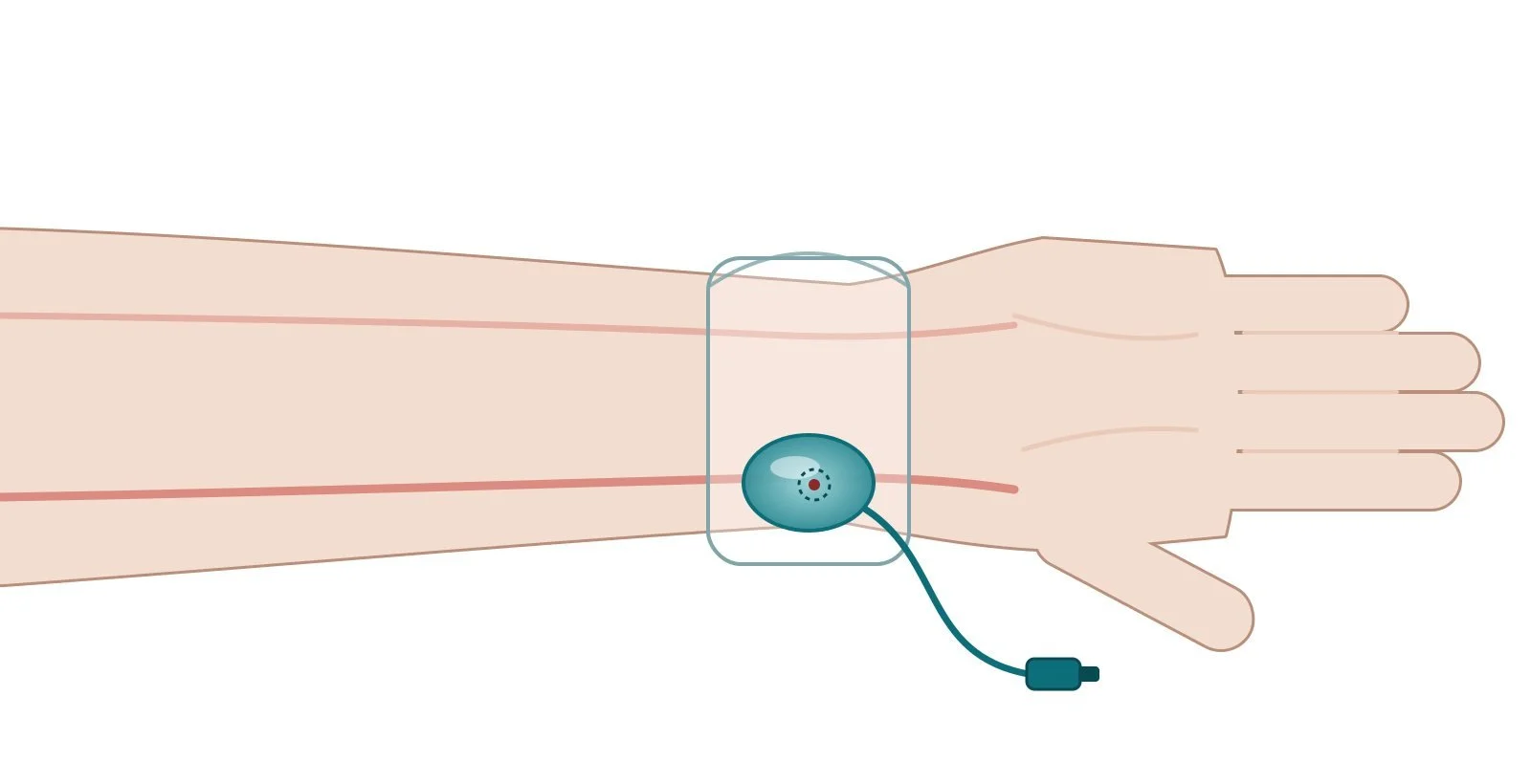
A compression band presses on the wrist puncture while the artery seals. The air in the band is then let out gradually.
Recovery and aftercare
Same day or next day: after a planned PCI, most people go home the same day or the next day. After a heart attack, the stay is usually a few days longer. Arrange a lift home. You shouldn't drive yourself.
The access site: a sore bruise for a few days is common. Watch for signs of infection.
Chest: some tightness or tenderness in the first day or so after PCI can be normal and usually settles. New or worsening chest pain is not normal. Call 999.
Activity and work: avoid heavy lifting and strenuous activity for about a week. After a planned PCI, many people are back at work in about a week. After a heart attack it takes longer.
Driving (UK): with a car or motorcycle licence you don't need to tell the DVLA, but you should stop driving for 1 week after a successful angioplasty, and for 4 weeks if angioplasty after a heart attack wasn't successful. Bus, coach and lorry drivers must tell the DVLA and stop driving for 6 weeks. Check with your doctor before you start driving again. See GOV.UK: Heart attacks, angioplasty and driving.
Medicines: after a stent you'll take antiplatelet medicines, often two together for a period of time (many people take them for up to a year), and usually aspirin long term. Never stop them early unless your cardiologist tells you to. Stopping early raises the risk of a clot in the stent.
Cardiac rehabilitation: ask about it, especially after a heart attack.
Seek urgent help (999) for chest pain like your angina or heart attack pain, especially with sweating, sickness or breathlessness. Also seek help for bleeding at the wrist or groin that won't stop with firm pressure, or for a hand or leg that turns cold, pale or numb.
Risks and complications
PCI is one of the most common heart procedures and is generally safe, but every procedure carries some risk. Your individual risk depends on your age, general health, kidney function and whether the procedure is planned or an emergency.
Common and usually minor
bruising, bleeding or soreness at the wrist or groin
brief chest discomfort during balloon inflation
Less common but serious
damage to the artery at the access site, or to a coronary artery
bleeding that needs treatment or transfusion
allergic reaction to the contrast dye
kidney injury (higher risk if kidney function is already reduced)
abnormal heart rhythms
heart attack, stroke or, rarely, death, or the need for emergency bypass surgery
Later risks
stent thrombosis: a clot blocking the stent, which is more likely if blood thinners are stopped early
restenosis: re-narrowing inside the stent. Drug-eluting stents reduce this risk.
For context, the British Heart Foundation puts the risk of major complications during angioplasty at usually less than one per cent. BCIS points out that many serious events around the time of PCI are caused by the heart disease itself rather than the procedure.
Cath lab kit list (printable)
Drugs, doses and dilutions: always as per centre protocol. Equipment, sizes and catheter shapes also vary by unit and operator preference. Device names below are generic. Use your unit's preferred products.
Example: Barts Heart Centre kit list — always follow your own centre protocol
Typical setup for a radial PCI, shared as one real-world example. It is not a protocol for your unit.
Sterile table
angiography pack, saline spike, 2 × skin-prep applicators
500 mL sterile saline
transparent film dressing (to secure a radial sheath)
6 Fr radial sheath (or 6 Fr femoral sheath for groin access)
haemostasis valve with short extension, balloon inflation device
balloon contrast mix 50:50 (contrast : saline), 30 mL Luer-lock syringe
Diagnostic catheters (check shots)
Left coronary: 6 Fr JL3.5 (radial) or JL4 (femoral). Alternatives: JL3, JL5, JL6, AR, MP
Right coronary: 6 Fr JR4. Alternatives: WR (Williams right), AL1–2, AR1–3, MP
Single-catheter option: 5 Fr dual-coronary diagnostic catheter for both coronaries
Tortuosity or spasm: 0.035" hydrophilic guidewire 150 cm, 0.035" exchange wire 260 cm, 0.014" coronary guidewire
Guide catheters (intervention)
Left coronary: 6 Fr extra-backup 3.5 curve
Right coronary: 6 Fr JR4
Coronary devices (sized to the vessel)
0.014" workhorse coronary guidewire (operator choice)
semi-compliant balloon (pre-dilation) and non-compliant balloon (post-dilation)
drug-eluting stent
IV drug line: 500 mL saline, long extension line, 3-way tap, 20 mL Luer-lock syringe, labelled 2 mL or 5 mL Luer-lock syringe for sedation, blunt drawing-up needle, giving set
Medications (scrub nurse):
lidocaine 1% (5 mL ampoule for radial, 20 mL for femoral)
heparin 10,000 units/10 mL
isosorbide dinitrate 10 mg/10 mL ampoule (2 mg/2 mL diluted with 8 mL saline = 200 micrograms/mL)
verapamil (radial anti-spasm): dose and dilution as per centre protocol
Sedation and analgesia (circulating nurse, as prescribed):
midazolam 2 mg/2 mL, given in 0.5 mg increments
morphine 10 mg/2 mL, given in 2.5 mg increments
Aortogram or femoral angiogram: 6 Fr pigtail catheter, high-pressure tubing and syringe, power injector, forceps (tapping out air, securing connections)
Closure: radial compression band (regular or large) · femoral vascular closure device (6 Fr or 8 Fr) or manual compression
Common views: RCA: LAO 30 · LAO 20/CRAN 20 or PA CRAN 30 · RAO 30. LCA: PA CAUD 40 · LAO 40/CAUD 40 ("spider") · LAO 10/CRAN 40 · RAO 40/CRAN 40 · RAO 30/CAUD 30
First time in the cath lab?
💡 First-time tip
Lead goes on before you go in. Find a lead apron and thyroid collar that fit, and wear your dosimeter where your unit says. Learn the sterile field first. Ask the scrub nurse where you can stand. If you're unsure, keep your hands to yourself and stay a step back. Watch the patient, not just the screen. Pain, nausea, and changes in rhythm or pressure are often noticed first by the nurse at the head end. Speak up. In a good lab "Can I ask why…?" is always welcome, and "Stop" from anyone is always heard. Expect the jargon. Keep the abbreviations glossary open on your phone.
FAQ
Is PCI painful?
You're usually awake, with local anaesthetic at the wrist or groin, so you shouldn't feel sharp pain. Many people feel pressure, and brief chest tightness while the balloon is inflated. Tell the team. A mild sedative can be given.
How long does it take?
Usually 30 minutes to 2 hours. Complex arteries can take longer. Add time for preparation and recovery.
Is PCI the same as angioplasty?
Mostly, yes. Angioplasty means opening an artery with a balloon. PCI is the umbrella term for treating the artery from inside, usually balloon plus stent.
Can a stent move or be felt?
A stent is pressed into the artery wall, and over time the artery lining grows over it. It can't be felt.
How long will I take blood thinners?
This varies with the reason for the PCI and your bleeding risk. Your cardiologist decides, and many people take two antiplatelets for up to a year. Don't stop them without speaking to your cardiology team.
Can I have an MRI scan with a stent?
Yes, in almost all cases. NHS MRI safety guidance notes that no coronary stent has ever been shown to be a hazard in 1.5 T or 3 T MRI scanners, and its policy allows scanning even straight after a stent is fitted. Always tell the MRI department as soon as possible that you have a stent, and bring your stent card if you were given one, because local policies can vary. (NHS GGC MRI Physics; BHF)
Related Cath Lab 101 guides
Sources
All checked on 8 October 2026. Each page's own date is shown where it gives one.
NHS. Coronary angioplasty and stent insertion: overview, how it's performed, recovery, risks. nhs.uk/tests-and-treatments/coronary-angioplasty (page last reviewed 4 Oct 2022)
British Heart Foundation. Coronary angioplasty and stents (PCI). bhf.org.uk (published 27 May 2025, next review May 2028)
British Cardiovascular Intervention Society. The PCI procedure. bcis.org.uk (no date shown)
Mayo Clinic. Coronary angioplasty and stents. mayoclinic.org (20 May 2026)
Cleveland Clinic. Percutaneous coronary intervention (PCI). my.clevelandclinic.org (updated 10 Aug 2026)
MedlinePlus (US National Library of Medicine). Angioplasty and stent placement – heart. medlineplus.gov (reviewed 14 Jul 2024)
GOV.UK (DVLA). Heart attacks, angioplasty, and driving. gov.uk/heart-attacks-and-driving (no review date shown)
NHS Greater Glasgow and Clyde MRI Physics. MRI safety implant policy for patients with coronary stents. mriphysics.scot.nhs.uk (no date shown)
British Heart Foundation. Cardiac MRI scan. bhf.org.uk (published 25 Sep 2023)
Disclaimer
Education only. This page is for general education for healthcare students, cath lab staff and curious readers. It isn't medical advice and doesn't replace advice from your own doctor, cardiology team or local unit protocols. Equipment and medicines are described generically. No product or manufacturer is recommended. If you have chest pain now, call 999.
Disclosure: I work in the medical device industry. This page is independent education and isn't sponsored or reviewed by any company.
All diagrams on this page are original illustrations created for rodnieoro.com.