TCPC (Fontan) ballooning and stenting: a Cath Lab 101 guide
Reviewed by Rodnie Oro, cath lab charge nurse · Last reviewed: October 2026 · Education only, not medical advice
TCPC stenting in one minute
What it is: a total cavopulmonary connection (TCPC) is the final stage of the Fontan operation for people born with only one working pumping chamber. Blue blood from the body flows straight into the lung arteries without passing through a pump. If part of this pathway narrows, a balloon can stretch it and a stent (a metal mesh tube, sometimes covered with fabric) can hold it open. This is done with catheters passed up a vein.
Who it is for: children and adults with a Fontan circulation where tests show a narrowing in the conduit, the lung arteries or the upper (Glenn) connection that is slowing blood flow, decided by a specialist congenital heart team.
Main risks: bleeding or bruising at the vein, a tear in the vessel, the stent moving, clots and irregular heart rhythms. Your team will explain your own risk.
Usually: under general anaesthetic, guided by X-ray and often echo, about 1 to 3 hours, usually one night in hospital.
On this page: What is a TCPC? · Who is it for? · When is it done? · Step by step · Recovery · Risks · Kit list · First time in the lab? · FAQ
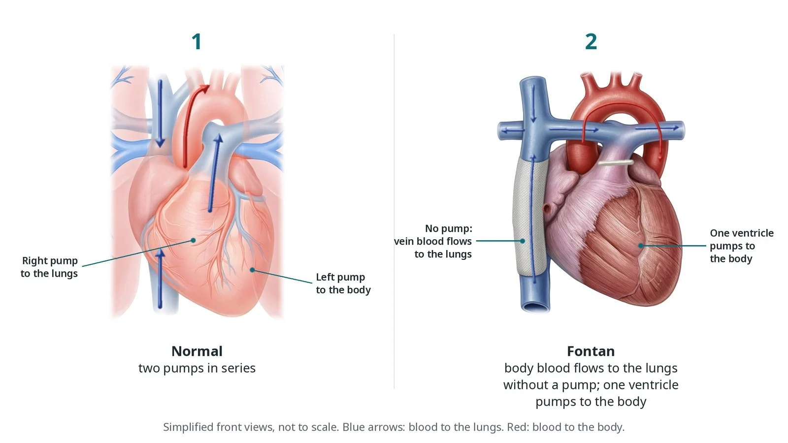
(1) Normal: two pumps in series. (2) Fontan: blood from the body flows passively to the lungs; one ventricle pumps to the body.
What is a TCPC?
In a normal heart, two pumps work in series. The right ventricle pumps blue blood to the lungs, and the left ventricle pumps red blood to the body.
Some babies are born with only one ventricle big enough to pump, for example with hypoplastic left heart syndrome or tricuspid atresia. Surgeons rebuild the circulation in stages so the single ventricle pumps to the body, while blue blood reaches the lungs passively, without a pump:
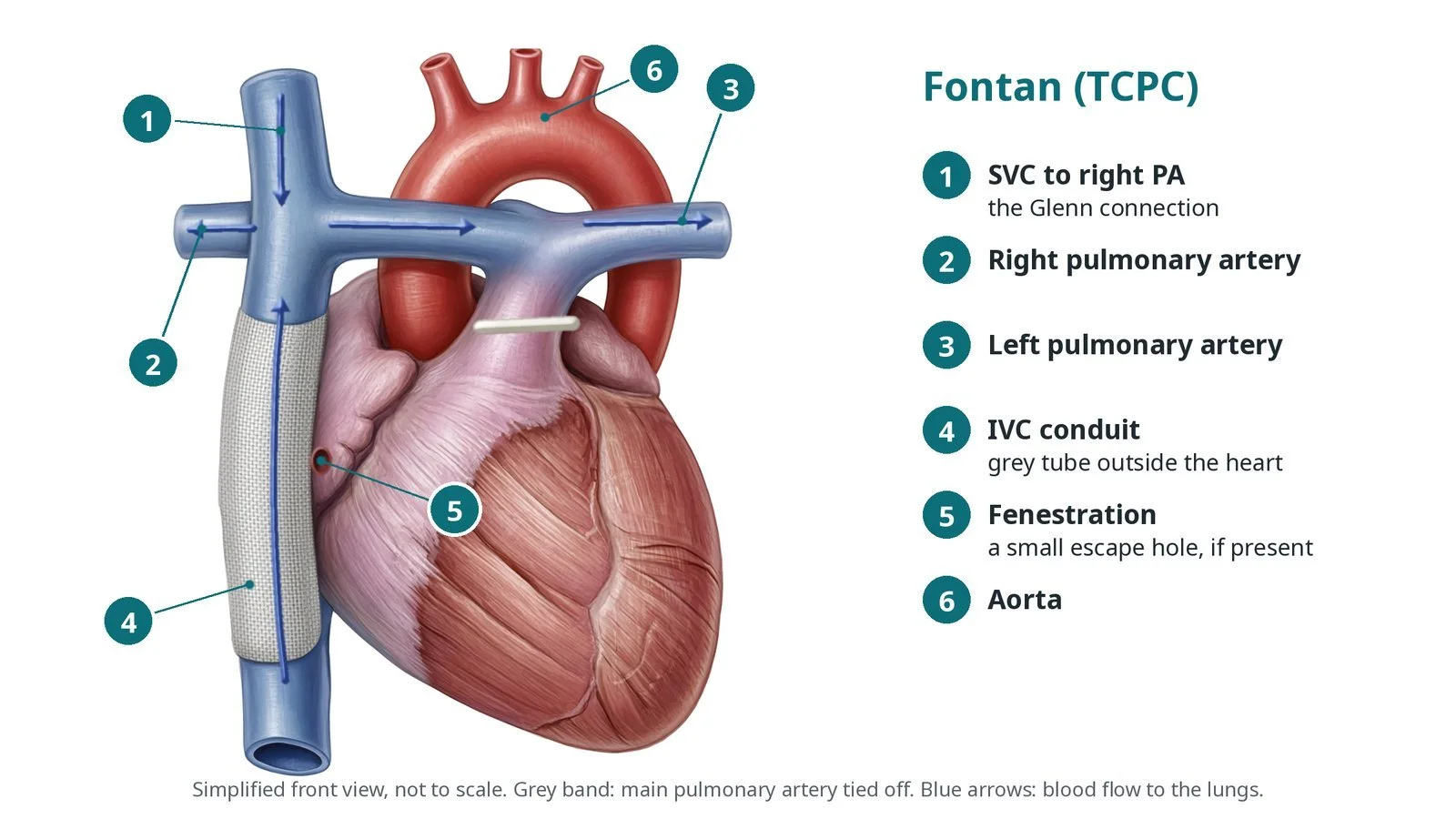
Glenn (bidirectional cavopulmonary connection): the superior vena cava (SVC), which drains the head and arms, is joined to the right pulmonary artery.
Fontan / TCPC: blood from the inferior vena cava (IVC), which drains the lower body, is routed to the pulmonary arteries too, usually through a tube called an extracardiac conduit, or through a lateral tunnel made inside the right atrium.
Many people have a small hole called a fenestration between the conduit and the atrium. It acts as a pressure "pop-off" valve, at the cost of a little blue blood reaching the body.
Because nothing pumps blood through the lungs, even a mild narrowing raises the pressure in the veins and lowers the flow to the lungs. That is why a narrowing that would not matter elsewhere can matter in a Fontan circulation.
New to the acronyms? See the Cath Lab Abbreviations A–Z.
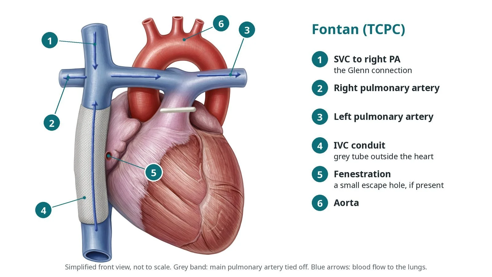
Fontan (TCPC) anatomy: (1) SVC to right PA (Glenn); (2) right and (3) left pulmonary arteries; (4) IVC conduit; (5) fenestration; (6) aorta.
Who is it for?
People with a Fontan circulation are followed for life by a specialist adult or paediatric congenital heart disease (ACHD/CHD) team. A catheter procedure may be considered when:
imaging (echo, MRI or CT) shows a narrowing of the conduit or lateral tunnel, the Glenn connection or the pulmonary arteries
a cardiac catheter study confirms a meaningful pressure difference (gradient) across the narrowing, or the narrowing looks likely to limit flow
there are signs of a failing Fontan, such as tiredness, swelling, fluid in the abdomen, liver changes, or protein loss from the gut, where a narrowing could be contributing
The same catheter study may also include other treatments, such as closing a fenestration or blocking abnormal collateral vessels, as decided by the team. The ESC 2020 guidelines for adult congenital heart disease recommend that people with a Fontan circulation are cared for in specialist centres, with regular imaging and catheter assessment when there are concerns.
When is it done?
It is a planned procedure in most cases. Before it, the team usually:
reviews recent echo, MRI or CT to plan the route and stent size
checks blood tests, including clotting and liver and kidney function
reviews anticoagulant or antiplatelet medicines, which most Fontan patients take, and gives instructions on when to stop them, as per centre protocol
plans the anaesthetic carefully. Breathing on a ventilator changes Fontan blood flow, so an anaesthetist experienced in congenital heart disease is involved
discusses the aims, the risks and the possibility of needing surgery instead
Step by step
1. Anaesthetic and set-up
The procedure is usually done under general anaesthetic, often with transoesophageal echo (TOE). Biplane X-ray (two cameras at once) helps show the 3D anatomy with less contrast. Antibiotics are given as per centre protocol.
2. Venous access and pressures
Access is from the femoral vein in the groin, under ultrasound. Sheath sizes are per device and operator. Sometimes the jugular vein in the neck is used as well, to reach the Glenn connection from above.
From the femoral vein, catheters pass up the IVC into the conduit and on to the pulmonary arteries. There is no right heart to pass through.
Pressures are measured along the pathway (a "pullback"), and angiograms (contrast pictures) show the narrowing. Anticoagulation (heparin) is given, dose as per centre protocol.
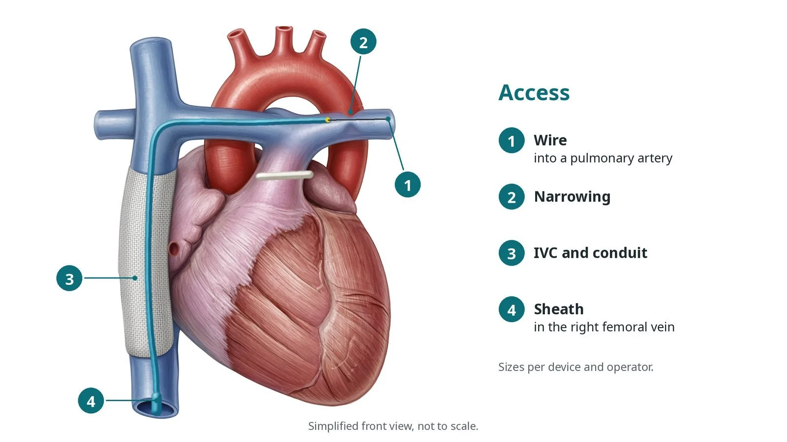
Access: (1) wire into a pulmonary artery; (2) narrowing; (3) IVC and conduit; (4) sheath in the right femoral vein.
3. Ballooning and stenting
A. Cross the narrowing. A catheter and wire are steered across the narrowing into a pulmonary artery branch. The wire is exchanged for a stiff exchange wire that supports the larger equipment.
B. Size it. The operator measures the vessel on the angiogram and imaging. A soft sizing balloon may be gently inflated to see the "waist" of the narrowing. Balloon sizes are per device and operator.
C. Place the stent. A long sheath is passed across the narrowing. The stent, often a covered stent to seal the vessel wall if it tears, is mounted on a two-layer (inner and outer) balloon and positioned using angiograms through the sheath. The inner balloon is inflated first to open the stent partly, so its position can be checked and adjusted, then the outer balloon expands it fully. Inflation volumes are per device and operator.
D. Post-dilate and check. A high-pressure balloon may be used to fully expand the stent. Pressures and angiograms are repeated to check the narrowing has gone and that there is no leak or tear.
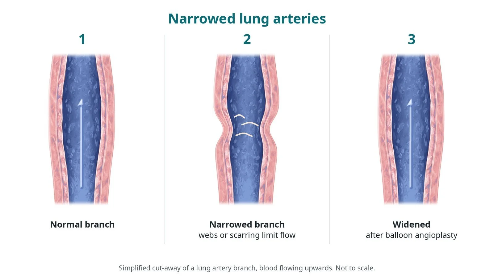
If a narrowing is treated with a balloon alone, the vessel is simply stretched without leaving a stent. The choice depends on the type of narrowing and the person's size.
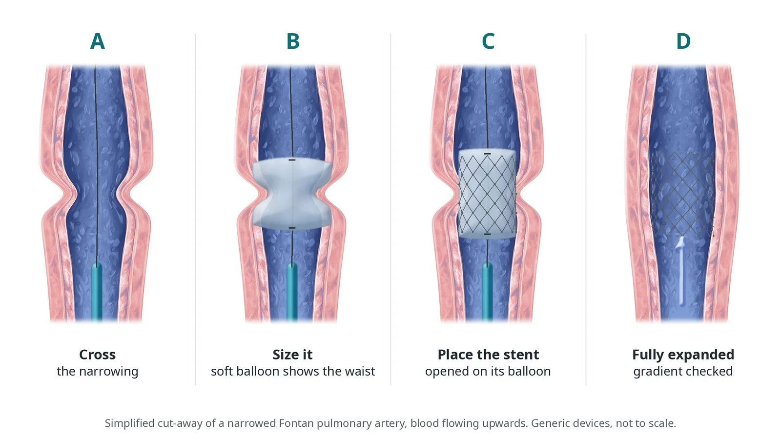
A: cross the narrowing. B: size it with a balloon. C: open the stent on its balloon. D: stent fully expanded.
4. Finishing
The sheaths are removed, and the vein is closed with pressure or a skin stitch, per operator. The person wakes up in recovery.
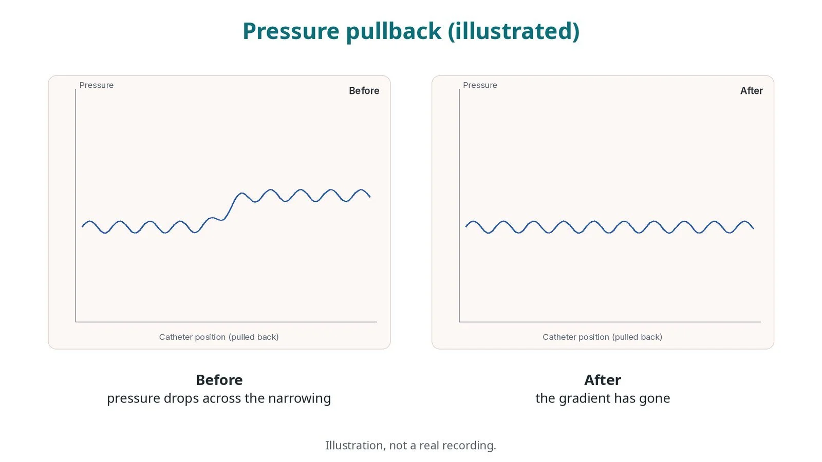
Illustrated pressure pullback, not a real recording. Before: pressure drops across the narrowing. After: the gradient has gone.
Recovery and aftercare
First hours: bed rest for a few hours with checks of the groin, blood pressure, oxygen levels and heart rhythm, as per centre protocol.
Going home: usually the next day after a check, which may include a chest X-ray or echo.
Blood thinners: most people restart their anticoagulant or antiplatelet medicine, sometimes with an extra antiplatelet for a while after a stent, as advised by your team.
Activity: avoid heavy lifting and strenuous exercise for about a week, or as advised.
Follow-up: with the congenital heart team, usually including imaging to check the stent.
Driving: DVLA rules depend on the underlying heart condition. Ask your team before driving, especially after a general anaesthetic.
Call 999 if you have chest pain, sudden breathlessness, fainting, or heavy bleeding from the groin that does not stop with firm pressure.
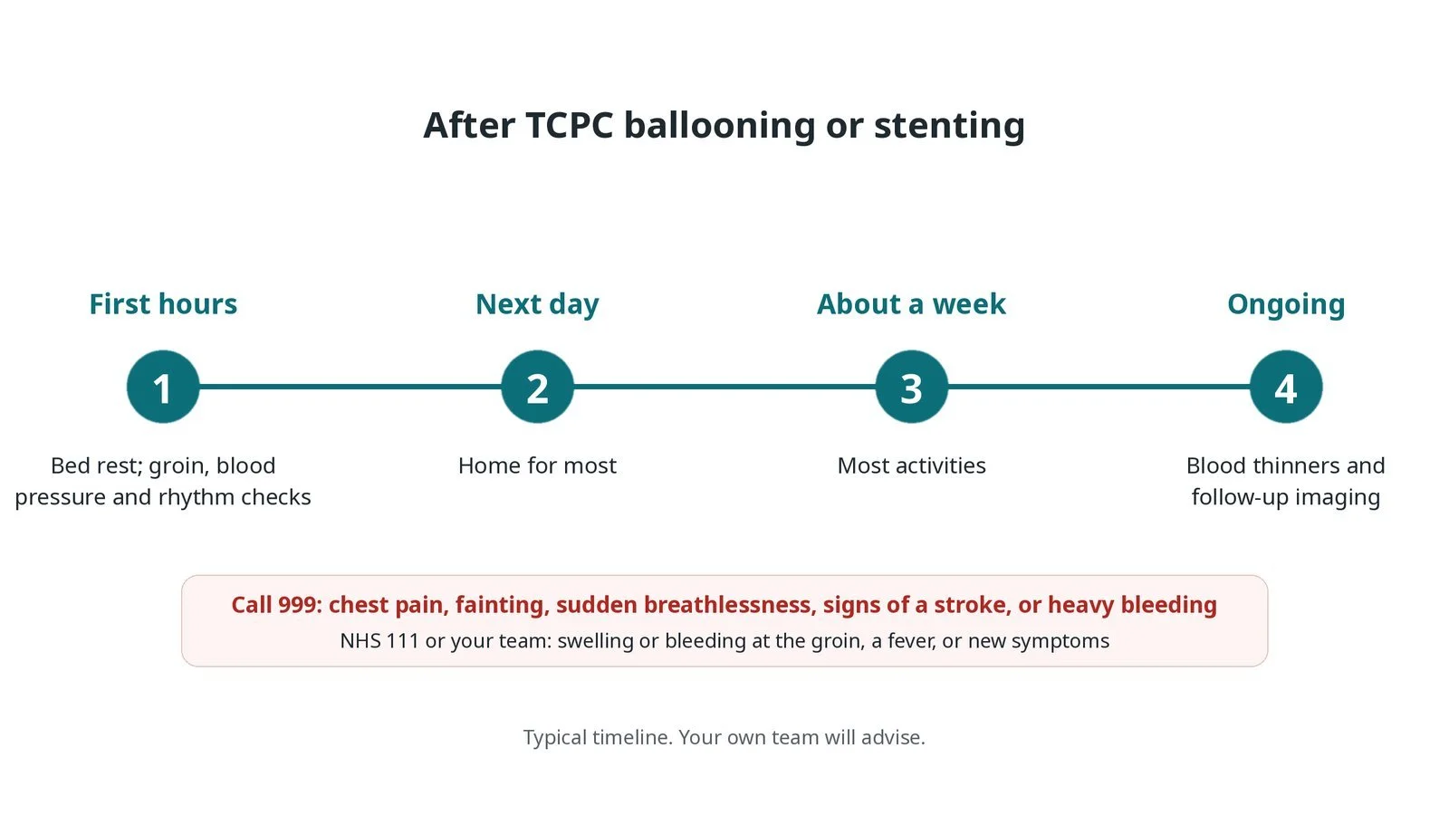
After TCPC ballooning or stenting: (1) bed rest and checks; (2) home for most; (3) most activities after about a week; (4) blood thinners and follow-up imaging.
Risks and complications
Your team will discuss your own risks. Possible problems include:
bleeding, bruising or a clot at the vein in the groin
a tear in the conduit or vessel; this is one reason covered stents are often used
the stent moving out of position (embolisation), which may need retrieval
irregular heart rhythms, usually short-lived
clots on the stent or in the Fontan pathway
reaction to contrast or the anaesthetic, and kidney strain from contrast
rarely, the need for urgent surgery
Cath lab kit list (printable)
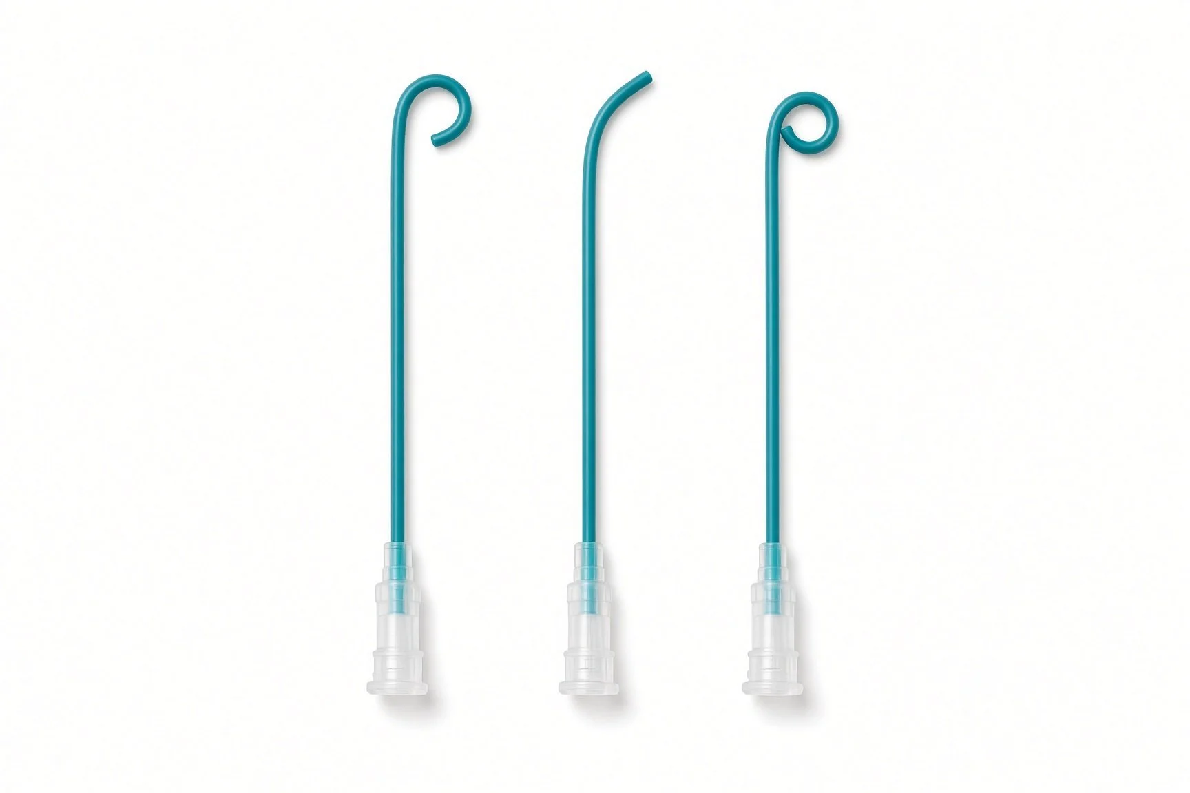
Diagnostic catheters: right coronary-shaped, multipurpose-shaped and pigtail catheters, per operator (from the guide kit list; no published kit-page picture)
Barts example kit list
An example from one UK centre, written generically. Every lab has its own protocol, and sizes and doses vary.
Access and set-up
femoral venous sheaths, sizes per device and operator
ultrasound probe and sterile probe cover
local anaesthetic, as per centre protocol
saline flush bag and flush set
heparin, as per centre protocol
non-absorbable suture for a figure-of-eight (Z) skin stitch, per operator
Catheters and wires
diagnostic catheters: right coronary-shaped, multipurpose-shaped and pigtail catheters, per operator
stiff exchange guidewire, J-tip, length per operator
Sizing
soft compliant sizing balloon, size per device and operator
contrast and saline mix for balloon inflation, as per centre protocol
3-way taps and Luer-lock syringes of several sizes
Stenting
long sheath, size per device and operator
two-layer (inner and outer) stent-delivery balloon, size per device and operator, with labelled syringes for the inner balloon, outer balloon and flush
covered stent, size per device and operator
high-pressure balloon for post-dilatation, size per device and operator
Imaging
biplane X-ray, TOE probe and echo machine, power injector for angiograms
Procedural steps (Barts example)
General anaesthetic; TOE probe placed
Femoral venous access under ultrasound; anticoagulation as per centre protocol
Pressures and angiograms along the Fontan pathway
Narrowing crossed; stiff exchange wire placed in a pulmonary artery branch
Sizing balloon, if used
Long sheath placed; stent positioned with angiograms through the sheath
Inner balloon then outer balloon inflated; position checked
Post-dilatation with a high-pressure balloon, if needed
Repeat pressures and angiograms
Sheaths removed; vein closed per operator; transfer to recovery
First time in the cath lab?
💡 First-time tip
Fontan patients are different. There is no pump to the lungs. Low blood pressure, dehydration and high ventilator pressures all reduce flow, so tell the team about changes straight away. Label the balloon syringes. Two-layer balloons need separate syringes for the inner and outer balloons. Label them clearly before the case. Have the covered-stent backup ready. Know where covered stents and retrieval snares are kept before the case starts. Watch the oxygen levels. Many Fontan patients normally run oxygen saturations in the low 90s or below. Know their usual level. Keep air out. Air in the venous lines can cross a fenestration to the body.
FAQ
Is this open-heart surgery?
No. It is a catheter procedure through a vein in the groin, so there is no cut in the chest.
Will the stent need replacing?
Stents stay in for life. In children, a stent can sometimes be stretched further with a balloon as they grow, depending on the stent.
Can I have an MRI scan afterwards?
Many stents are labelled MR conditional, which means a scan is possible under set conditions. Always tell the scanning team about your stent.
Will I still need blood thinners?
Most people with a Fontan circulation already take an anticoagulant or antiplatelet. Your team will tell you if anything changes after the stent.
Related Cath Lab 101 guides
Sources
All checked on 10 October 2026. Each page's own date is shown where it gives one.
Baumgartner H, et al. 2020 ESC Guidelines for the management of adult congenital heart disease. Eur Heart J 2021;42(6):563–645. PubMed 32860028
NHS. Congenital heart disease. nhs.uk/conditions/congenital-heart-disease
British Heart Foundation. Congenital heart conditions. bhf.org.uk
DVLA / GOV.UK. Cardiovascular disorders: assessing fitness to drive. gov.uk
Disclaimer
Education only. This page is for general education for healthcare students, cath lab staff and curious readers. It isn't medical advice and doesn't replace advice from your own doctor, heart team or local unit protocols. Equipment and medicines are described generically. No product or manufacturer is recommended. If you think someone is having a heart emergency, call 999.
Disclosure: I work in the medical device industry. This page is independent education and isn't sponsored or reviewed by any company.
All diagrams on this page are original illustrations created for rodnieoro.com.