Pulmonary vein venoplasty and stenting: a Cath Lab 101 guide
Reviewed by Rodnie Oro, cath lab charge nurse · Last reviewed: October 2026 · Education only, not medical advice
Pulmonary vein venoplasty in one minute
What it is: the four pulmonary veins carry oxygen-rich blood from the lungs into the left atrium. Pulmonary vein stenosis is a narrowing of one or more of them. Venoplasty widens the narrowing with a balloon, often followed by a stent, passed up a vein from the groin and across the wall between the atria.
Who it is for: people with significant pulmonary vein stenosis causing symptoms, which can be congenital, follow heart surgery, or (in adults) follow AF ablation, as decided by a specialist team. It is a rare condition.
Main risks: the vein narrowing again, a tear in the vein, bleeding into the lung, fluid around the heart, stroke, and the stent moving. Your team will explain your own risk.
Usually: under general anaesthetic, about 2 to 3 hours, often home after 1 to 2 nights.
On this page: What is pulmonary vein venoplasty? · Who is it for? · When is it done? · Step by step · Recovery · Risks · Kit list · First time in the lab? · FAQ
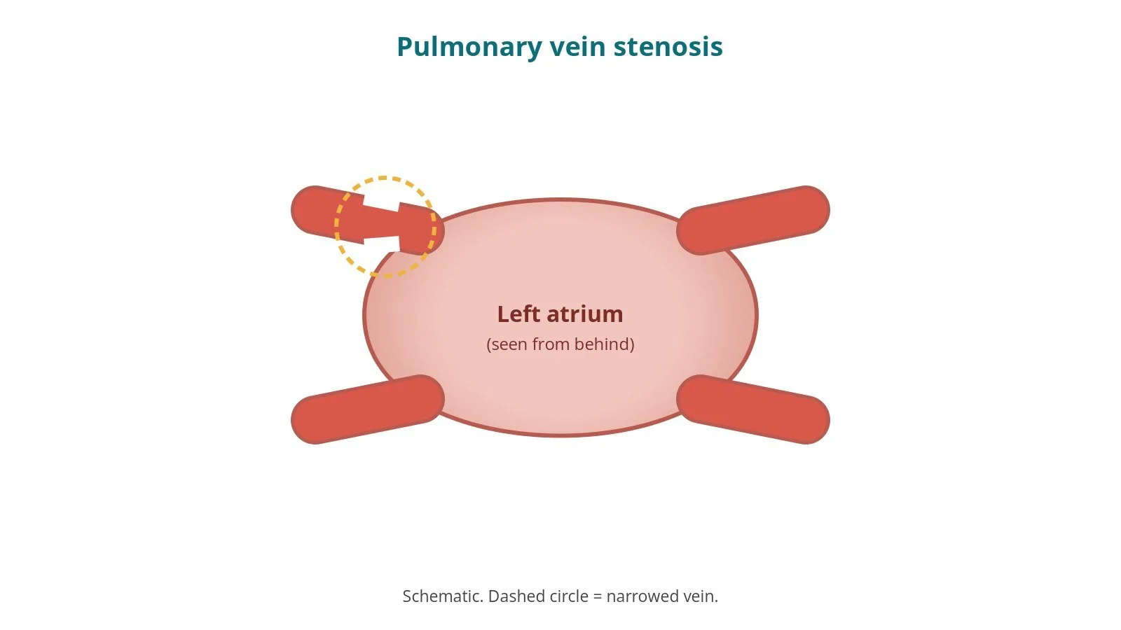
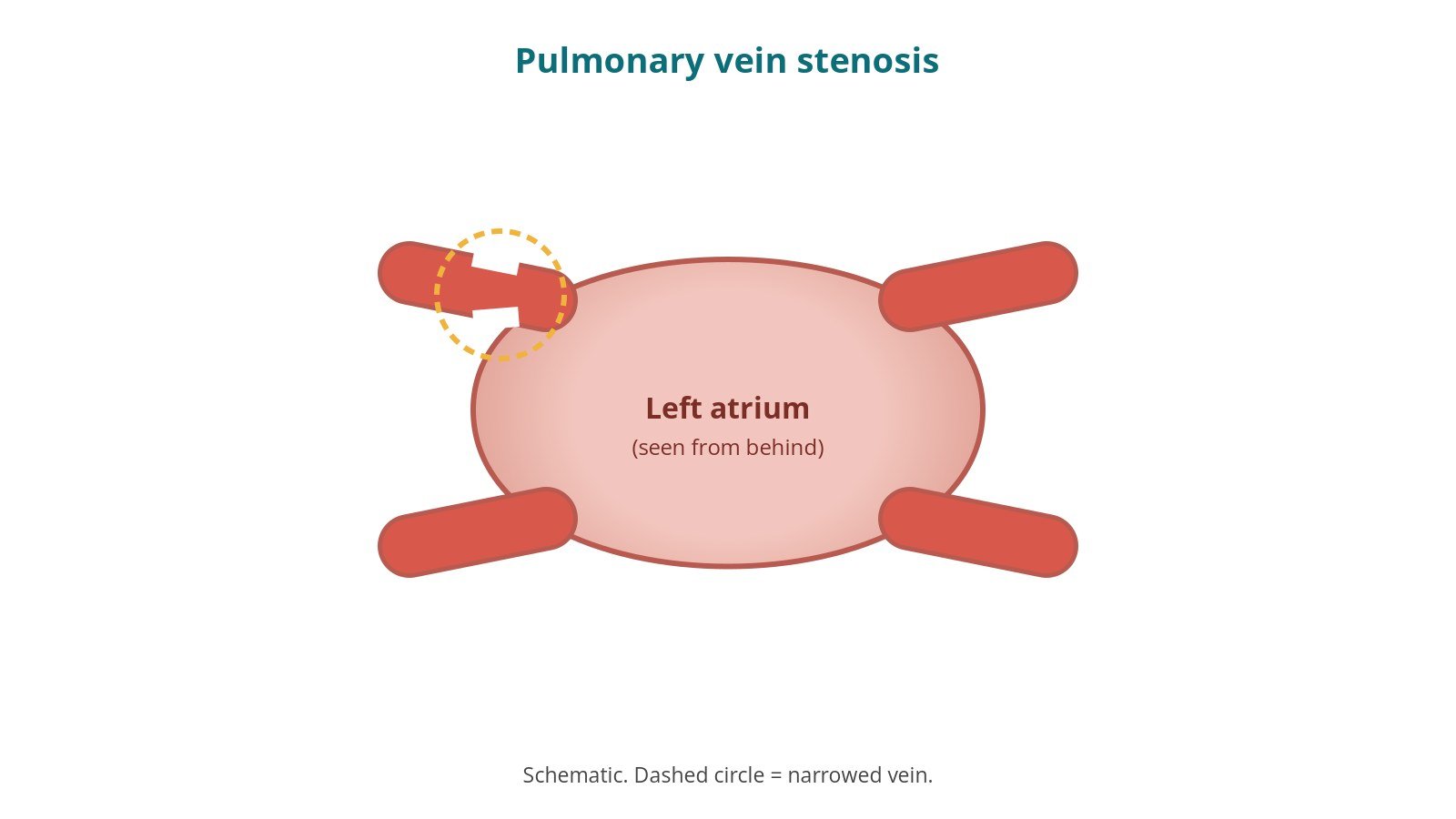
Pulmonary vein stenosis: one vein (circled) is narrowed where it joins the left atrium.
What is pulmonary vein venoplasty?
Normally, two pulmonary veins from each lung drain into the left atrium (LA). In pulmonary vein stenosis (PVS), one or more veins become narrowed, so blood backs up in that part of the lung. This raises pressure in the lung's blood vessels and can cause breathlessness, cough, chest pain, coughing up blood or repeated chest infections. In children it can lead to pulmonary hypertension.
Causes include:
congenital narrowing, present from birth, sometimes with other heart defects
narrowing after surgery for congenital heart disease involving the pulmonary veins
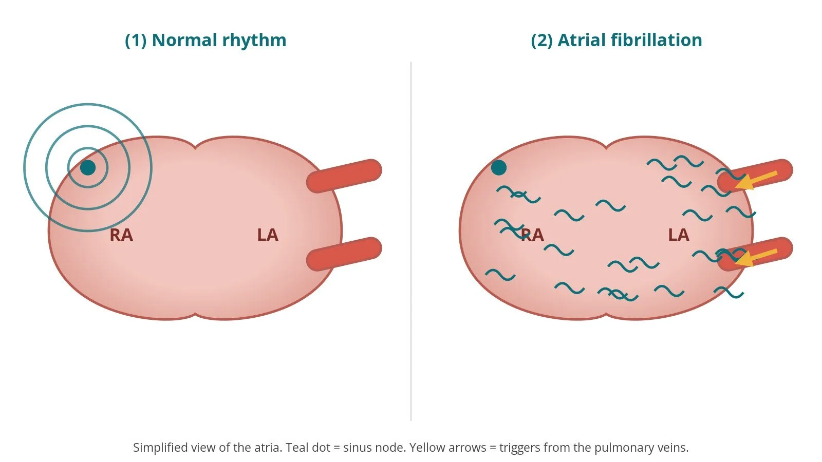
in adults, scarring after catheter ablation for AF, now uncommon
rarely, compression from outside, for example by scarring in the chest
Venoplasty widens the narrowed vein from the inside using a balloon. Because veins can narrow again, a stent is often placed to hold it open. The procedure may be a long-term treatment or a bridge to further treatment, and repeat procedures are sometimes needed.
New to the acronyms? See the Cath Lab Abbreviations A–Z.
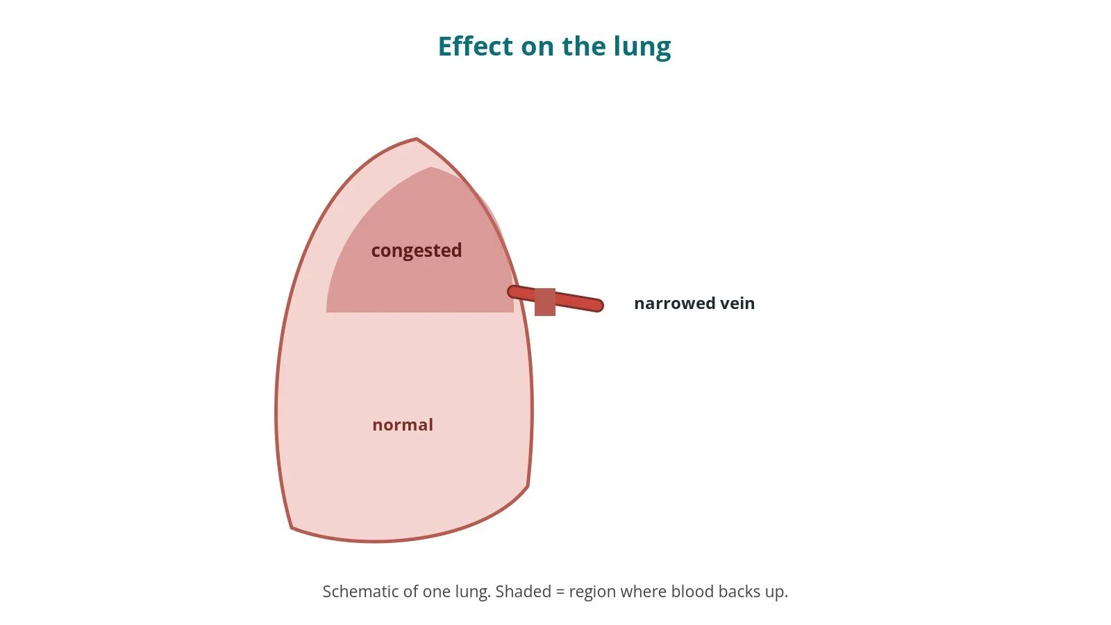
A narrowed vein causes blood to back up in that part of the lung.
Who is it for?
The ESC adult congenital heart disease guidelines (2020) and the ESC 2024 AF guidelines describe pulmonary vein stenosis as a condition for specialist assessment; catheter treatment with balloon and stenting is used for significant, symptomatic narrowing, while surgery is considered in some cases. Treatment is planned by a specialist team, often combining congenital, structural and electrophysiology expertise.
When is it done?
It is planned. Before it, the team usually arranges:
CT or MRI to map the veins and narrowings
lung perfusion scan in some cases, to see how much blood each lung region receives
echo, ECG and blood tests
a review of blood thinners, as per centre protocol
Step by step
1. Anaesthetic and access
Done under general anaesthetic, often with echo. Sheaths are placed in the femoral vein (and sometimes the femoral artery) under ultrasound; sizes per device and operator.
2. Transseptal puncture
A transseptal needle and sheath cross the atrial septum into the LA, guided by X-ray and echo. Heparin is given, dose as per centre protocol. A long sheath is placed in the LA.
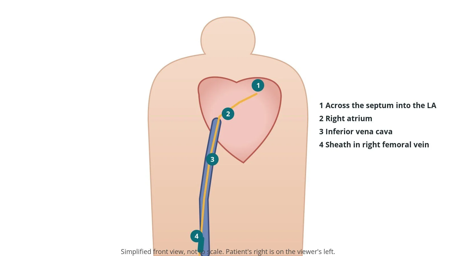
Access: (1) across the septum into the left atrium; (2) right atrium; (3) inferior vena cava; (4) sheath in the right femoral vein.
3. Opening the vein
A. Wire across. A catheter and soft wire are passed through the narrowing into the vein beyond, then exchanged for a supportive wire. Pressures are measured on both sides and a venogram (contrast injection) shows the narrowing.
B. Balloon. A balloon is inflated in the narrowing, size per device and operator.
C. Stent (often). A stent may be placed to hold the vein open, sized to the vein.
D. Check. A repeat venogram and pressures check the result. Other narrowed veins may be treated in the same procedure.
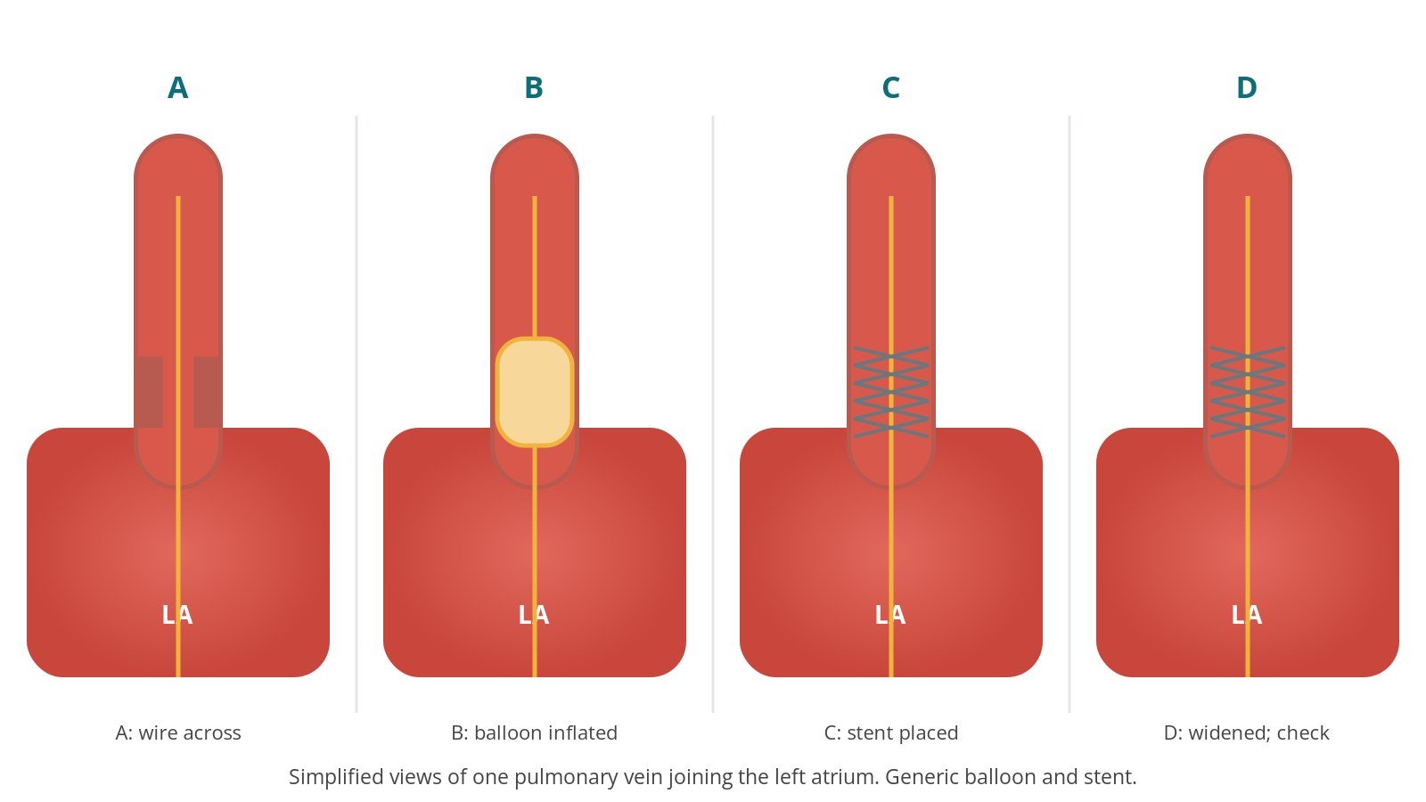
A: wire across. B: balloon inflated. C: stent placed. D: vein widened; check.
4. Finishing
The sheaths are removed. The septum is checked on echo. The groin is closed with pressure or a stitch, per operator.
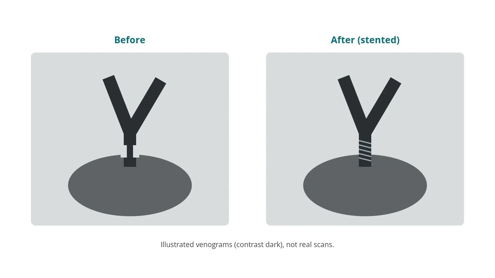
Illustrated, not real scans. Before: narrowing. After: widened vein with stent.
Recovery and aftercare
First hours: bed rest with checks of the groin, breathing, heart rhythm and blood pressure, as per centre protocol.
Going home: often after 1 to 2 nights.
Medicines: antiplatelet and/or anticoagulant medicines are usually given after stenting, as per your team's plan.
Follow-up: CT or other scans, because veins can narrow again; repeat procedures are sometimes needed.
Activity: avoid heavy lifting for about a week, or as advised.
Driving: ask your team.
Call 999 if you have chest pain, sudden breathlessness, coughing up blood, fainting, signs of a stroke, or heavy groin bleeding.
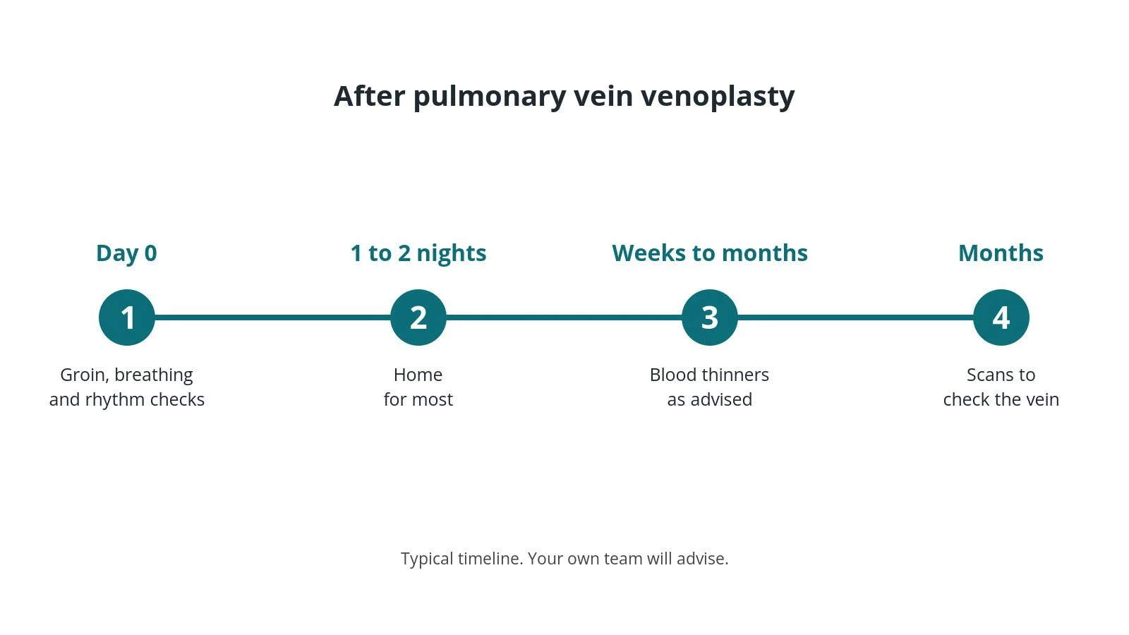
After venoplasty: (1) checks; (2) home after 1 to 2 nights; (3) blood thinners as advised; (4) scans to check the vein.
Risks and complications
Your team will discuss your own risks. Possible problems include:
the vein narrowing again (restenosis), which is common
a tear in the vein, bleeding into the lung or coughing up blood
fluid around the heart (tamponade)
stroke from clot or air
the stent moving
bleeding at the groin
a remaining hole in the atrial septum
Cath lab kit list (printable)
Barts example kit list
An example from one UK centre, written generically. Every lab has its own protocol, and sizes and doses vary.
angiography pack
femoral sheaths; long transseptal-length sheath, sizes per device and operator
diagnostic catheters (pigtail, multipurpose and a rapid-exchange catheter), per operator
stiff-tipped 0.014" or 0.018" wire, super-stiff short-taper wire, stiff pre-shaped wire and long hydrophilic wire, per operator
diathermy pen, machine and pad, if radiofrequency or diathermy-assisted transseptal puncture is used
Luer-lock syringes, 3-way tap, saline bag and spike
transseptal needle and transseptal sheath, per operator
venoplasty balloons (sizes on request), per device and operator; contrast/saline mix for the balloon, as per centre protocol; inflation device
stents, sizes per device and operator
heparin as per centre protocol
Procedural steps (Barts example)
General anaesthetic; femoral venous access
Transseptal puncture; heparin as per centre protocol; long sheath into the LA
Catheter to the target vein; wire across the narrowing; pressures and venogram
Balloon inflation; stent if needed
Repeat venogram and pressures; treat other veins if planned
Sheaths out; haemostasis
First time in the cath lab?
💡 First-time tip
Ask for sizes early. Balloons and stents are chosen from the CT and venogram; have a range available. Mix the balloon fluid correctly. Use the contrast mix as per centre protocol. Record the pressures. Pressures before and after show whether the narrowing has been relieved. Respect the air. Long sheaths in the left atrium must be kept bubble-free. Watch for coughing up blood. It can mean a vein injury; tell the operator straight away.
FAQ
Is pulmonary vein stenosis common?
No, it is rare.
Will the vein stay open?
Veins can narrow again, so regular scans are needed and repeat procedures are sometimes required.
Is surgery an option?
In some cases. Your team will discuss which treatment suits you.
Will I need blood thinners?
Usually after stenting, for a period set by your team.
Related Cath Lab 101 guides
Sources
All checked on 10 October 2026. Each page's own date is shown where it gives one.
Baumgartner H, et al. 2020 ESC Guidelines for the management of adult congenital heart disease. Eur Heart J 2021;42(6):563–645. PubMed 32860028
Van Gelder IC, et al. 2024 ESC Guidelines for the management of atrial fibrillation. Eur Heart J 2024;45(36):3314–3414. PubMed 39210723
British Heart Foundation. Congenital heart disease. bhf.org.uk
Disclaimer
Education only. This page is for general education for healthcare students, cath lab staff and curious readers. It isn't medical advice and doesn't replace advice from your own doctor, heart team or local unit protocols. Equipment and medicines are described generically. No product or manufacturer is recommended. If you think someone is having a heart emergency, call 999.
Disclosure: I work for a company that makes a device used in this procedure. This page is independent education, isn't sponsored or reviewed by any company, and doesn't recommend any product.
All diagrams on this page are original illustrations created for rodnieoro.com.