Transcatheter tricuspid valve implantation (TTVI): a Cath Lab 101 guide
Reviewed by Rodnie Oro, cath lab charge nurse · Last reviewed: October 2026 · Education only, not medical advice
TTVI in one minute
What it is: the tricuspid valve sits between the right atrium and right ventricle. Transcatheter tricuspid valve implantation (TTVI) places a new valve through a vein: either inside a worn-out surgical tricuspid valve (valve-in-valve, often through the neck), or as a replacement of a person's own leaking valve using a dedicated device through the groin.
Who it is for: people with severe tricuspid regurgitation or a failed surgical tricuspid valve causing symptoms, for whom open-heart surgery is high risk and (for a native valve) repair with a catheter is unsuitable, as decided by a heart team.
Main risks: bleeding, heart rhythm problems including heart block needing a pacemaker, the valve moving or leaking, and clot on the valve. Your team will explain your own risk.
Usually: under general anaesthetic with transoesophageal echo (TOE), about 1.5 to 3 hours, often home after a few nights.
On this page: What is TTVI? · Who is it for? · When is it done? · Step by step · Recovery · Risks · Kit list · First time in the lab? · FAQ
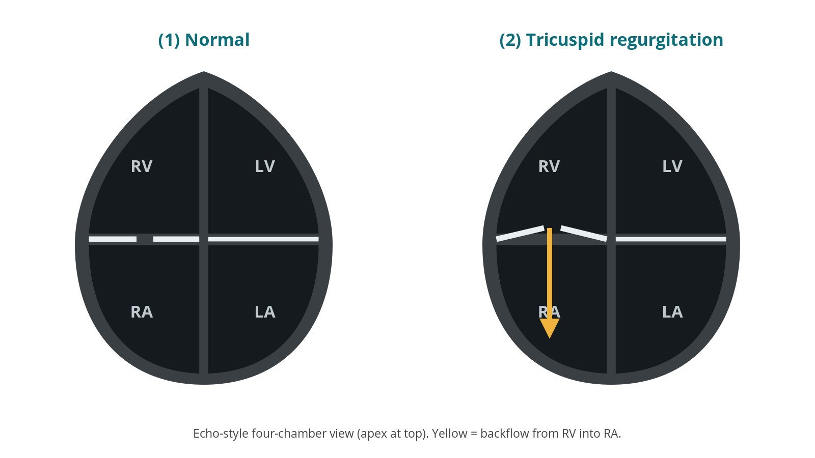
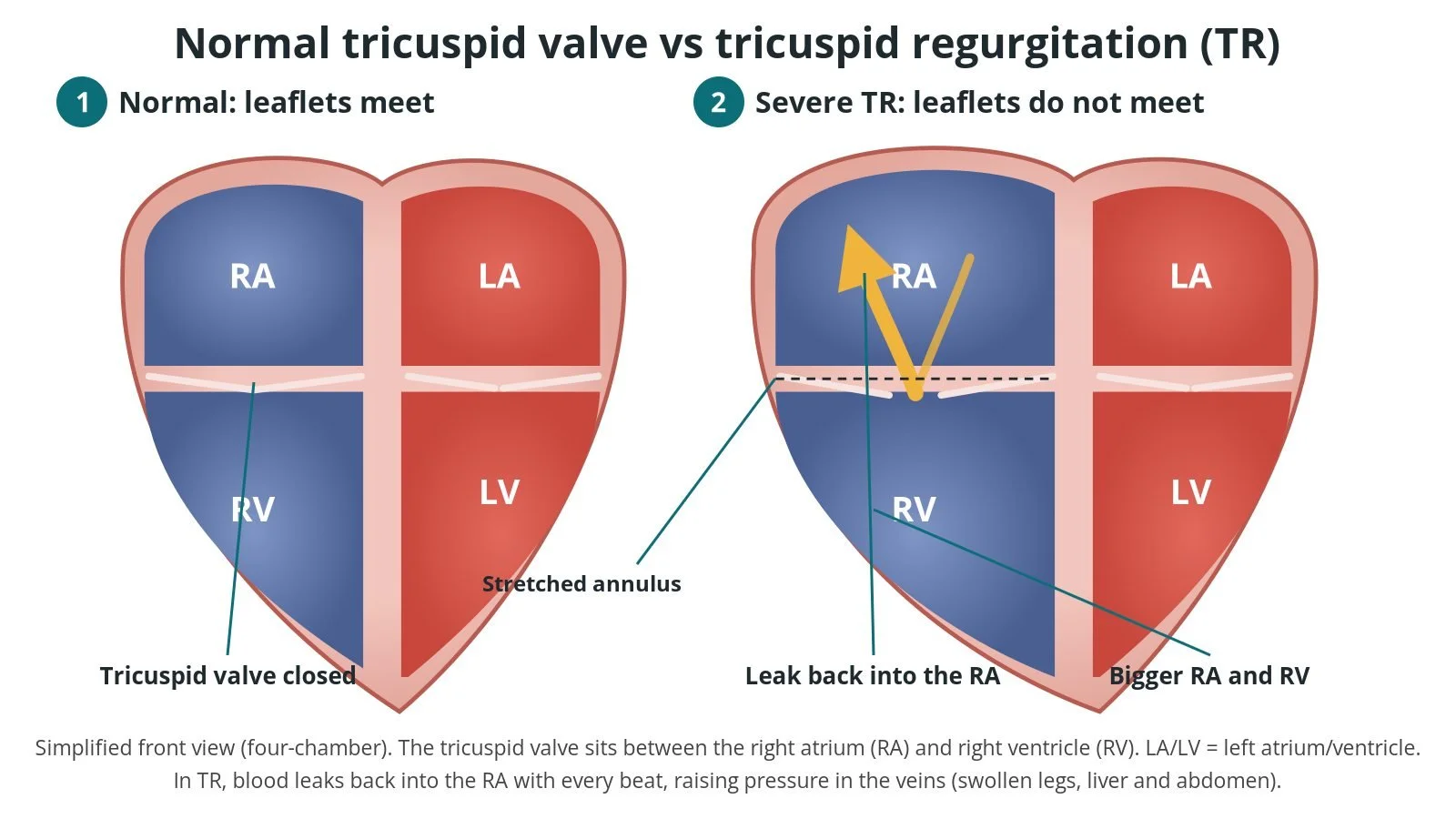
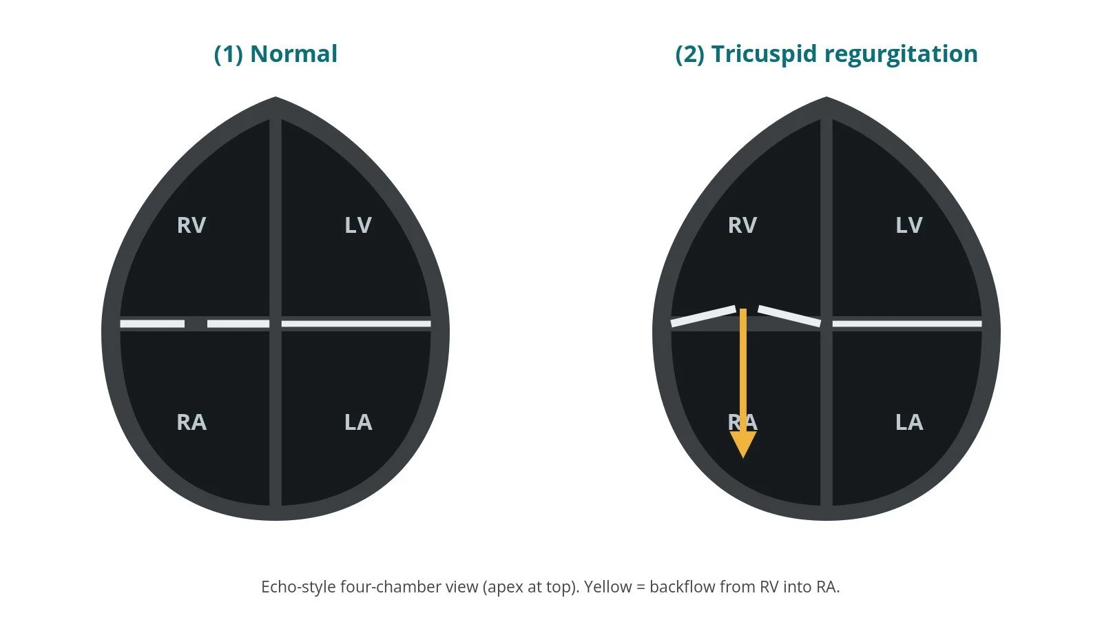
(1) Normal: the tricuspid valve seals. (2) Tricuspid regurgitation: blood leaks back into the right atrium.
What is TTVI?
The tricuspid valve lets blood flow from the right atrium (RA) into the right ventricle (RV), which pumps it to the lungs. When it leaks (tricuspid regurgitation, TR), blood flows backwards into the RA and the veins. This can cause swollen legs and abdomen, tiredness, poor appetite and breathlessness. Severe TR is often linked to left-sided heart disease, AF, lung pressure problems or pacemaker leads.
Transcatheter tricuspid valve implantation (TTVI) places a new valve without open-heart surgery. There are two main situations:
Valve-in-valve (ViV): a previous surgical tissue valve (or sometimes a ring) in the tricuspid position has worn out. A balloon-expandable transcatheter valve is expanded inside it, using the old frame as an anchor. This is often done through the right internal jugular vein in the neck, which gives a straight route down to the tricuspid valve, or through the femoral vein.
Native valve replacement (TTVR): a dedicated, self-expanding transcatheter valve replaces the function of the person's own leaking valve, delivered from the femoral vein through the IVC.
When the leak can be reduced by repairing the valve with a clip, tricuspid TEER may be used instead. See the tricuspid TEER guide.
New to the acronyms? See the Cath Lab Abbreviations A–Z.
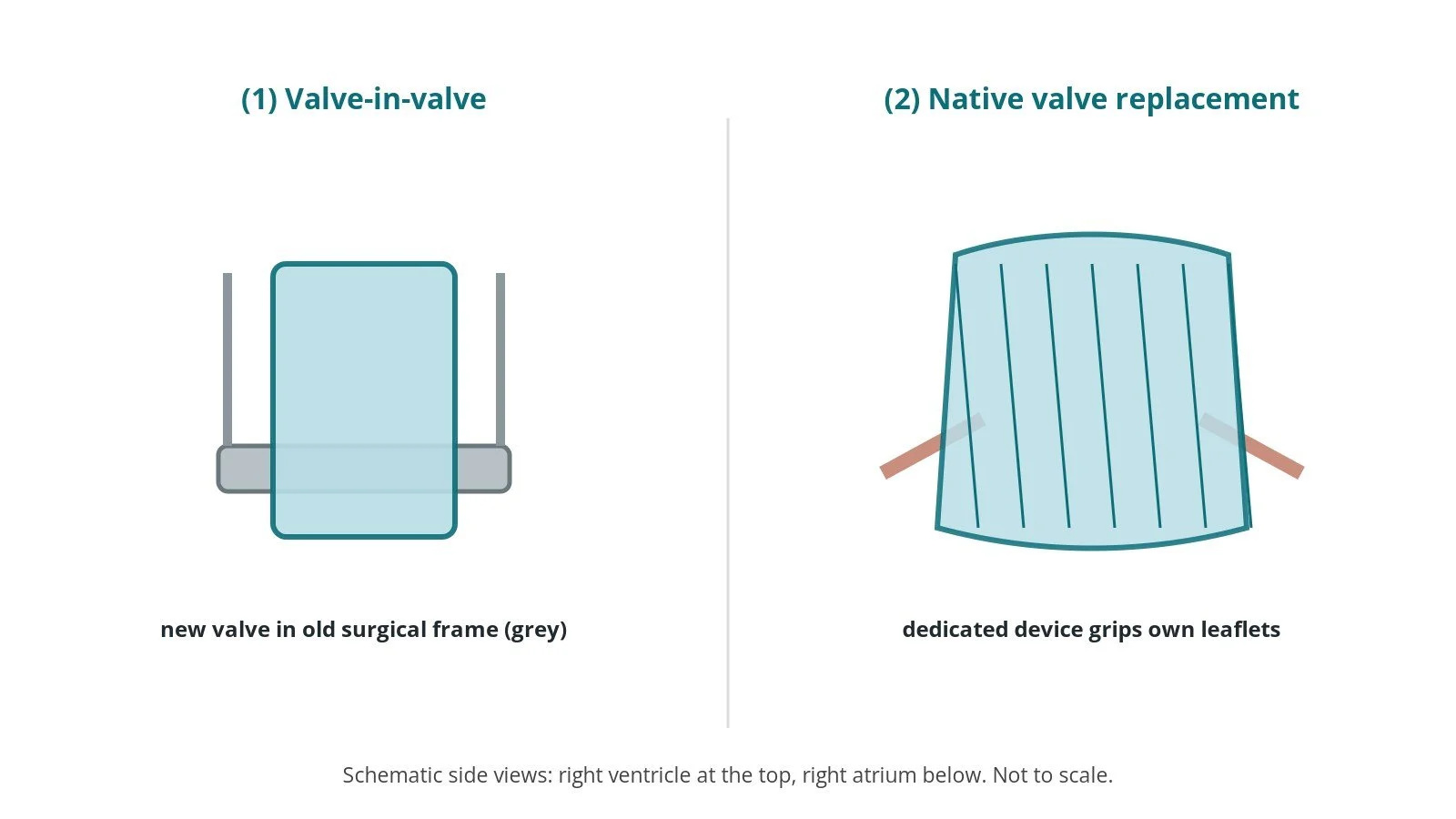
(1) Valve-in-valve inside a failed surgical valve. (2) Native valve replacement with a dedicated device.
Who is it for?
NICE (HTG771, February 2026) says transcatheter tricuspid valve implantation can be used in the NHS during an evidence generation period for symptomatic severe TR when open surgery is high risk and transcatheter repair is unsuitable, with enhanced informed consent and auditing of outcomes. The ESC/EACTS valvular heart disease guidelines support heart-team assessment of transcatheter treatment in selected people with symptomatic severe TR who are at high surgical risk. For failed surgical valves, valve-in-valve is an established alternative to repeat surgery in selected patients.
When is it done?
It is usually planned. Before it, the team usually arranges:
echo and TOE to assess the valve, the RV and lung pressures
CT to size the valve or old prosthesis and plan access
a check of any pacemaker leads that cross the valve
blood tests, including a group and save
a review of blood thinners, as per centre protocol
Step by step
1. Anaesthetic and set-up
TTVI is done in a cath lab or hybrid theatre under general anaesthetic with TOE. Local anaesthetic is used at the access sites, as per centre protocol.
2. Access
Valve-in-valve via the neck: a large sheath is placed in the right internal jugular vein under ultrasound. A femoral vein sheath is used for a pacing wire and a femoral artery for pressure monitoring. Sizes are per device and operator.
Native replacement via the groin: a large sheath goes in the right femoral vein, often after a suture-based closure device is placed in advance, with a steerable delivery system.
Heparin is given, dose as per centre protocol.
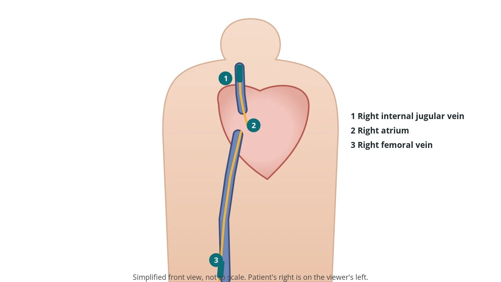
Access: (1) right internal jugular vein (often for valve-in-valve); (2) right atrium; (3) right femoral vein (native replacement and pacing).
3. Implanting the valve
A. Cross the valve. A catheter crosses the tricuspid valve into the RV, taking care not to pass through the RV chordae, and a stiff wire is placed in the RV or pulmonary artery, per operator.
B. Position. For valve-in-valve, the balloon-expandable valve, mounted the opposite way round to a TAVI valve (outer skirt towards the RA), is aligned with the old valve frame on X-ray and TOE. For native replacement, the dedicated device is steered into position across the leaflets.
C. Deploy. For valve-in-valve, the balloon is slowly inflated, often with rapid pacing. Native devices are released in stages to capture the leaflets, per device instructions.
D. Check. TOE, angiography and pressures confirm position, function and any leak.
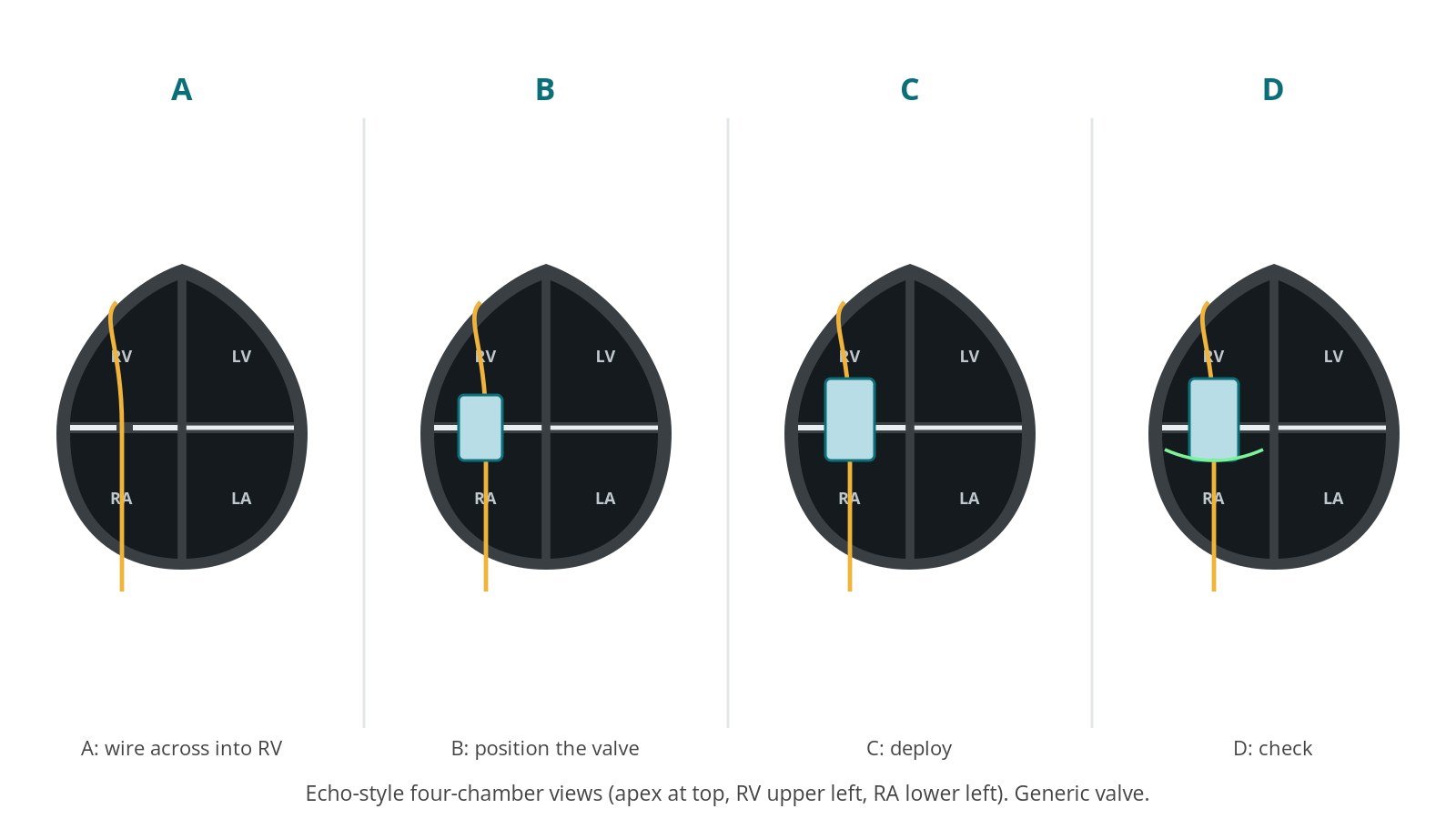
A: cross the valve. B: position. C: deploy. D: check.
4. Finishing
The delivery system and wire are removed. Sheaths are removed. The neck is closed with pressure or a stitch, and the groin with the pre-placed suture or pressure, per operator.
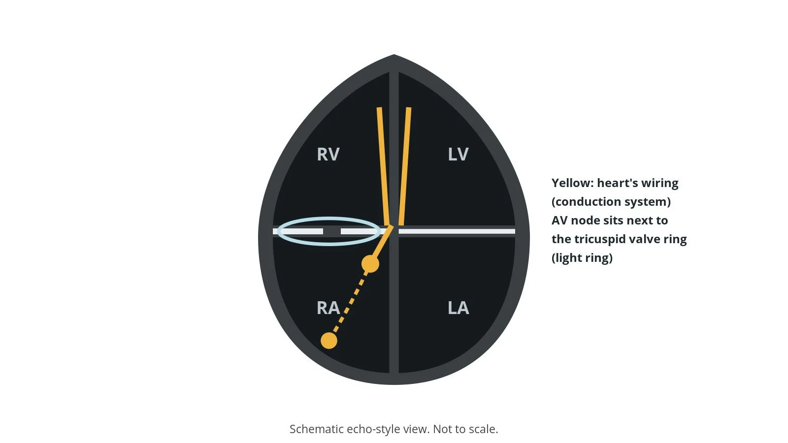
The heart's wiring (yellow) runs close to the tricuspid valve, so a new valve can cause heart block needing a pacemaker.
Recovery and aftercare
First hours: recovery, often in a high-dependency area, with heart rhythm monitoring, because heart block can occur after native valve replacement.
Echo: before discharge to check the valve.
Going home: often after a few nights.
Medicines: a blood thinner (anticoagulant) is usually needed for a time, as per your team's plan. Diuretics are adjusted as swelling improves.
Activity: avoid heavy lifting for about a week, or as advised.
Follow-up: clinic and echo checks.
Driving: DVLA guidance after a percutaneous valve procedure for a car licence is not to drive for 2 weeks; bus and lorry drivers must tell the DVLA. Ask your team.
Call 999 if you have chest pain, sudden breathlessness, fainting, a very slow pulse, or heavy bleeding from the neck or groin.
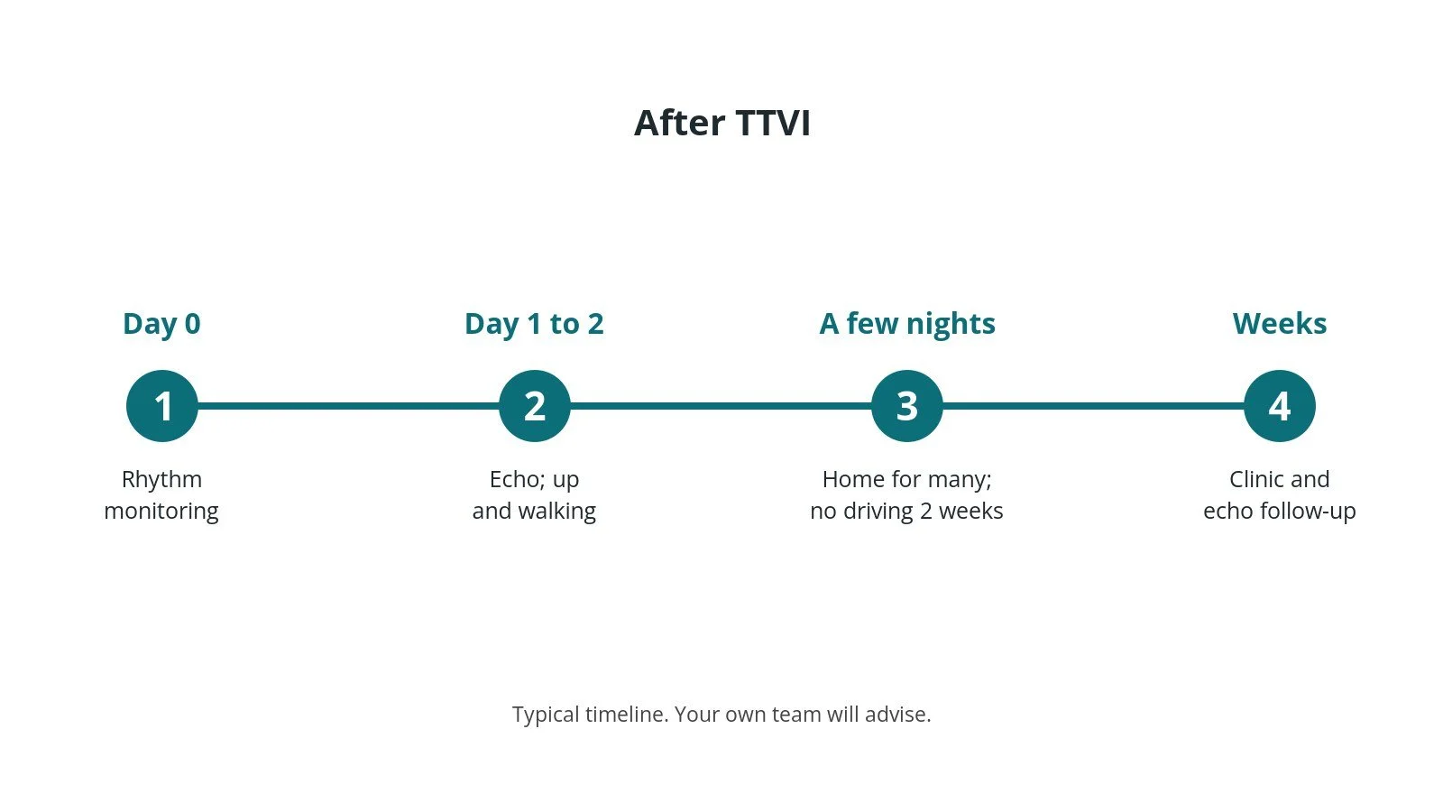
After TTVI: (1) rhythm monitoring; (2) echo; (3) home after a few nights for many; (4) clinic and echo follow-up.
Risks and complications
Your team will discuss your own risks. Possible problems include:
bleeding or vein injury at the neck or groin
heart block needing a permanent pacemaker
the valve moving out of position or a leak around its edge
clot forming on the valve (valve thrombosis)
injury to the right coronary artery, the RV or surrounding structures
fluid around the heart (tamponade)
problems with existing pacemaker leads
Cath lab kit list (printable)
Barts example kit list (valve-in-valve via the right internal jugular vein)
An example from one UK centre, written generically. Every lab has its own protocol, and sizes and doses vary.
Scrub (small) trolley
structural heart procedure pack
femoral venous sheath for pacing; diagnostic catheters (right coronary shape and pigtail)
large-bore long sheath for the jugular vein, size per device and operator
suture-based closure device, per operator
stiff pre-shaped wire and long J-tip exchange wire, per operator
non-absorbable suture for a skin (figure-of-eight) stitch, per operator
local anaesthetic, heparin and other drugs, as per centre protocol
Valve (crimping) trolley
balloon-expandable transcatheter valve, size per device and operator
crimping kit; valve mounted the opposite way round to TAVI, with the outer skirt towards the RA, crimping per device instructions
3-way tap; large syringe for de-airing; syringe for flushing
contrast/saline inflation mix, per device instructions
saline bowls for rinsing the valve, per device instructions
scissors and forceps
Native valve replacement extras
steerable guide catheter; stiffer exchange wire; dedicated device kit, per device and operator
Procedural steps (Barts example)
General anaesthetic and TOE; femoral vein pacing wire; arterial line
Ultrasound-guided right internal jugular access; large sheath
Heparin as per centre protocol; cross the valve; stiff wire placed
Valve crimped the opposite way round to TAVI (checked by two people)
Aligned with the old valve frame; deployed with slow balloon inflation under rapid pacing
TOE, angiography and pressures; system removed
Sheaths out; neck stitch and groin haemostasis
First time in the cath lab?
💡 First-time tip
Orientation check. For valve-in-valve the valve is crimped the opposite way round to a TAVI valve, skirt towards the RA. Two people check it. Neck access set-up. The head is turned and the drapes placed to keep the jugular site sterile and accessible. Watch the rhythm. Heart block can happen; keep pacing ready. Know the old valve. Each surgical valve looks different on X-ray. Have its details and CT plan to hand. De-air. Everything is flushed and de-aired; the right heart still matters.
FAQ
Is this open-heart surgery?
No. It is done through veins in the neck or groin.
Will I need a pacemaker?
Some people do, especially after native valve replacement. Your team will monitor your rhythm.
Will my swelling improve?
Many people feel less swollen and less tired, but this varies. Your medicines will be reviewed.
How long will the valve last?
Transcatheter valves are tissue valves and can wear out over time. Regular echo checks are needed.
Related Cath Lab 101 guides
Sources
All checked on 10 October 2026. Each page's own date is shown where it gives one.
NICE. Transcatheter tricuspid valve implantation for symptomatic severe tricuspid regurgitation (HTG771). nice.org.uk/guidance/htg771 (published 25 Feb 2026)
NICE. Heart valve disease presenting in adults: investigation and management (NG208). nice.org.uk/guidance/ng208
Vahanian A, et al. 2021 ESC/EACTS Guidelines for the management of valvular heart disease. Eur Heart J 2022;43(7):561–632. PubMed 34453165
NHS. Heart valve disease. nhs.uk/conditions/heart-valve-disease
British Heart Foundation. Heart valve disease. bhf.org.uk
DVLA / GOV.UK. Cardiovascular disorders: assessing fitness to drive (percutaneous valve intervention). gov.uk
Disclaimer
Education only. This page is for general education for healthcare students, cath lab staff and curious readers. It isn't medical advice and doesn't replace advice from your own doctor, heart team or local unit protocols. Equipment and medicines are described generically. No product or manufacturer is recommended. If you think someone is having a heart emergency, call 999.
Disclosure: I work for a company that makes a device used in this procedure. This page is independent education, isn't sponsored or reviewed by any company, and doesn't recommend any product.
All diagrams on this page are original illustrations created for rodnieoro.com.