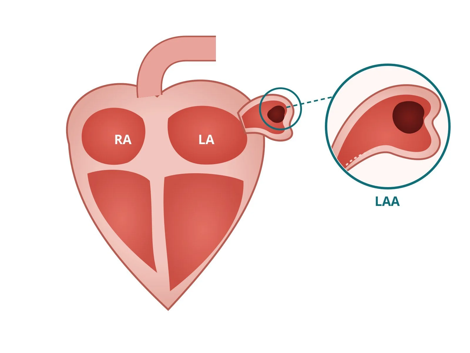
Left atrial appendage occlusion (LAAO): a Cath Lab 101 guide
Reviewed by Rodnie Oro, cath lab charge nurse · Last reviewed: October 2026 · Education only, not medical advice
LAAO in one minute
What it is: left atrial appendage occlusion (LAAO) is a keyhole heart procedure that seals off the left atrial appendage, a small pouch on the heart's upper left chamber where most stroke-causing clots in atrial fibrillation (AF) are thought to form. A thin tube goes in through a vein in the groin, and a small occluder (a fabric-covered metal plug) is left in the mouth of the pouch.
Who it's for: people with AF who are at risk of stroke but can't take long-term anticoagulants (blood thinners), for example because of serious bleeding. It isn't a replacement for anticoagulants in people who can take them.
Main risks: bruising or bleeding at the groin is the most common. Serious problems, such as fluid or bleeding around the heart, the device moving out of place, stroke, or a clot forming on the device, are uncommon. Your heart team will explain your own risk.
Usually: general anaesthetic or deep sedation, about 1 to 2 hours, home the same or next day.
On this page: What is LAAO? · Who is it for? · When is it done? · Step by step · Recovery · Risks · Kit list · First time in the lab? · FAQ
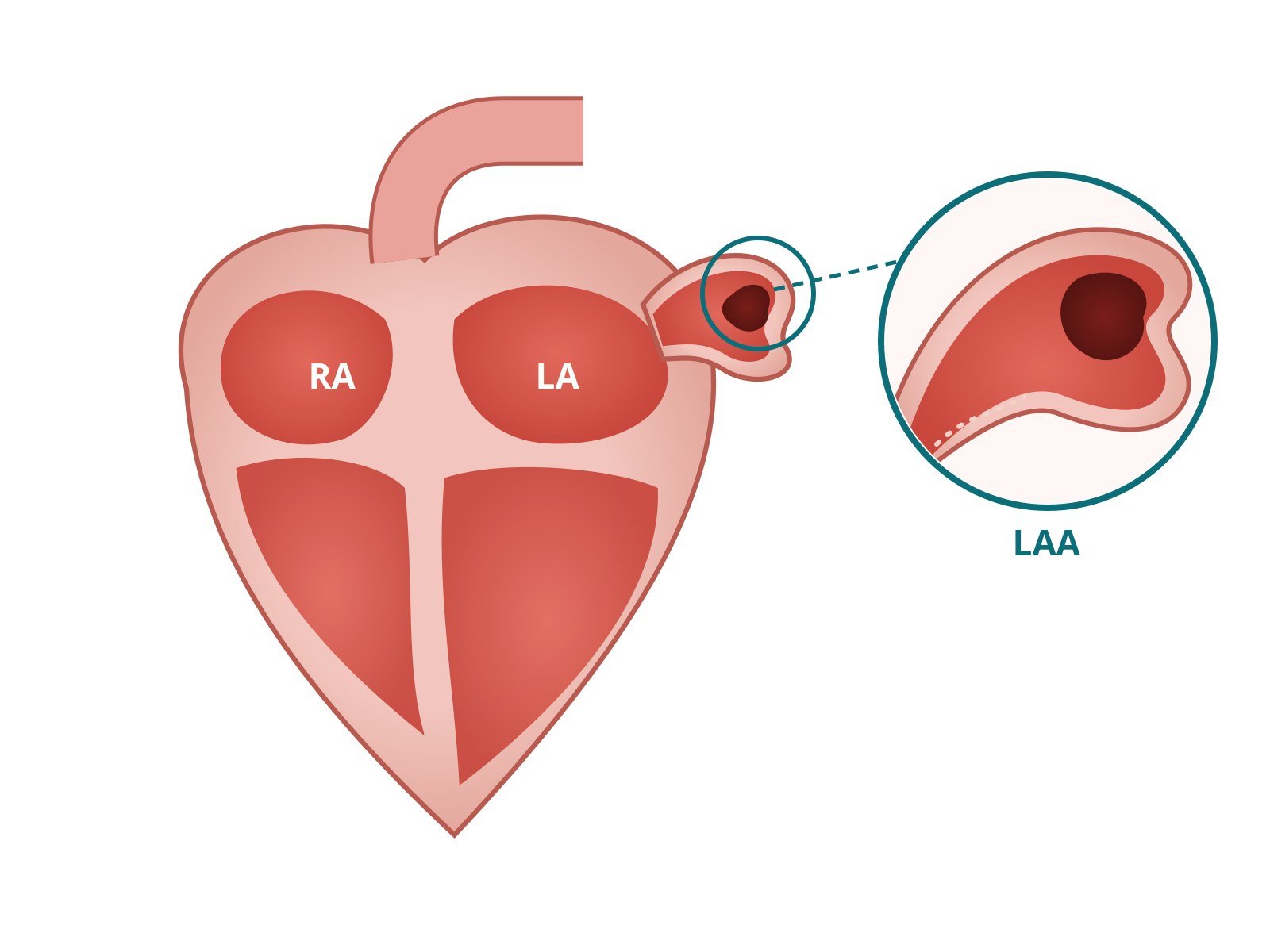
The left atrial appendage (LAA) is a small pouch off the left atrium (LA). In atrial fibrillation, blood can pool and clot here. The circle magnifies a clot inside the pouch.
What is LAAO?
In atrial fibrillation the top chambers of the heart (the atria) quiver instead of squeezing properly. Blood can then pool and clot. If a clot breaks off and travels to the brain, it causes a stroke. AF is one of the main conditions that make a stroke more likely.
The left atrial appendage (LAA) is a small, finger-shaped pouch off the left atrium. Blood moves slowly there in AF, and in non-valvular AF it's where most clots that cause strokes are thought to start (about 90% in a classic surgical study).
Most people with AF lower their stroke risk with anticoagulant tablets. LAAO is a different approach. Instead of thinning the blood, it closes the pouch from the inside with a small implanted occluder, so clots can't form in it and escape. Over the following weeks the heart's own lining grows over the device.
You may also hear it called LAA closure, LAAC or left atrial appendage closure. New to the acronyms? See the Cath Lab Abbreviations A–Z.
Who is it for?
UK and European guidance reserve LAAO for a specific group:
People with AF and a raised stroke risk (usually assessed with a score such as CHA₂DS₂-VASc), and
who can't take, or can't tolerate, long-term anticoagulants, for example after a major or life-threatening bleed.
NICE says to consider LAAO only in this situation, and not to offer it as an alternative to anticoagulation for people who can take anticoagulants. The 2024 ESC AF guidelines take a similar position. The decision is made by a heart team with the patient, weighing the procedure's risks against the person's stroke and bleeding risk.
LAAO lowers stroke risk from the appendage. It doesn't treat AF itself, so symptoms such as palpitations still need their own treatment.
When is it done?
LAAO is a planned procedure, not an emergency one. Before it, the team usually:
images the appendage with a CT scan or a transoesophageal echo (TOE), to check its size and shape and to make sure there's no clot already in it
reviews medicines, including any blood thinners, and plans the medicines needed after the procedure
discusses it at a heart team meeting, often together with the cardiologist who looks after the person's AF
Step by step
1. Preparation
The team checks bloods, allergies (especially to contrast dye) and kidney function. LAAO is usually done under a general anaesthetic with a TOE probe in the oesophagus, or under sedation with an ultrasound catheter inside the heart (intracardiac echo, ICE). Which one is used depends on the centre.
2. Access through the groin vein
After local anaesthetic, the operator uses ultrasound to place a sheath in the femoral vein at the top of the leg. Catheters travel up the vein to the right atrium. Because the device is delivered through a large-bore sheath, the vein is often "pre-closed" with a suture-based closure device at the start.
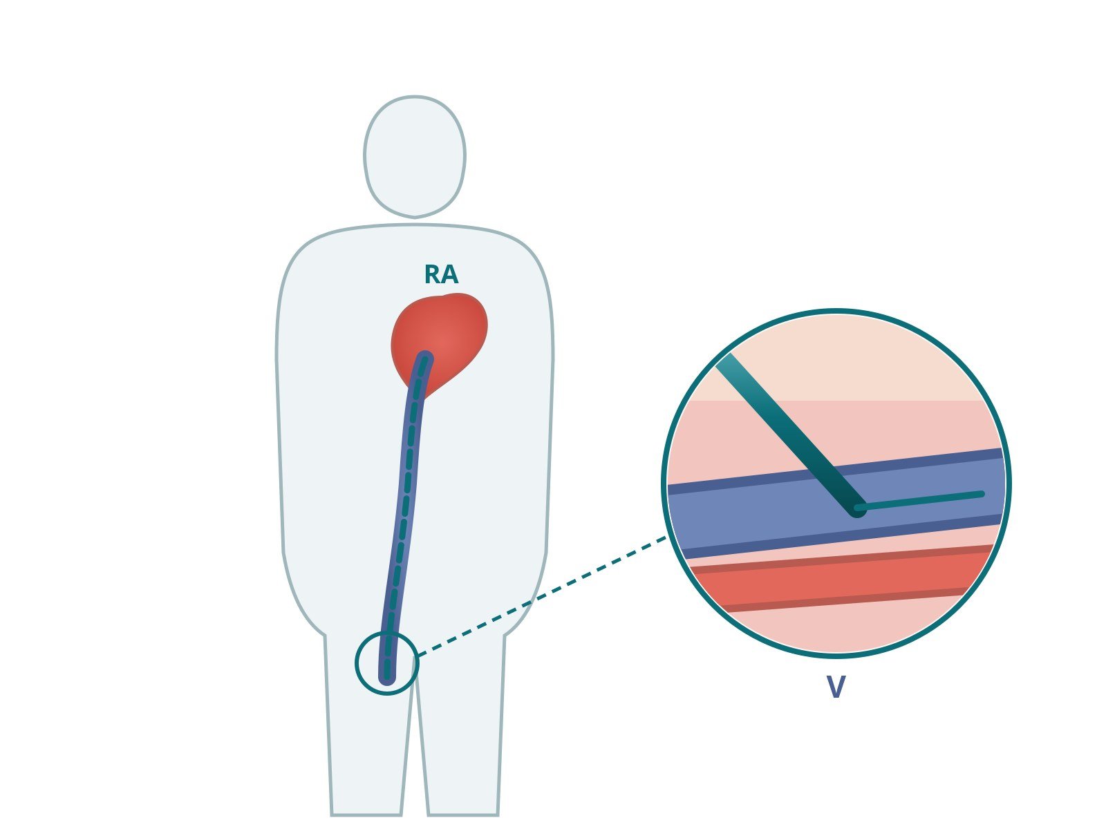
A sheath is placed in the femoral vein (V) at the groin, and catheters travel up the vein to the right atrium (RA). The close-up shows the sheath entering the vein, with the femoral artery just below it.
3. Transseptal puncture
The appendage is on the left side of the heart, so the team crosses the thin wall between the two atria (the interatrial septum) with a transseptal needle and sheath. Echo guides the exact spot, usually low and towards the back of the septum, to give a good angle into the appendage. Heparin is given once the left atrium is reached, as per centre protocol, with ACT checks.
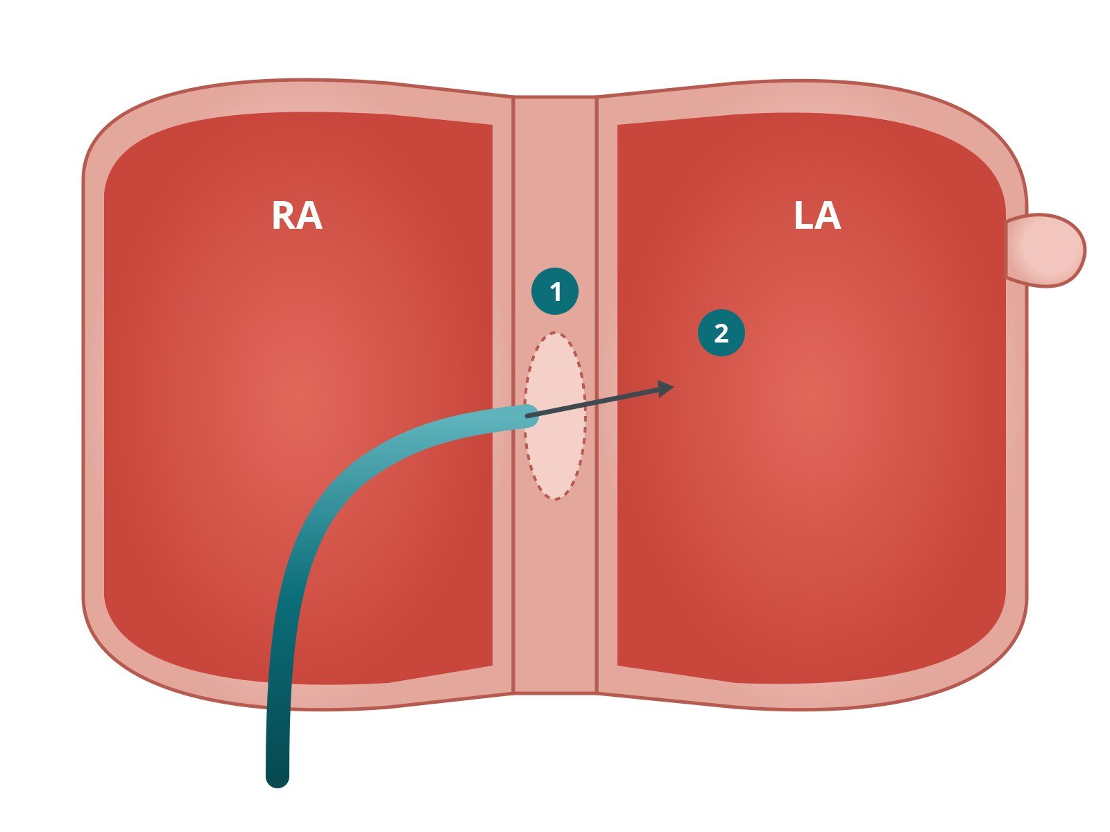
A needle and sheath cross the thin wall between the right atrium (RA) and left atrium (LA): (1) the puncture site in the septum, (2) the needle tip entering the left atrium.
4. Sizing and placing the device
A stiff 0.035" exchange wire is placed in the left atrium (often in the left upper pulmonary vein) and the large delivery sheath is advanced over it. A pigtail catheter is placed in the appendage, and contrast pictures (angiography) and echo are used to measure the opening and depth.
The operator then chooses a device size, unsheathes it in the mouth of the appendage, checks its position and seal, and only then releases it. Occluders come in a few designs, such as single-lobe plugs and disc-and-lobe devices, and the choice depends on the anatomy and the operator.
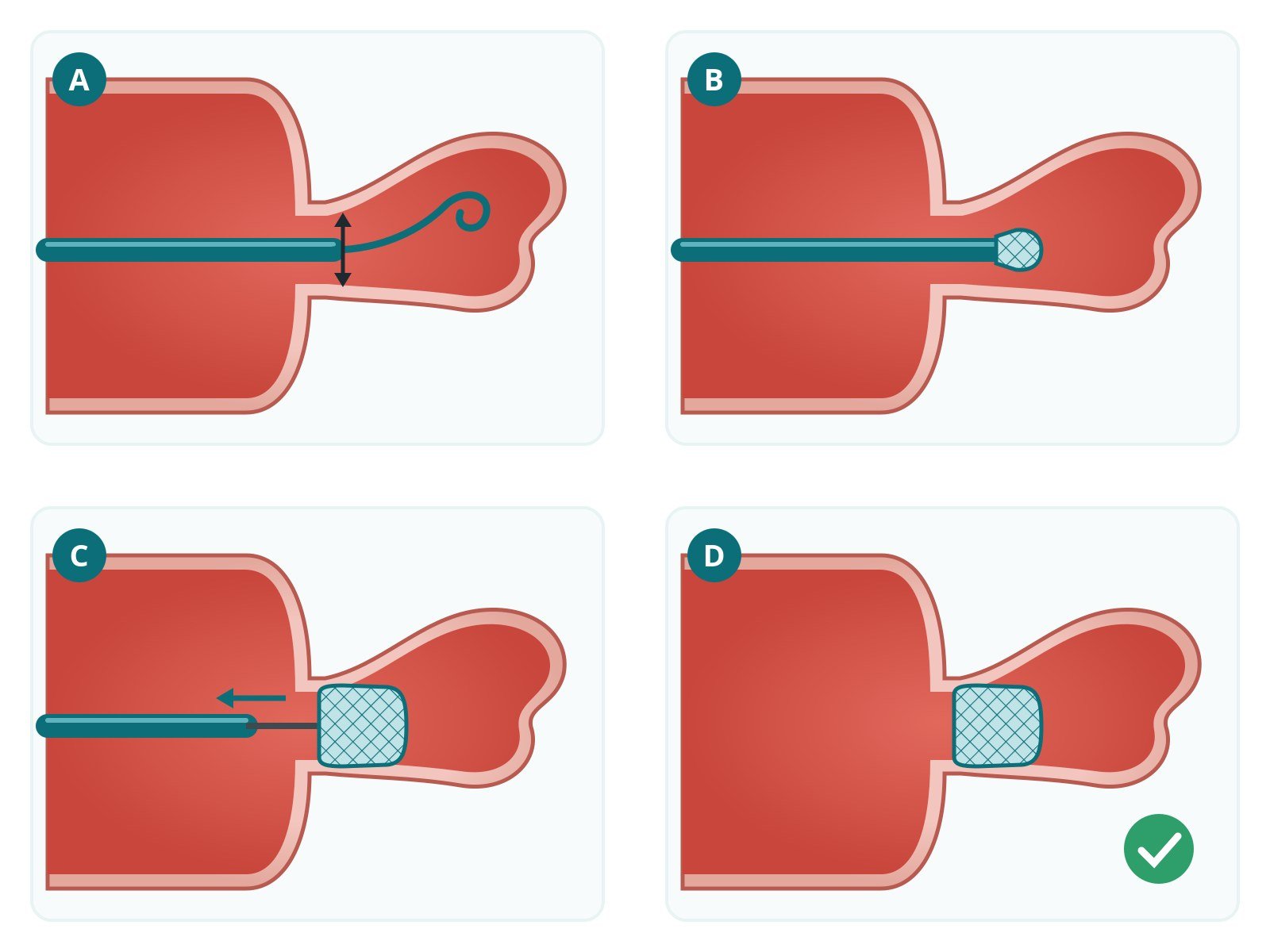
The left atrium (left) and the finger-shaped appendage (right). A: the sheath sits in the appendage and the opening (ostium) is measured. B: the occluder starts to open from the sheath inside the appendage. C: fully opened, it seals the opening and its position is checked with a gentle tug on the delivery cable. D: once the seal is confirmed, the cable is released and the device stays in place.
5. Checking the seal and finishing
Before release, echo and contrast confirm the device is stable (a gentle "tug test"), covers the opening and has little or no leak around it. The team also checks there's no new fluid around the heart. The delivery system comes out, and the groin vein is closed with the pre-placed sutures, a figure-of-eight stitch or pressure, as per centre protocol.
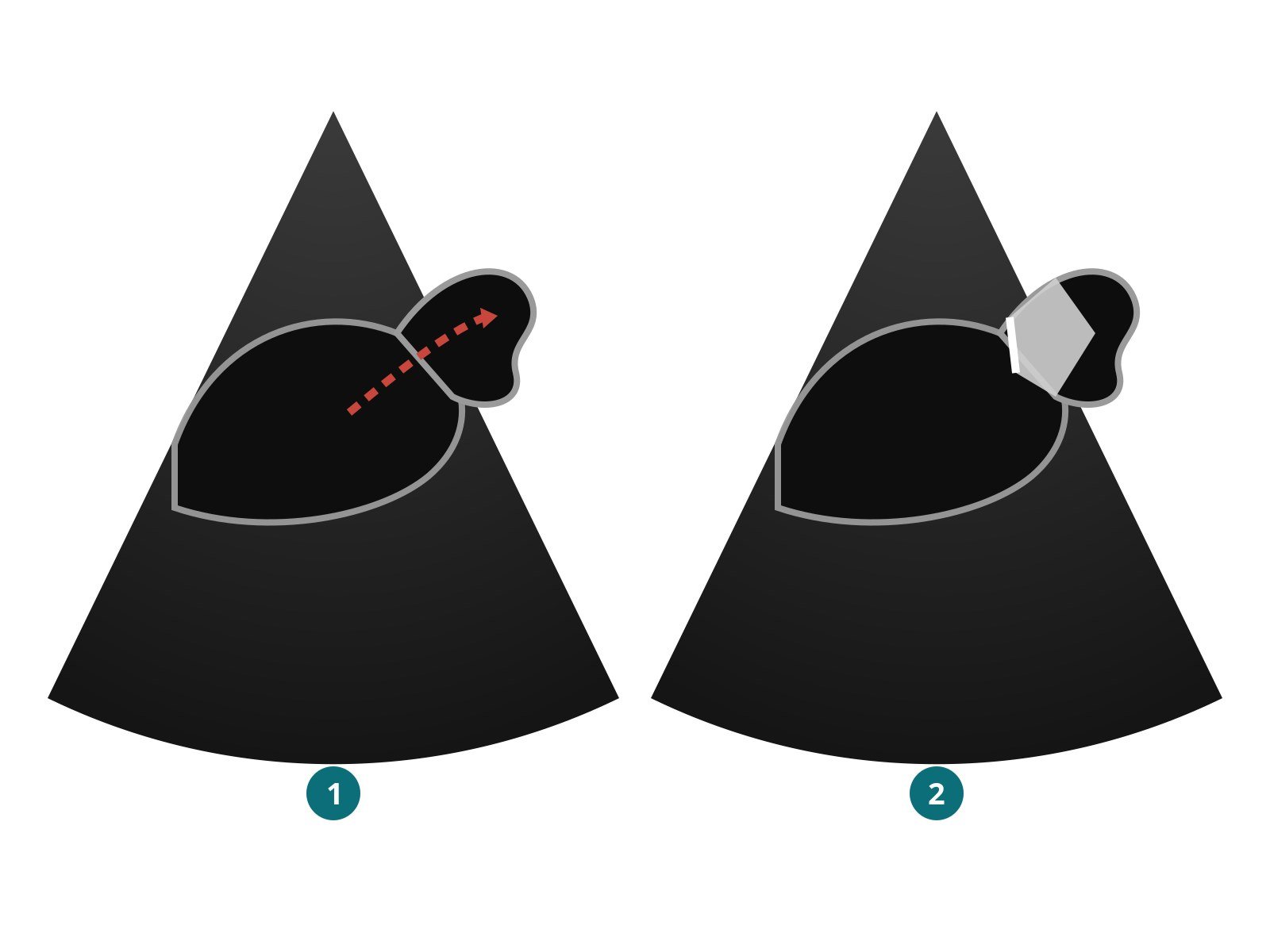
Illustration of the echo pictures: (1) before, blood can flow into the open appendage; (2) after, the occluder seals its opening.
Recovery and aftercare
Same day or next day home. Most people have an echo before discharge to check the device and look for fluid around the heart.
Groin care. Rest the leg for a few hours. Avoid heavy lifting and strenuous exercise for about a week, or as your team advises.
Medicines. Antiplatelet and/or anticoagulant medicines are usually needed for a period while the device heals in. The plan depends on the device, the centre and your bleeding risk. Don't stop or change them without speaking to your heart team.
Follow-up scan. A CT or TOE is usually repeated a few weeks to months later to check the seal and look for a clot on the device.
Call 999 for stroke symptoms (face drooping, arm weakness, speech problems), chest pain or collapse. Call your team for groin swelling, bleeding or new breathlessness.
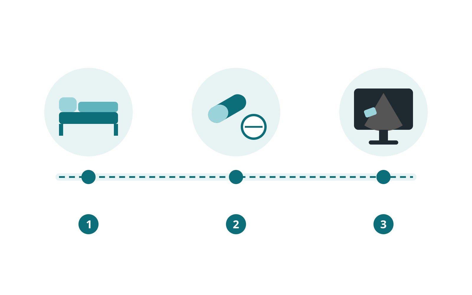
After LAAO: (1) home the same or next day after an echo check, (2) medicines for a period while the device heals in, (3) a follow-up scan a few weeks to months later.
Risks and complications
Most procedures go smoothly. Possible problems include:
Bleeding or bruising at the groin, the most common problem
Fluid or bleeding around the heart (pericardial effusion or tamponade), which may need draining
Device embolisation: the device moves out of place and has to be retrieved, sometimes surgically
Stroke or air embolism during the procedure
Device-related thrombus: a clot forming on the device surface, usually managed with medicines
Peri-device leak: a small gap around the edge of the device
Problems from the TOE probe or anaesthetic, such as a sore throat
Reaction to contrast dye or effects on the kidneys
Your heart team will explain how these apply to you.
Cath lab kit list (printable)
Drugs, doses and dilutions: always as per centre protocol. Equipment, sizes and catheter shapes also vary by unit, operator and device. Device names below are generic. Always check the sheath and wire required in the chosen device's instructions for use.
Example: Barts Heart Centre kit list — always follow your own centre protocol
Typical setup for a femoral-vein LAAO under general anaesthetic with TOE guidance, shared as one real-world example. It is not a protocol for your unit.
Sterile table
angiography pack, 500 mL saline bag and spike
fluid waste container
ultrasound probe cover (for ultrasound-guided venous access)
haemostasis valve or Y-connector
Access and closure
8 Fr femoral venous sheath (initial access)
suture-based vascular closure device (pre-close technique)
LAAO delivery/access sheath, size as specified for the chosen device
Transseptal puncture
transseptal needle
fixed-curve transseptal sheath
Catheters and wires
6 Fr pigtail catheter (LAA angiography and sizing)
6 Fr MPA1 (multipurpose) diagnostic catheter
0.035" × 260 cm extra-stiff exchange wire (tip shape and position per operator and device)
Device
LAA occluder, size chosen in the lab from TOE and angiography measurements (often with the next size up available)
Medications (scrub nurse):
lidocaine 1% 10 mL (local anaesthetic for access)
heparin 10,000 units/10 mL: dose and ACT target as per centre protocol
Imaging: TOE (general anaesthetic) or ICE (sedation), fluoroscopy and contrast, as per centre protocol.
Procedural steps (Barts example)
Right femoral venous access with ultrasound, 8 Fr sheath, pre-close with a suture-based closure device
Transseptal puncture with the transseptal needle and sheath under TOE guidance
Once in the left atrium, remove the needle; heparin as per centre protocol
Introduce the MPA1 catheter over a guidewire, then exchange for the extra-stiff 0.035" × 260 cm wire
Upsize to the LAAO delivery sheath over the stiff wire; pigtail into the appendage for angiography and sizing
Prepare and deliver the chosen occluder; check position, stability and seal with TOE and contrast before release
Remove the delivery system and close the groin with the pre-placed sutures
First time in the cath lab?
💡 First-time tip
Lead goes on before you go in. Find a lead apron and thyroid collar that fit, and wear your dosimeter where your unit says. Know who's at the head end. In LAAO the anaesthetist and the echo operator share the top of the table. Keep the TOE probe and airway lines clear when you move around. Watch the pressure and the echo. A falling blood pressure or new fluid on the echo can be the first sign of a problem around the heart. Say so straight away. Flush everything, twice. Air in a left-sided sheath can travel to the brain. Watch how the scrub nurse de-airs the delivery sheath and device. Speak up. In a good lab "Can I ask why…?" is always welcome, and "Stop" from anyone is always heard. Expect the jargon. Keep the abbreviations glossary open on your phone.
FAQ
Is LAAO open-heart surgery?
No. It's a keyhole (catheter) procedure through a vein in the groin. There are no large cuts. A different, surgical way of closing the appendage is sometimes done during open-heart surgery for other reasons.
Will I be awake?
That depends on the centre. Many use a general anaesthetic because the TOE probe is uncomfortable for a long time. Others use sedation with an ultrasound catheter inside the heart instead.
Will I come off blood thinners?
That's often the goal, but not straight away. Most people need some antiplatelet or anticoagulant medicine for a period after the procedure while the device heals in. Your heart team will plan this with you. Don't stop any medicine without speaking to them.
Does LAAO cure AF?
No. It reduces the risk of stroke from clots in the appendage. AF itself, and any symptoms, still need their own treatment.
Can the device be felt?
No. It sits inside the heart and the heart's lining grows over it in the weeks after the procedure.
Related Cath Lab 101 guides
Sources
All checked on 10 October 2026. Each page's own date is shown where it gives one.
NICE. Atrial fibrillation: diagnosis and management (NG196), recommendations 1.6.18–1.6.19 on left atrial appendage occlusion. nice.org.uk/guidance/ng196 (published 2021; LAAO recommendations dated 2014)
NICE. Percutaneous occlusion of the left atrial appendage in non-valvular atrial fibrillation for the prevention of thromboembolism (IPG349). nice.org.uk/guidance/ipg349 (published 23 Jun 2010)
Van Gelder IC, et al. 2024 ESC Guidelines for the management of atrial fibrillation. Eur Heart J 2024;45(36):3314. PubMed 39210723
Glikson M, et al. EHRA/EAPCI expert consensus statement on catheter-based left atrial appendage occlusion – an update. Europace 2020;22(2). PubMed 31504441
Blackshear JL, Odell JA. Appendage obliteration to reduce stroke in cardiac surgical patients with atrial fibrillation. Ann Thorac Surg 1996;61(2):755–9. PubMed 8572814
NHS. Atrial fibrillation. nhs.uk/conditions/atrial-fibrillation (page last reviewed 13 Jan 2025)
NHS. Stroke: causes. nhs.uk/conditions/stroke/causes (reviewed 12 Sep 2024)
British Heart Foundation. Atrial fibrillation. bhf.org.uk
Disclaimer
Education only. This page is for general education for healthcare students, cath lab staff and curious readers. It isn't medical advice and doesn't replace advice from your own doctor, heart team or local unit protocols. Equipment and medicines are described generically. No product or manufacturer is recommended. If you think someone is having a stroke, call 999.
Disclosure: I work in the medical device industry. This page is independent education and isn't sponsored or reviewed by any company.
All diagrams on this page are original illustrations created for rodnieoro.com.