PFO closure (patent foramen ovale): a Cath Lab 101 guide
Reviewed by Rodnie Oro, cath lab charge nurse · Last reviewed: October 2026 · Education only, not medical advice
PFO closure in one minute
What it is: a patent foramen ovale (PFO) is a small flap-like opening between the two upper chambers of the heart, the right and left atria. It is left over from before birth and is present in about 1 in 4 adults. PFO closure is a keyhole (catheter) procedure that places a small double-disc occluder across the opening, passed up a vein from the groin.
Who it is for: mainly selected adults who have had an ischaemic stroke or TIA with no other cause found (a cryptogenic stroke), where a specialist team, including a stroke doctor (neurologist) and a heart doctor, thinks the PFO was the likely route for a clot. Most people with a PFO never need treatment.
Main risks: an irregular heart rhythm called atrial fibrillation (AF), usually short-lived, bleeding or bruising at the groin and, rarely, the device moving or wearing through the heart wall. Your team will explain your own risk.
Usually: under general anaesthetic or sedation, guided by echo and X-ray, about 1 hour, often home the same or the next day.
On this page: What is a PFO? · Who is it for? · When is it done? · Step by step · Recovery · Risks · Kit list · First time in the lab? · FAQ
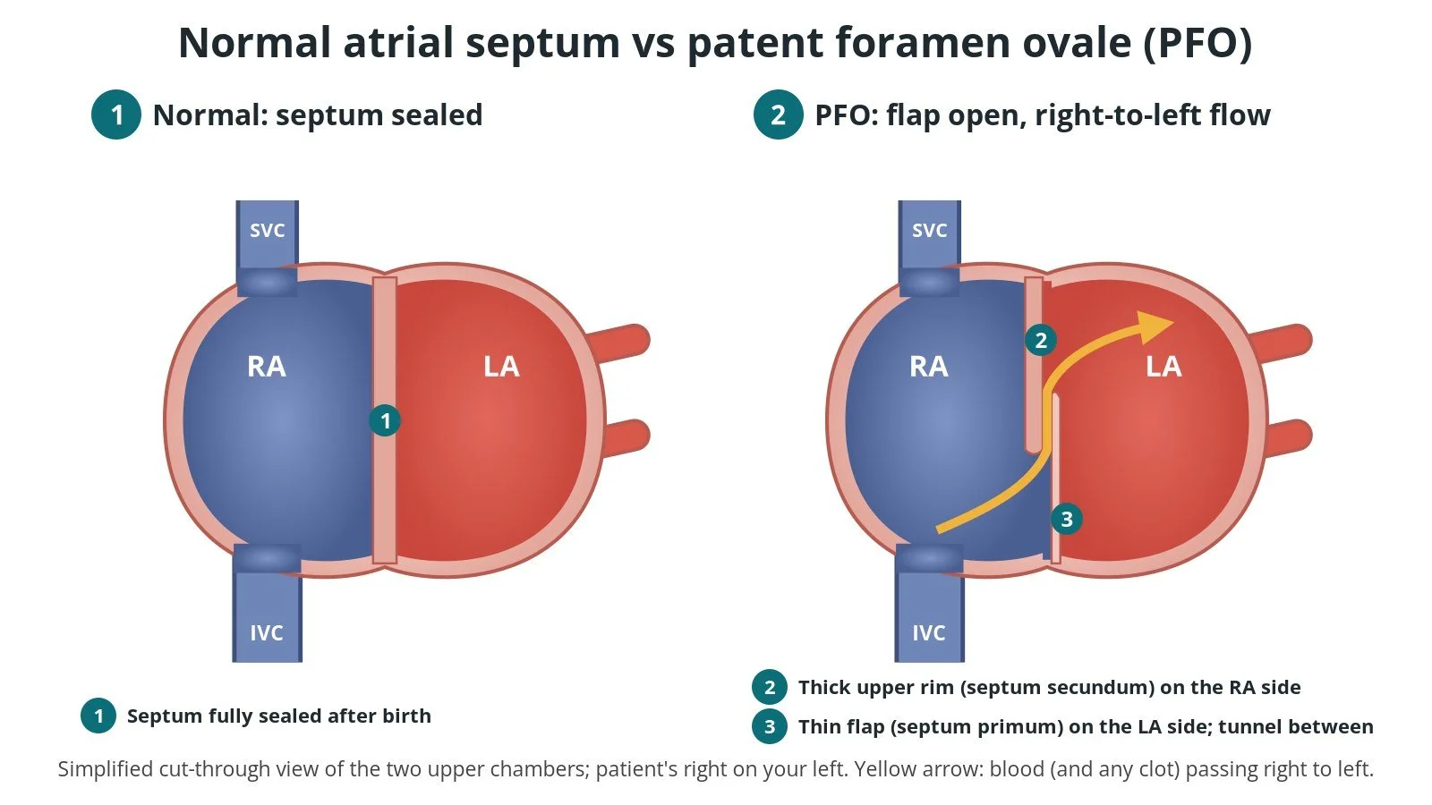
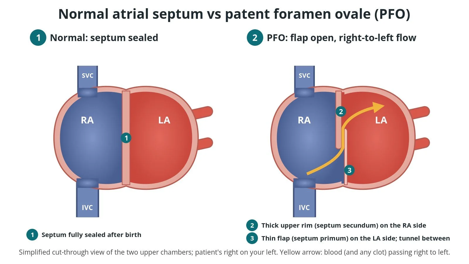
Normal septum vs PFO. (1) Normal: the septum is sealed. (2) PFO: the thin flap and thick rim overlap but do not seal, so blood can pass right to left through the tunnel.
What is a PFO?
The atrial septum is the wall between the right atrium (RA), which receives blue blood from the body through the two big veins (the inferior and superior vena cava, IVC and SVC), and the left atrium (LA), which receives red blood from the lungs.
Before birth, the lungs are not used, so blood needs to cross from the right side to the left. It does this through the foramen ovale, a gap between two overlapping parts of the septum: a thin flap on the left side (the septum primum) and a thicker rim on the right side (the septum secundum). After birth the pressure in the LA rises, the flap is pushed shut and, in most people, it seals over.
In about a quarter of adults the flap never fully seals. This is a patent (open) foramen ovale. Most of the time the higher pressure in the LA keeps it closed. But when the pressure on the right side briefly rises, for example when coughing, straining or lifting, the flap can open and a little blood can pass right to left. If that blood carries a small clot, usually from a vein, it can bypass the lungs' filter and travel to the brain. This is called a paradoxical embolism.
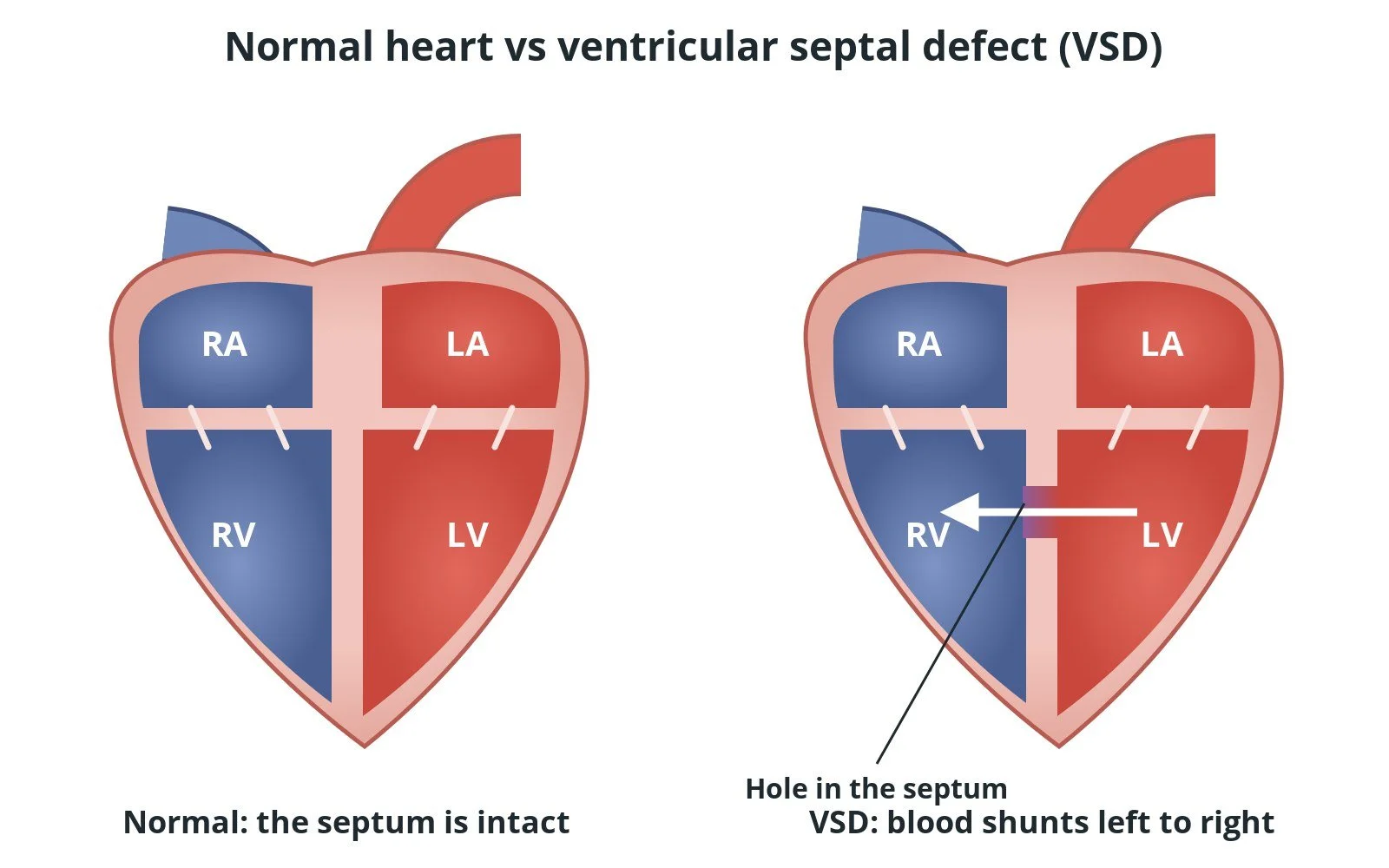
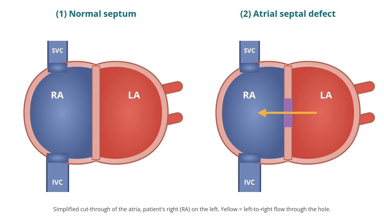
A PFO is different from an atrial septal defect (ASD), which is a true hole in the septum with no flap. Some PFOs sit next to a floppy, bulging septum, an atrial septal aneurysm, or have a large shunt. These features make it more likely that the PFO played a part in a stroke.
PFO closure seals the tunnel from inside the heart. A double-disc occluder, a small device made of a fine metal mesh frame covered with fabric, is folded into a thin tube, passed up from a vein in the groin and opened so that one disc sits on each side of the septum. Over the following months, the body's own tissue grows over it.
New to the acronyms? See the Cath Lab Abbreviations A–Z.
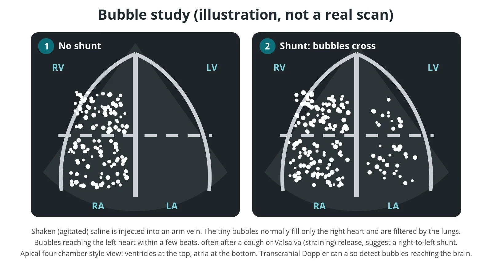
Illustrated bubble study, not a real scan. (1) No shunt: bubbles stay in the right heart. (2) Shunt: bubbles appear in the left heart within a few beats.
Who is it for?
The decision is made after a full stroke work-up, usually by a multidisciplinary team (MDT) that includes a stroke neurologist and a cardiologist (a "heart–brain team"). UK (NICE), European (ESC/EAPCI position paper) and American (AHA/ASA 2021) guidance broadly agree that:
closure is considered in adults who have had an ischaemic stroke or TIA (or other embolism) with no other cause found after thorough tests, such as brain imaging, heart rhythm monitoring for AF, imaging of the neck and brain arteries, echo and blood tests
the team weighs how likely it is that the PFO caused the event. Younger age, a stroke pattern that looks embolic, a large shunt and an atrial septal aneurysm all make a causal role more likely
age limits differ between guidelines. The AHA/ASA 2021 guideline describes closure as reasonable for people aged 18 to 60 with a non-lacunar stroke, no other cause and high-risk PFO features. The ESC/EAPCI position paper discusses roughly 18 to 65. Outside these ranges, decisions are individual
the person's own wishes, after a balanced discussion of benefits and risks, are central
NICE (interventional procedures guidance IPG472, now listed as HealthTech guidance HTG325) states that the evidence supports PFO closure to prevent recurrent cerebral embolic events with normal arrangements, and that patients should be selected by a team including a neurologist and a cardiologist. In England, NHS England's clinical commissioning policy sets out who is eligible for NHS-funded closure.
PFO closure is not recommended just because a PFO is found by chance. It is also not routine treatment for migraine, and closure for diving or other reasons is a specialist decision outside this page. Other causes, such as AF, must be looked for and treated on their own terms.
When is it done?
PFO closure is a planned procedure, usually a few weeks or months after the stroke or TIA once the work-up is complete. Before it, the team usually:
confirms the PFO and the shunt with a bubble study: harmless agitated saline is injected into an arm vein during an echo, often with a cough or Valsalva (straining) manoeuvre, and bubbles seen in the left heart within a few beats suggest a right-to-left shunt. Transcranial Doppler (TCD) can also detect bubbles reaching the brain
looks at the anatomy in detail with a transoesophageal echo (TOE): the length of the tunnel, the rims, any atrial septal aneurysm, other holes, and clot in the heart
checks for AF with heart rhythm monitoring, and for other heart or artery causes of stroke
reviews blood thinners, allergies (including nickel, as most device frames contain nickel-titanium) and the plan for antiplatelet medicines, as per centre protocol
talks through the options and the person's goals before consent
Step by step
1. Anaesthetic and set-up
PFO closure takes about an hour. Some centres use a general anaesthetic so a TOE probe can guide the procedure. Others use local anaesthetic with sedation and an intracardiac echo (ICE) catheter, placed through a second vein, so no probe goes down the throat. X-ray (fluoroscopy) is used throughout. Antibiotics are given as per centre protocol.
2. Femoral venous access
Access is from the femoral vein at the top of the leg, usually the right, under ultrasound guidance. A sheath is inserted; the delivery sheath is sized per device and operator.
From the femoral vein, catheters pass up the iliac vein and the IVC into the RA. Blood from the IVC naturally flows towards the septum, which is why the PFO is easiest to cross from below.
For ICE-guided cases a second venous sheath is placed for the ICE catheter, size per device and operator.
Anticoagulation (usually heparin) is given before the PFO is crossed, dose as per centre protocol.
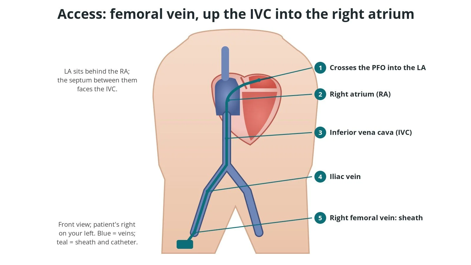
Access: (1) catheter across the PFO into the LA; (2) right atrium; (3) inferior vena cava; (4) iliac vein; (5) sheath in the right femoral vein.
3. Crossing, sizing, deploying and releasing
A. Crossing the PFO. A soft-tipped catheter and wire are steered from the IVC across the tunnel into the LA, and the wire is parked in a pulmonary vein, usually the left upper. It is exchanged for a stiff exchange wire that supports the delivery sheath.
Sizing. The team decides the device size from the TOE or ICE measurements. Some operators also gently inflate a soft sizing balloon across the tunnel to see its stretched size and check that the true PFO, rather than a separate small hole, has been crossed. Balloon use and sizes are per device and operator.
B. Left disc. The delivery sheath is passed over the wire into the LA and de-aired carefully, as air must not enter the left heart. The folded device is pushed out until the left-atrial disc opens in the LA. The sheath and disc are then pulled back gently until the disc sits flat against the left side of the septum.
C. Right disc. Holding gentle tension, the sheath is withdrawn further so the right-atrial disc opens on the right side, sandwiching the septum between the two discs.
Push–pull check. Before release, the operator gently pushes and pulls the device to make sure it is stable. Echo checks that the discs grip the septum on both sides, are clear of nearby structures such as the aorta and the valves, and that there is little or no leak.
D. Release. If everything looks right, the device is unscrewed or released from its delivery cable. Most devices can be pulled back into the sheath and repositioned or swapped before release.
A final echo, often with another bubble study, checks the device position and any remaining shunt, and looks for fluid around the heart.
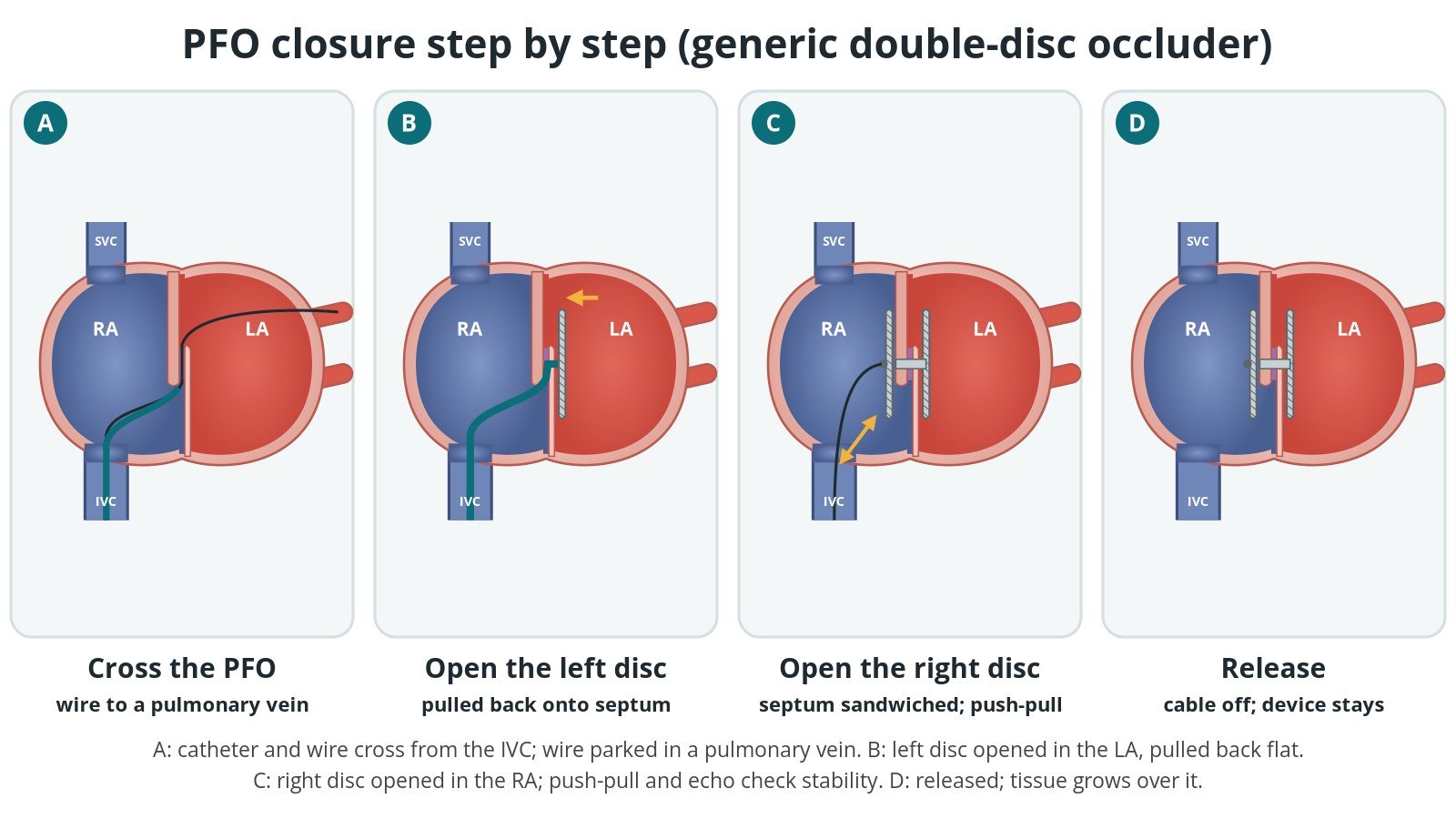
A: cross the PFO. B: open the left disc and pull it back onto the septum. C: open the right disc; push-pull and echo check. D: release the device.
4. Finishing
The delivery sheath is removed and the vein is closed with pressure, a temporary skin stitch or a closure device, per operator. The TOE probe is removed and the person is woken up and taken to recovery.
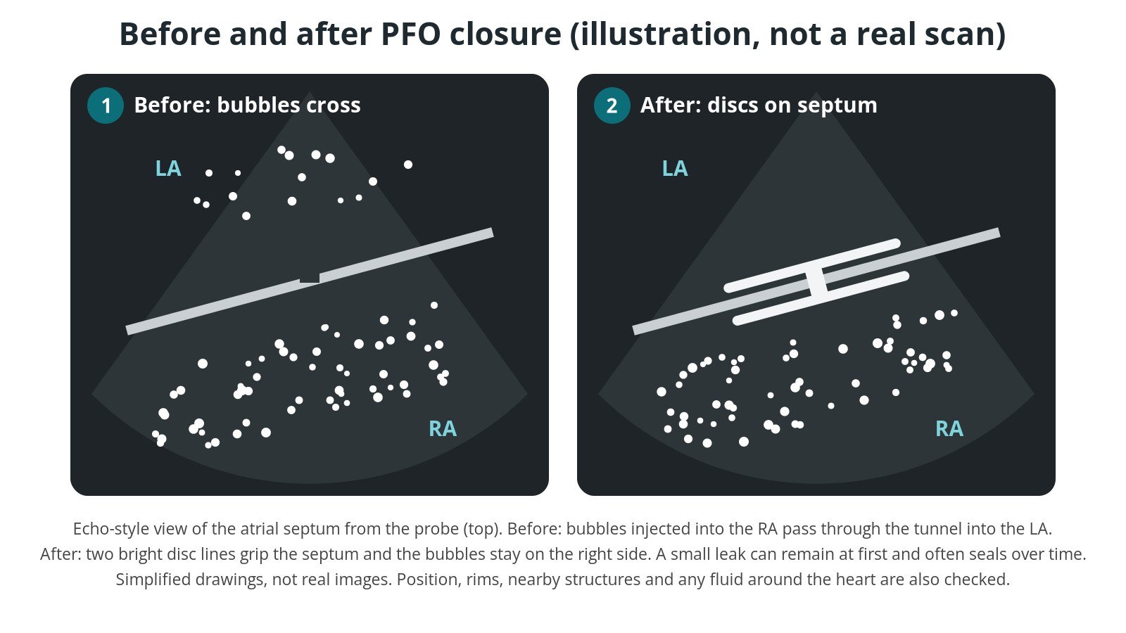
Illustrated echo, not a real scan. Before: bubbles cross the septum. After: the two discs grip the septum and bubbles stay on the right side.
Recovery and aftercare
First hours: bed rest for a few hours, with checks of the groin, blood pressure, heart rhythm and the leg pulses, as per centre protocol. An ECG and often an echo are done before going home.
Going home: many people go home the same day or the next day.
Groin: keep it clean and dry for a few days. Avoid heavy lifting and strenuous exercise for about a week, or as advised by your team.
Antiplatelet medicines: you will usually take antiplatelet medicines (such as aspirin, often with a second one for a time) while tissue grows over the device. The ESC/EAPCI position paper suggests two antiplatelet medicines for 1 to 6 months, then one for at least 5 years. Your own plan is set by your team as per centre protocol; don't stop these medicines without advice.
Infection prevention: antibiotic cover for dental work in the first months is decided by your team. Good dental care is always important with any heart device.
Follow-up: an echo, often with a bubble study, is usually done a few months later to check the device and any remaining shunt.
Driving (UK): the DVLA rules after a stroke or TIA apply first: car and motorcycle (Group 1) drivers must not drive for 1 month after a stroke or TIA, and only return if recovery is satisfactory. Bus and lorry (Group 2) drivers have stricter rules and must tell the DVLA. The DVLA cardiovascular guidance has no separate rule for PFO closure itself. Ask your team when it is safe to drive after the procedure, and check the DVLA guidance for your licence.
Get help: contact your team if the groin swells, bleeds or becomes painful, or if you notice a fast or irregular heartbeat or palpitations. Call 999 for chest pain, fainting, severe breathlessness, or signs of stroke such as face drooping, arm weakness or slurred speech.
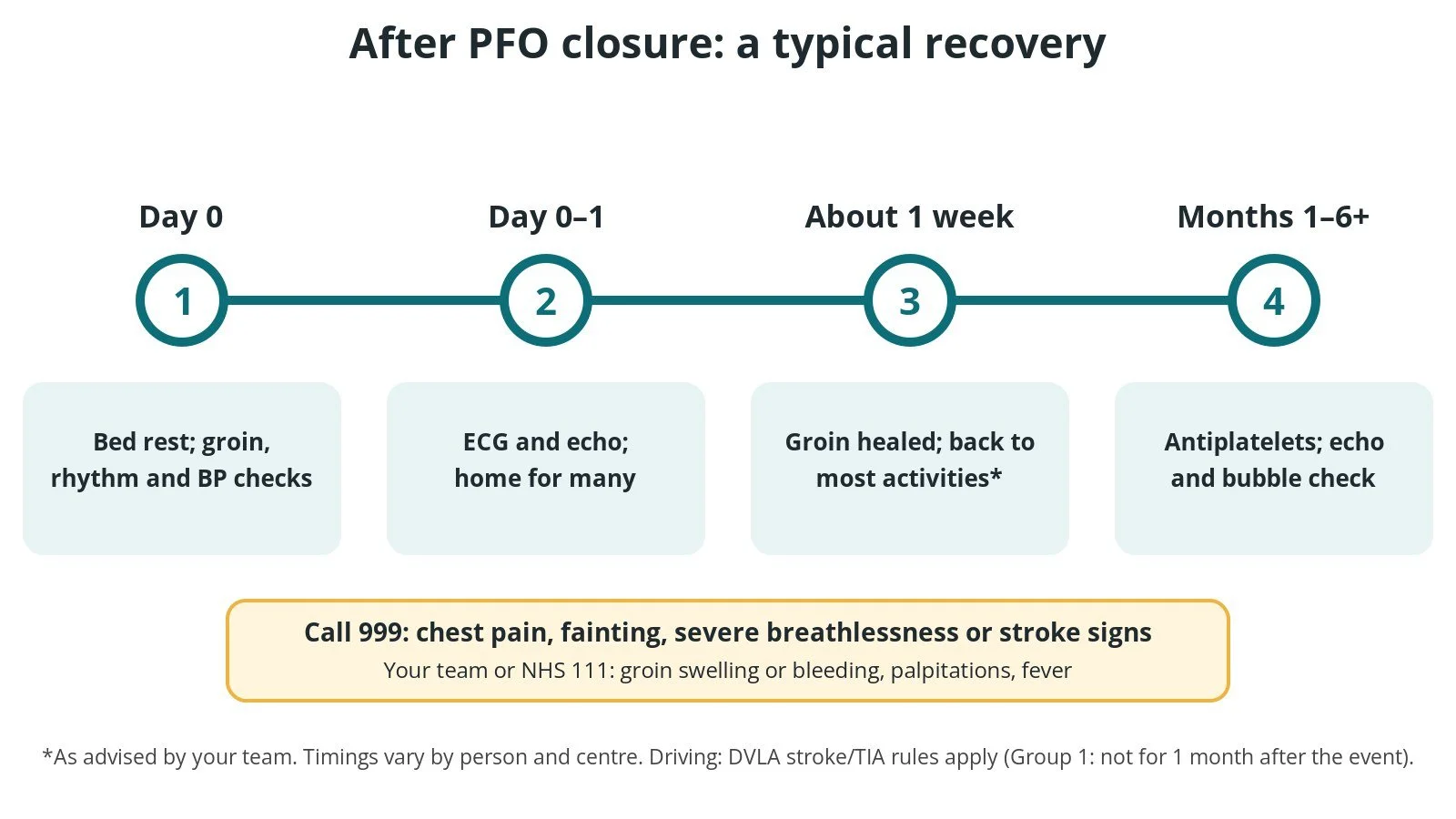
After PFO closure: (1) bed rest and checks; (2) ECG, echo, home for many; (3) most activities after about a week, as advised; (4) antiplatelets and a follow-up echo and bubble check.
Risks and complications
PFO closure is a relatively short procedure, but every procedure carries risk. Possible problems include:
Atrial fibrillation (AF) or other irregular rhythms, most often in the first weeks and usually short-lived
Bleeding, bruising or damage to the groin vein, sometimes with an abnormal connection between an artery and vein
A remaining small leak (residual shunt) around or through the device
Clot forming on the device, which is one reason for antiplatelet medicines
Fluid around the heart (pericardial effusion or tamponade), rare
The device moving out of place (embolisation), rare, usually retrieved with a catheter or, very rarely, surgery
Device erosion, where the device wears through the heart wall, is rare but serious
Stroke or air embolism during the procedure, rare
Allergic reaction, including to nickel, or a reaction to the anaesthetic
Infection of the device (endocarditis), rare
Your team will explain how these apply to you.
Cath lab kit list (printable)
Drugs, doses and dilutions: always as per centre protocol. Equipment, sizes and choices also vary by unit and operator. Device and product names below are generic. Always check the sheath, wire and delivery system required in each product's instructions for use.
Example: Barts Heart Centre kit list — always follow your own centre protocol
Typical setup for PFO closure under general anaesthetic with TOE guidance, shared as one real-world example. It is not a protocol for your unit.
Scrub trolley
angiography pack
femoral venous sheath, size per device and operator
second venous sheath for an ICE catheter, if ICE-guided, per device and operator
multipurpose-shaped diagnostic catheter for crossing, per operator
standard J-tip guidewire, and a stiff exchange guidewire (long length), per operator
double-disc PFO occluder, size per imaging and operator
delivery sheath and delivery system matched to the chosen device, per its instructions for use
Luer-lock syringes for flushing and de-airing the device and sheath
3-way tap with extension tubing
heparinised saline flush bag with a giving set, as per centre protocol
non-absorbable suture for a temporary skin stitch, if used, per operator
ultrasound probe cover(s): one for vascular access, and one for the ICE catheter if used
Drugs: as per centre protocol
local anaesthetic for the groin
anticoagulation (heparin) before crossing the PFO
antibiotics
agitated saline for the bubble study
Sizing balloon (if used)
soft compliant sizing balloon, size per operator
contrast and saline mix for the balloon, as per centre protocol
3-way tap and large Luer-lock syringes
Imaging
TOE probe and echo machine, or ICE catheter and console
manifold for contrast injections to confirm device position, if used
Procedural steps (Barts example)
Right femoral venous access under ultrasound; venous sheath inserted (plus a second venous sheath if ICE-guided)
Anticoagulation as per centre protocol
Multipurpose catheter and wire used to cross the PFO from the IVC
Catheter parked in a pulmonary vein (usually left upper); wire exchanged for a stiff exchange wire; catheter removed
PFO sized on TOE or ICE, with a sizing balloon if the operator uses one
Delivery sheath advanced into the LA and de-aired
Left disc opened in the LA and pulled back to the septum; right disc opened in the RA
Push–pull check and echo check of position, rims and leak
Device released; final echo and bubble study
Everything removed; vein closed per operator
End of case; transfer to recovery
First time in the cath lab?
💡 First-time tip
Respect the air. The delivery sheath sits in the left atrium. Follow the de-airing steps exactly and keep flush lines free of bubbles: air on the left side can reach the brain. Know your echo views. Learn the TOE or ICE view that shows the tunnel and both discs. The echo operator calls whether the discs are flat and gripping the septum. Watch the rhythm. Wires in the atria often cause extra beats or short runs of AF. Have the defibrillator ready and tell the team about anything new. Check the device before it opens. Confirm the device type, size and expiry with the operator before it is loaded, and keep the packaging until the case is finished. Watch the groin and the pressure at the end. A falling blood pressure can mean fluid around the heart; let the team know straight away.
FAQ
Is a PFO the same as a hole in the heart?
It is one kind. A PFO is a flap-like tunnel left over from before birth, while an atrial septal defect (ASD) is a true hole in the septum. They are treated differently.
Does everyone with a PFO need it closed?
No. About 1 in 4 people have a PFO, and most never have a problem. Closure is considered mainly after a stroke or TIA where no other cause is found and the team thinks the PFO played a part.
Am I awake for it?
It depends on the centre. With TOE guidance you usually have a general anaesthetic. With ICE guidance it can be done under local anaesthetic and sedation.
Can I feel the device?
No. Some people notice palpitations in the first weeks, usually from short-lived extra beats or AF. Tell your team if you do.
Will I still need blood thinners?
Usually antiplatelet medicines, often for several years, as advised by your team. Some people also need other blood thinners for separate reasons, such as AF.
Can I have an MRI scan afterwards?
Most PFO occluders are labelled MR conditional, which means a scan is possible under set conditions. Always tell the scanning team about your device and carry your implant card.
Related Cath Lab 101 guides
Sources
All checked on 10 October 2026. Each page's own date is shown where it gives one.
NICE. Percutaneous closure of patent foramen ovale to prevent recurrent cerebral embolic events (IPG472, now HTG325). nice.org.uk/guidance/htg325 (published 1 Dec 2013)
Pristipino C, et al. European position paper on the management of patients with patent foramen ovale. General approach and left circulation thromboembolism. Eur Heart J 2019;40(38):3182–3195. PubMed 30358849
Kleindorfer DO, et al. 2021 Guideline for the Prevention of Stroke in Patients With Stroke and Transient Ischemic Attack (AHA/ASA). Stroke 2021;52(7):e364–e467. PubMed 34024117
NICE. Stroke and transient ischaemic attack in over 16s: diagnosis and initial management (NG128). nice.org.uk/guidance/ng128
NHS England. Clinical commissioning policy: percutaneous patent foramen ovale closure for the secondary prevention of cerebral embolic stroke in adults. england.nhs.uk
NHS. Transient ischaemic attack (TIA). nhs.uk/conditions/transient-ischaemic-attack-tia
British Heart Foundation. Congenital heart conditions. bhf.org.uk
Stroke Association. Causes of ischaemic stroke. stroke.org.uk
DVLA / GOV.UK. Neurological disorders: assessing fitness to drive (stroke and TIA). gov.uk (last updated 7 Nov 2025)
Disclaimer
Education only. This page is for general education for healthcare students, cath lab staff and curious readers. It isn't medical advice and doesn't replace advice from your own doctor, heart team or local unit protocols. Equipment and medicines are described generically. No product or manufacturer is recommended. If you think someone is having a heart emergency or a stroke, call 999.
Disclosure: I work for a company that makes a device used in this procedure. This page is independent education, isn't sponsored or reviewed by any company, and doesn't recommend any product.
All diagrams on this page are original illustrations created for rodnieoro.com.