Transcarotid TAVI (carotid artery access): a Cath Lab 101 guide
Reviewed by Rodnie Oro, cath lab charge nurse · Last reviewed: October 2026 · Education only, not medical advice
Transcarotid TAVI in one minute
What it is: TAVI replaces a narrowed aortic valve using a catheter. Transcarotid TAVI delivers the new valve through the common carotid artery in the neck, exposed through a small surgical incision, instead of through the groin.
Who it is for: people with severe aortic stenosis chosen for TAVI by a heart team, whose leg arteries are unsuitable, and whose carotid arteries and brain circulation are suitable on scans.
Main risks: stroke, bleeding or a haematoma in the neck, nerve injury causing hoarseness, heart block needing a pacemaker, and the usual TAVI risks. Your team will explain your own risk.
Usually: under general anaesthetic in a hybrid theatre, with surgical and cath lab teams, about 2 to 3 hours, often home after a few nights.
On this page: What is transcarotid TAVI? · Who is it for? · When is it done? · Step by step · Recovery · Risks · Kit list · First time in the lab? · FAQ
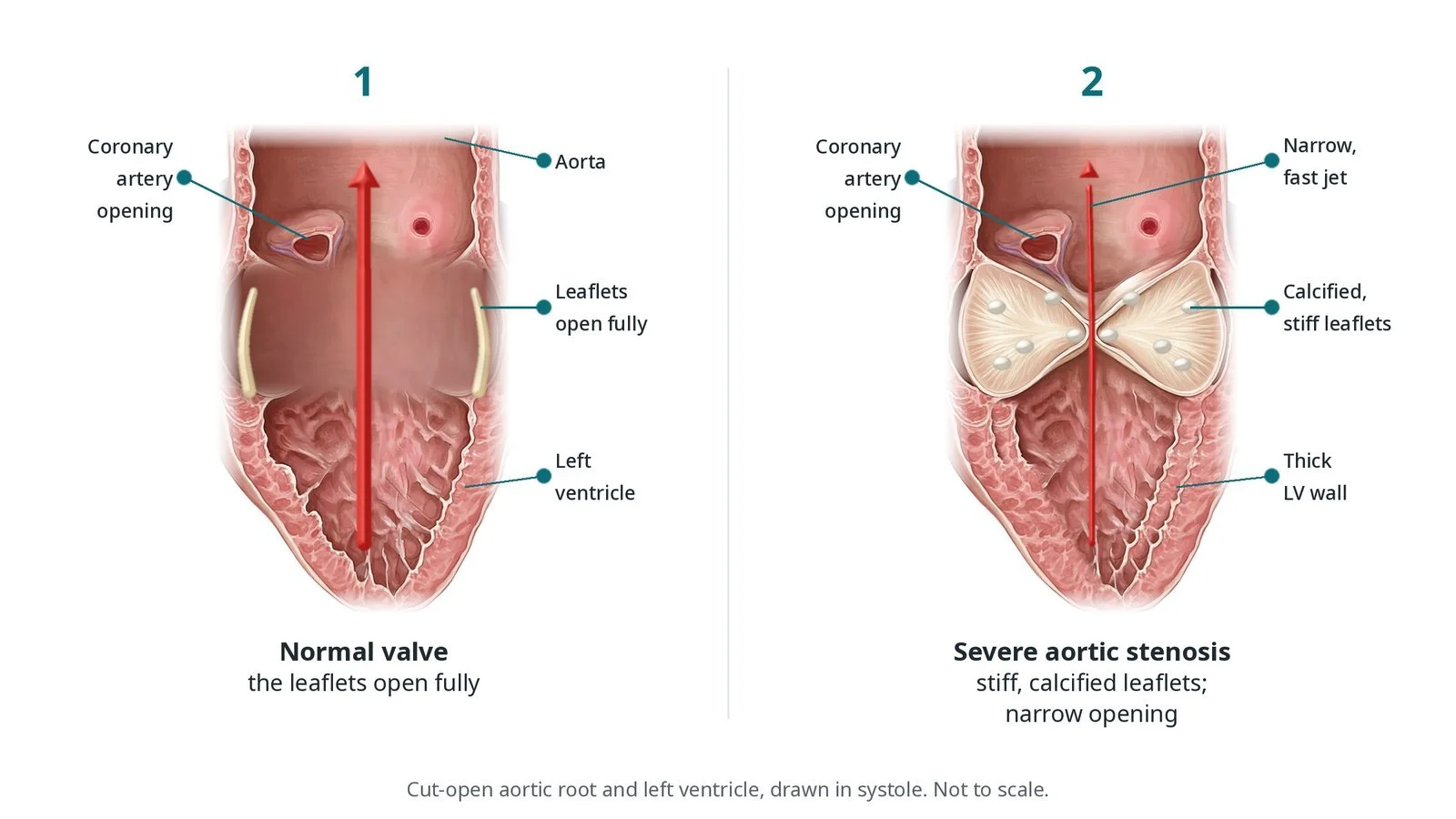
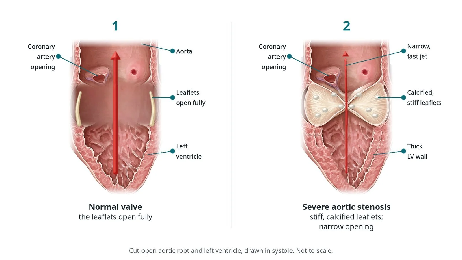
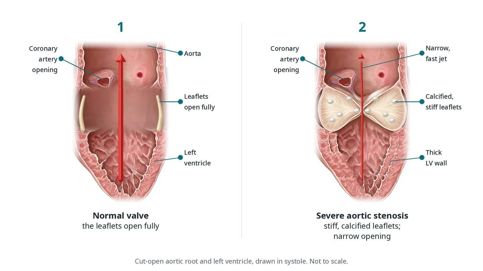
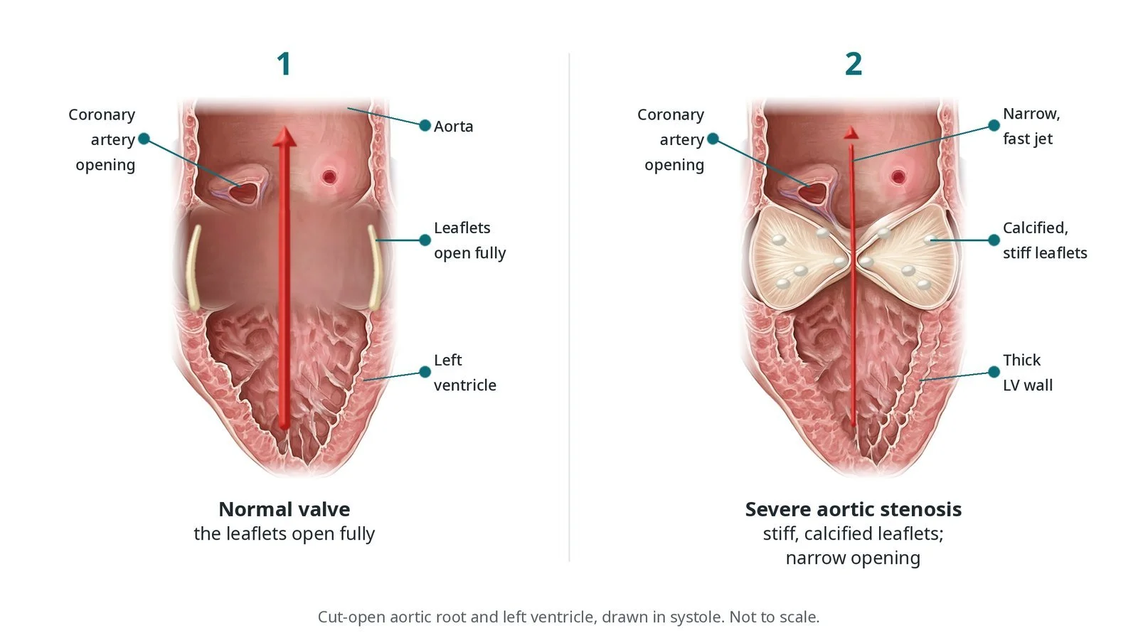
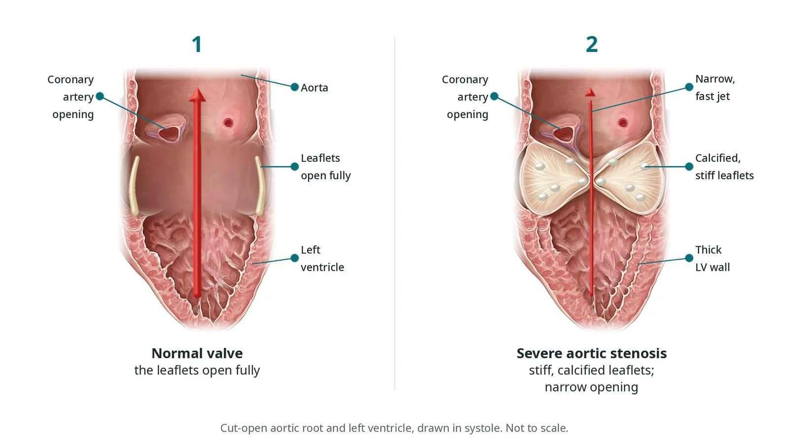
Normal valve vs severe aortic stenosis (AS), cut-open aortic root drawn in systole. Normal: the leaflets open fully against the wall. Severe AS: calcified, stiff leaflets, a narrow fast jet and a thick LV wall. The coronary arteries start from the sinuses just above the valve.
What is transcarotid TAVI?
The aortic valve lets blood leave the heart into the aorta. In aortic stenosis, it becomes stiff and narrowed, usually from calcium build-up with age, causing breathlessness, chest pain, dizziness or blackouts. TAVI (transcatheter aortic valve implantation) places a new valve inside the old one using a catheter, without open-heart surgery. See the main TAVI guide.
Most TAVIs are done through the femoral artery in the groin (transfemoral). In some people the leg and pelvic arteries are too narrow, diseased, twisted or heavily calcified for the large delivery system. An alternative access route is then needed. The main options are transaxillary/subclavian (under the collarbone), transcarotid (the neck), transcaval (from the femoral vein into the aorta), and surgical routes through the chest wall. The heart team chooses the route using CT.
In transcarotid TAVI, a surgeon exposes the common carotid artery in the neck through a small incision. A sheath is placed directly into the artery, giving a short, straight path down to the aortic valve. Either side can be used; the choice depends on CT, the other neck arteries and the brain circulation. It is usually a hybrid case, combining a vascular or cardiac surgical team with the cath lab team.
New to the acronyms? See the Cath Lab Abbreviations A–Z.
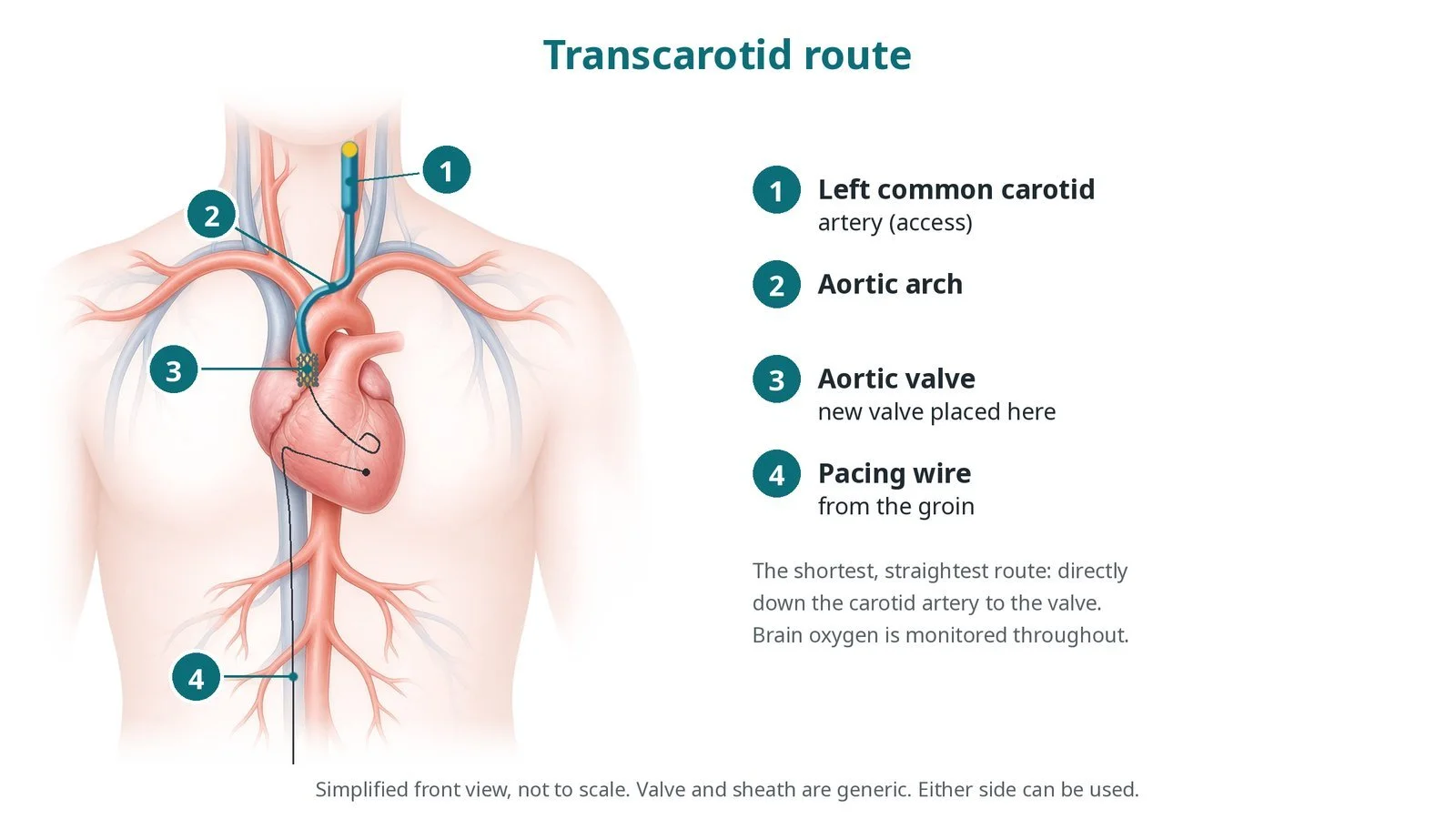
Route, front view: (1) left common carotid artery (access); (2) aortic arch; (3) aortic valve; (4) pacing wire from the groin. The shortest, straightest route to the valve.
Who is it for?
NICE guidance on heart valve disease (NG208) recommends TAVI or surgery for severe symptomatic aortic stenosis based on age, risk and a heart team decision, and NICE interventional guidance on TAVI (HTG446) supports its use. The ESC/EACTS valvular heart disease guidelines state that transfemoral access is preferred and that non-transfemoral access may be considered in people unsuitable for surgery and for transfemoral TAVI. The choice between alternative routes depends on the person's anatomy and the centre's experience.
For carotid access, scans must show a carotid artery without significant narrowing, and the circle of Willis (the connection of arteries at the base of the brain) must allow blood to reach the brain from the other side while the artery is clamped.
When is it done?
It is planned. Before it, the team usually arranges:
CT angiography of the heart, aorta, access arteries and neck vessels
carotid ultrasound and sometimes imaging of the brain arteries
echo, ECG, blood tests and a group and save
a review of blood thinners, as per centre protocol
Step by step
1. Anaesthetic and set-up
The case is done in a hybrid theatre under general anaesthetic. An arterial line is placed in the wrist before the anaesthetic. A femoral vein sheath carries a temporary pacing wire, and a femoral or radial artery sheath is used for angiography. Cerebral oximetry (sensors on the forehead) monitors brain oxygen levels. A cell saver may be prepared. The neck, chest and both groins are prepared and draped.
2. Carotid exposure and brain check
The surgeon makes a small incision in the lower neck and exposes the common carotid artery, tying off small veins in the way.
The artery is briefly clamped to check that brain oxygen levels stay acceptable.
Heparin is given, dose as per centre protocol.
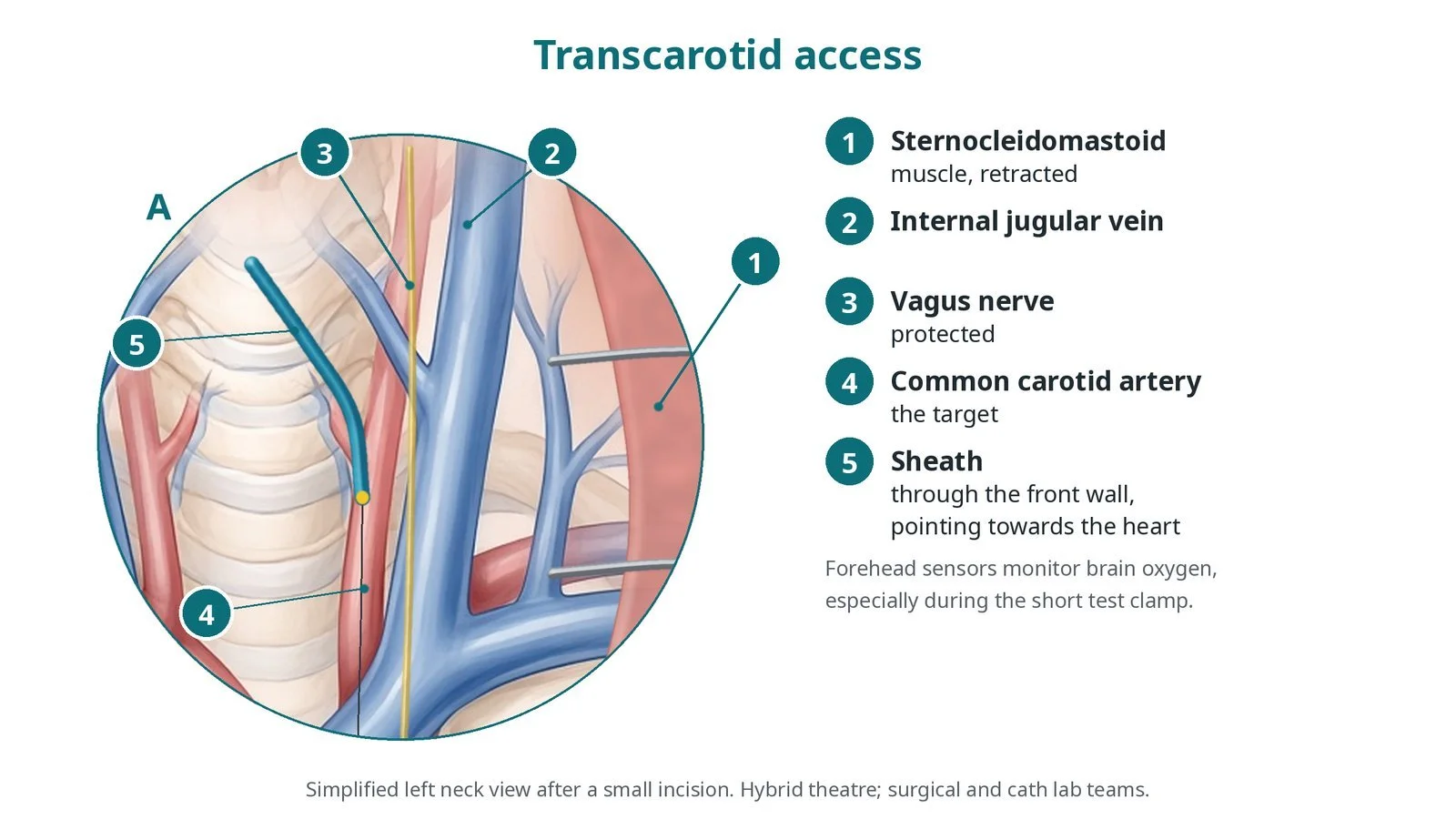
Close-up of the left neck: after a small incision the common carotid artery is exposed, with the internal jugular vein and vagus nerve beside it and the sternocleidomastoid muscle retracted. The sheath enters the front wall of the artery and points towards the heart. Forehead sensors monitor brain oxygen during the test clamp.
3. Access and valve delivery
A. Sheath in. The carotid artery is punctured and a short sheath placed. A catheter and soft wire cross the aortic valve, then a stiff wire is placed in the left ventricle.
B. Balloon (if used). The valve may be stretched first with a balloon under rapid pacing; size per operator.
C. Large sheath and valve. The sheath is exchanged for the TAVI sheath and the valve is positioned across the aortic valve.
D. Deploy. The valve is expanded under rapid pacing, as in transfemoral TAVI. Echo, angiography and pressures check the result.
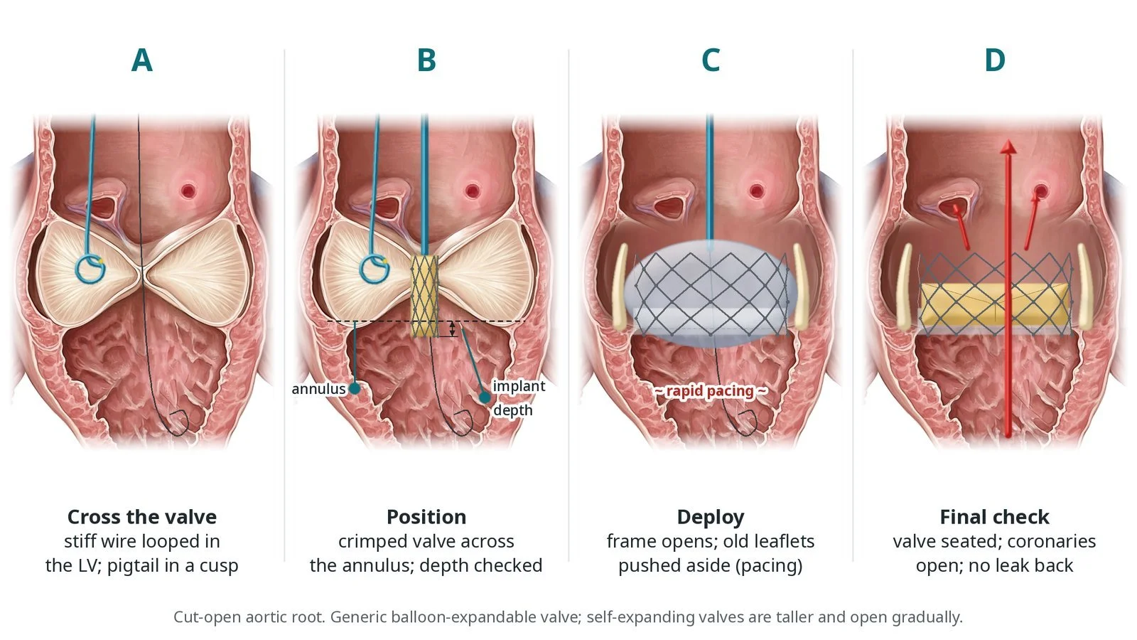
A: a stiff wire is looped in the LV and a pigtail sits in a cusp. B: the crimped valve is positioned across the annulus at the planned depth. C: the frame opens and the old leaflets are pushed aside, with pacing. D: the valve is seated, the coronaries are open and there is no leak back. The deployment steps are the same for every access route.
4. Closure
The sheath is removed with the artery controlled by clamps, and the surgeon repairs the carotid artery with stitches and closes the neck. The femoral access is closed with closure devices or pressure, per operator.
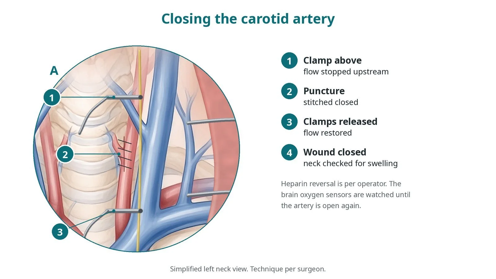
Closure: clamps above and below control the artery while the surgeon stitches the puncture closed; the clamps are then released and flow is restored.
Recovery and aftercare
First hours: recovery, often in a coronary care, high-dependency or intensive care area, with checks of the neck wound, brain function (speech, arm and leg strength), heart rhythm and blood pressure, as per centre protocol.
Echo and blood tests: before discharge.
Heart rhythm: some people develop heart block and need a permanent pacemaker.
Going home: often after 1 to 3 nights, sometimes longer after alternative access.
Neck: watch for swelling, difficulty swallowing or breathing, or hoarseness.
Activity: avoid heavy lifting for a few weeks, or as advised.
Medicines: antiplatelet or anticoagulant medicines, as per your team's plan.
Driving: DVLA guidance after a percutaneous valve procedure for a car licence is not to drive for 2 weeks; bus and lorry drivers must tell the DVLA. Ask your team.
Call 999 if you have chest pain, sudden breathlessness, fainting, signs of a stroke, or bleeding or swelling at the access site.
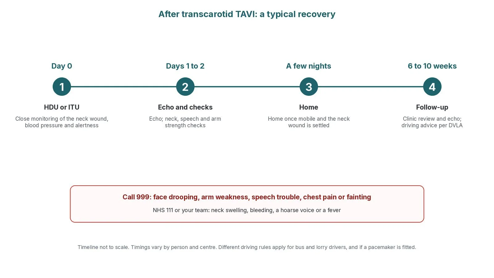
After transcarotid TAVI: (1) HDU/ITU monitoring; (2) echo, neck and brain checks; (3) home after a few nights; (4) follow-up at 6 to 10 weeks.
Risks and complications
Your team will discuss your own risks. Possible problems include:
stroke or TIA, including from reduced blood flow during clamping
bleeding or a haematoma in the neck, which can press on the airway
injury to nerves in the neck, causing hoarseness or swallowing problems (often temporary)
narrowing of the carotid artery later
stroke
bleeding or injury to the access artery or vein
heart block needing a permanent pacemaker
a leak around the new valve
kidney strain from contrast dye
rarely, blockage of a coronary artery, damage to the aortic root, or the valve moving
Cath lab kit list (printable)
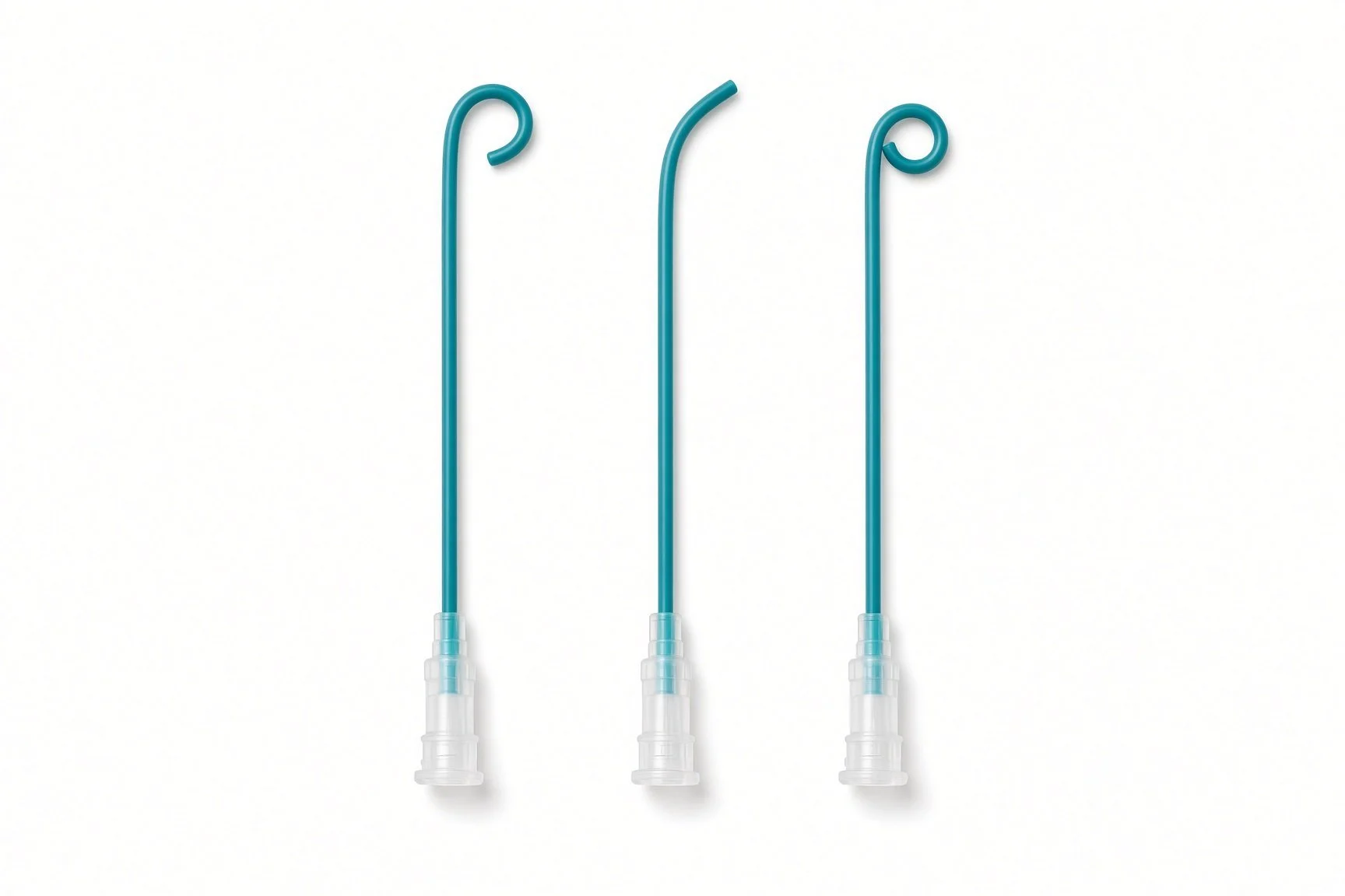
Diagnostic catheters (left coronary, right coronary and multipurpose shapes; pigtail) (from the guide kit list; no published kit-page picture)
Barts example kit list
An example from one UK centre, written generically. Every lab has its own protocol, and sizes and doses vary.
Set-up
hybrid theatre; theatre (surgical) team and cath lab team
arterial line before induction; cerebral oximetry; cell saver
temporary pacing via a femoral vein
Kit
TAVI procedure pack and surgical instrument set (provided by theatre)
micropuncture kit
carotid sheath, femoral arterial and venous sheaths, sizes per device and operator
temporary pacing wire (balloon-tipped if used)
diagnostic catheters (left coronary, right coronary and multipurpose shapes; pigtail)
standard and long exchange wires; stiff pre-shaped LV wire, per operator
valvuloplasty balloon, size per operator
TAVI valve, delivery system and large-bore sheath, per device and operator
femoral closure devices, per operator
crimping kit: rinsing bowls, saline and heparinised saline, per device instructions
Procedural steps (Barts example)
Arterial line; general anaesthetic; drapes expose neck, chest and both groins
Femoral arterial (or radial) and venous access; pacing wire; aortogram
Surgical cut-down to the common carotid artery; clamp test with cerebral oximetry
Heparin as per centre protocol; micropuncture and carotid sheath
Cross the valve; exchange wires; stiff wire in the LV
Balloon valvuloplasty under rapid pacing (if used)
Exchange to large sheath; valve deployed under rapid pacing
Sheath out under clamp control; surgical repair of the carotid; femoral closure
To HDU/ITU; echo and bloods
First time in the cath lab?
💡 First-time tip
Two teams, one plan. Agree roles with the theatre team at the briefing: who scrubs for the neck and who for the valve. Watch the oximetry. Report any fall in brain oxygen levels during clamping. Neck checks. After the case, a neck haematoma can threaten the airway; know the escalation plan. Count everything. It is a surgical case; instrument and swab counts apply. Pacing ready. Check rapid pacing before balloon and valve deployment.
FAQ
Why the neck?
Because the leg arteries are unsuitable, and the carotid gives a short, direct route to the aortic valve.
Will I have a scar?
Yes, a small scar in the lower neck.
Does clamping the artery harm the brain?
The team checks your brain oxygen levels during a test clamp before going ahead. Stroke remains a risk, which your team will discuss.
How long will I stay in hospital?
Often a few nights, sometimes longer.
Related Cath Lab 101 guides
Sources
All checked on 10 October 2026. Each page's own date is shown where it gives one.
NICE. Heart valve disease presenting in adults: investigation and management (NG208). nice.org.uk/guidance/ng208
NICE. Transcatheter aortic valve implantation for aortic stenosis (HTG446). nice.org.uk/guidance/htg446 (published 26 Jul 2017)
Vahanian A, et al. 2021 ESC/EACTS Guidelines for the management of valvular heart disease. Eur Heart J 2022;43(7):561–632. PubMed 34453165
British Heart Foundation. TAVI. bhf.org.uk
NHS. Aortic valve replacement. nhs.uk
DVLA / GOV.UK. Cardiovascular disorders: assessing fitness to drive (percutaneous valve intervention). gov.uk
Disclaimer
Education only. This page is for general education for healthcare students, cath lab staff and curious readers. It isn't medical advice and doesn't replace advice from your own doctor, heart team or local unit protocols. Equipment and medicines are described generically. No product or manufacturer is recommended. If you think someone is having a heart emergency, call 999.
Disclosure: I work for a company that makes a device used in this procedure. This page is independent education, isn't sponsored or reviewed by any company, and doesn't recommend any product.
All diagrams on this page are original illustrations created for rodnieoro.com.