Cath Lab Procedures: Complete Reference List
Reviewed by Rodnie Oro, cath lab charge nurse · Last reviewed: October 2026 · Education only, not medical advice
This list in one minute
What it is: a plain-English list of 51 procedures that can be done in or alongside a cardiac catheterisation (cath) lab, grouped into 7 categories.
How to use it: each item has a one-line description. Items in blue link to a full Cath Lab 101 guide. Others are listed for reference and may get their own guide later.
Note: which procedures are done, and where, varies between hospitals.
On this page: Diagnostic · Interventional Coronary · Structural Heart · Peripheral Vascular · Support and Rescue · Electrophysiology (often a separate lab) · Imaging Adjuncts
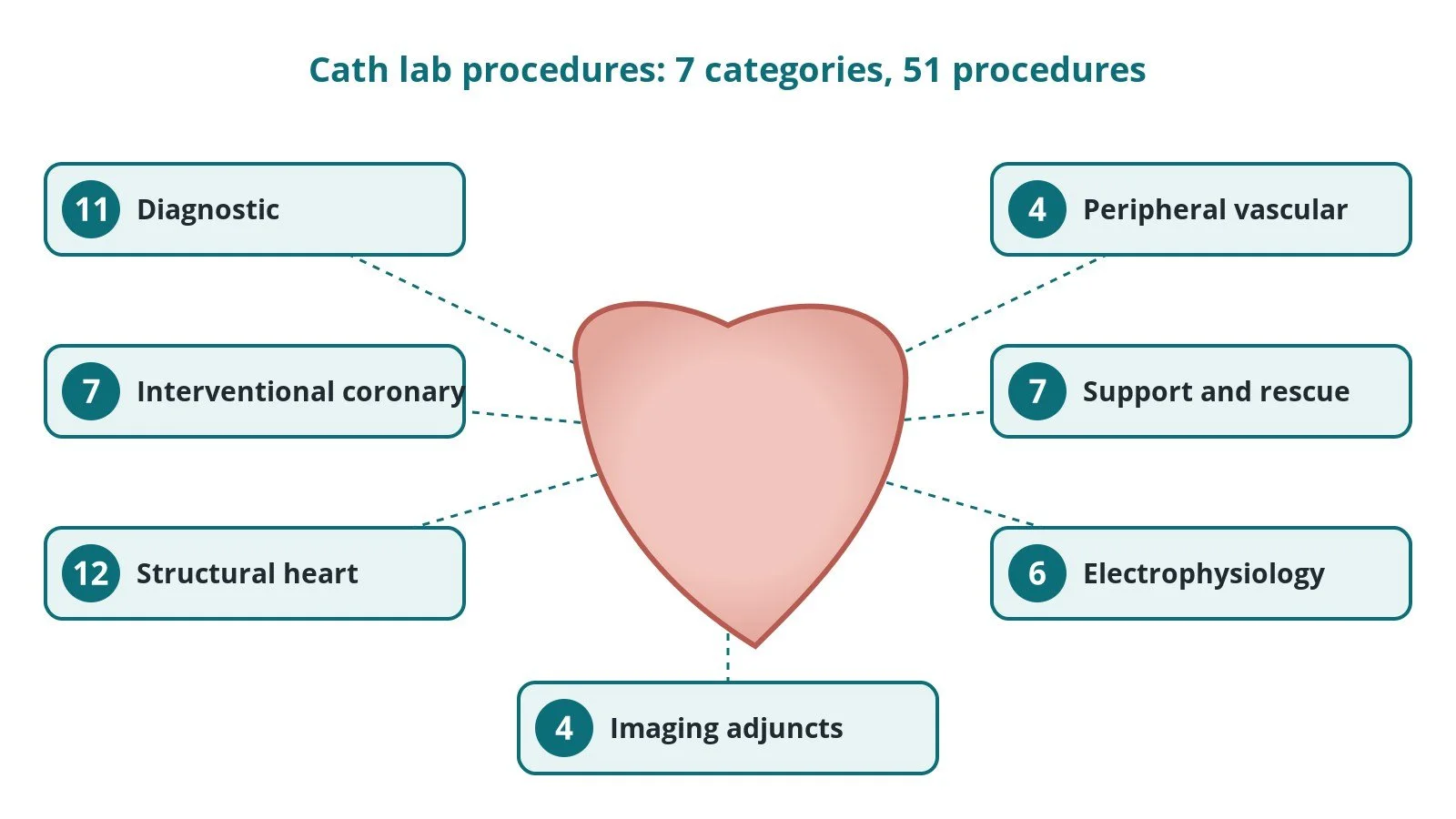
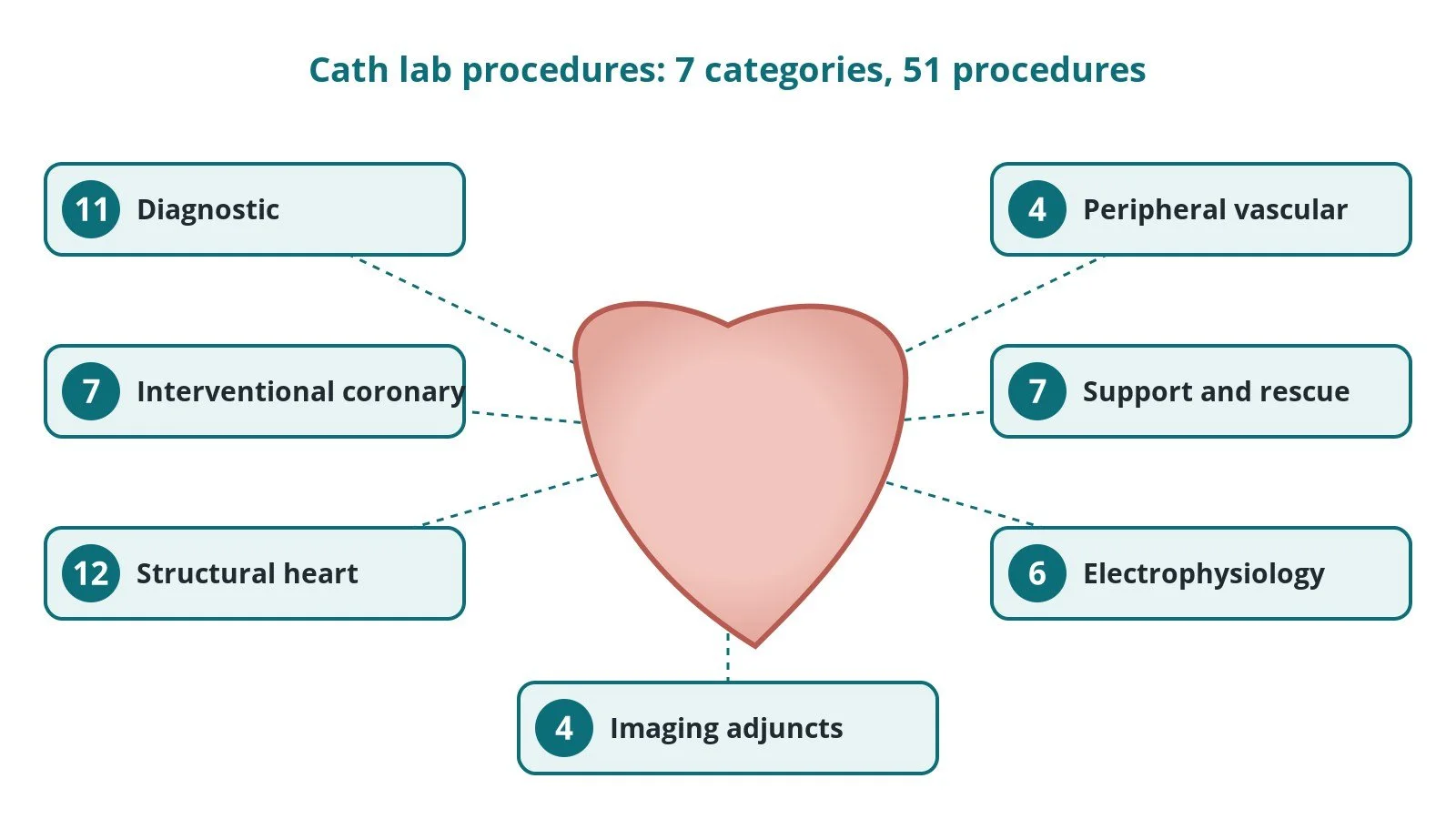
The 51 procedures on this page, grouped into seven categories. Original illustration for rodnieoro.com.
About this list
A cath lab is a room with X-ray imaging where doctors, nurses, physiologists and radiographers work through small tubes (catheters) passed into blood vessels, usually at the wrist or groin. Some procedures are diagnostic (taking pictures or measurements). Others are treatments (opening, closing, replacing or supporting). Electrophysiology work is often done in a separate, dedicated lab.
New to the acronyms? See the Cath Lab Abbreviations A–Z.
Diagnostic
 Coronary angiography: X-ray pictures of the heart arteries using contrast dye, to look for narrowings or blockages.
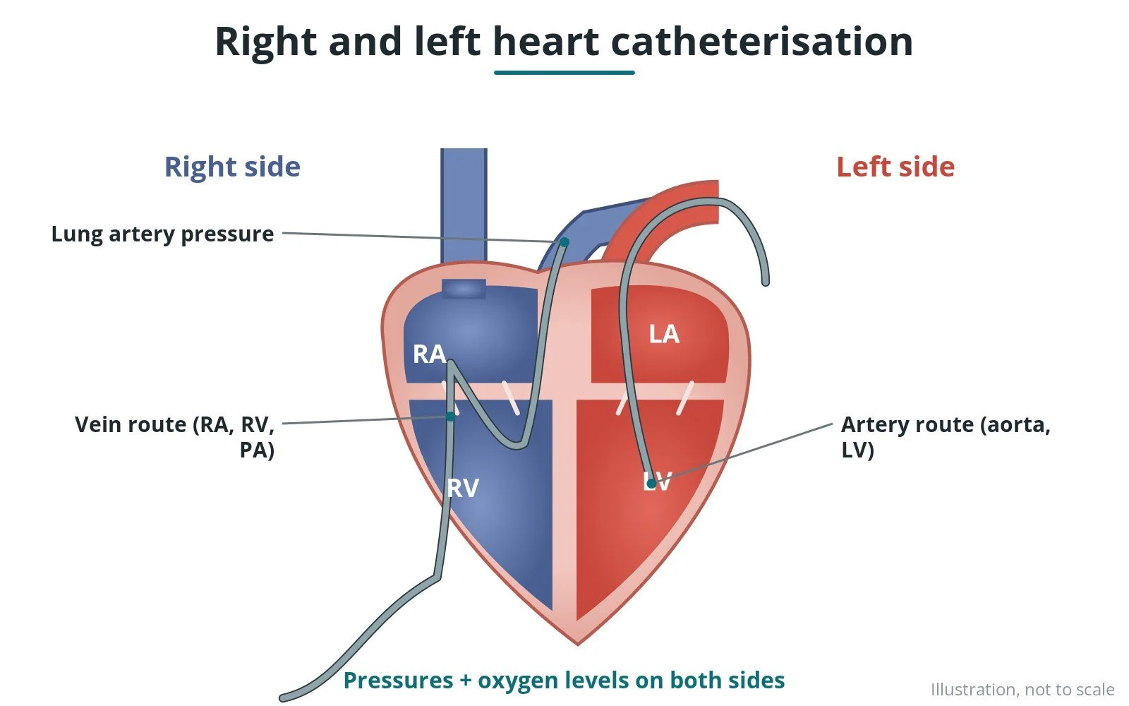
 Right and left heart catheterisation: Measuring pressures and oxygen levels inside the chambers on both sides of the heart.
 Aortography: Contrast pictures of the aorta, the main artery leaving the heart, and sometimes the aortic valve. Covered in the coronary angiogram guide.
 Ventriculography: Contrast pictures of a pumping chamber to look at its size, movement and any valve leak. Covered in the coronary angiogram guide.
 Pulmonary angiography: Contrast pictures of the arteries in the lungs, for example to look for clots or narrowings.
 Bypass graft angiography: Contrast pictures of bypass grafts from earlier heart surgery, to check they are open.
 Endomyocardial biopsy: Taking tiny samples of heart muscle with a catheter, to help diagnose heart muscle disease.
 Intracardiac and intravascular pressure measurement: Recording blood pressure inside the heart chambers and blood vessels through a catheter. Covered in the right and left heart catheterisation guide.
 Shunt detection: Checking for blood flowing abnormally between the two sides of the heart, often by sampling oxygen levels.
 Cardiac output measurement: Working out how much blood the heart pumps each minute. Covered in the right and left heart catheterisation guide.
 Myocardial metabolism studies: Research-type sampling of blood from the heart to study how the heart muscle uses energy.
Interventional Coronary
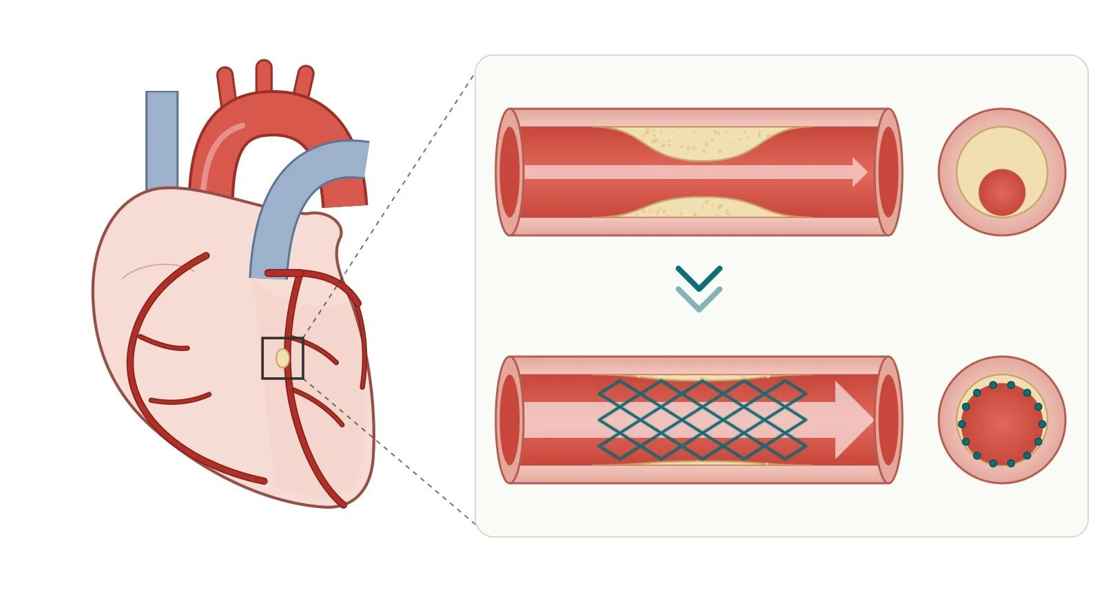
 Balloon angioplasty: Opening a narrowed heart artery by inflating a small balloon inside it.
 Coronary stenting: Placing a small mesh tube (stent) to hold a heart artery open after it has been widened.
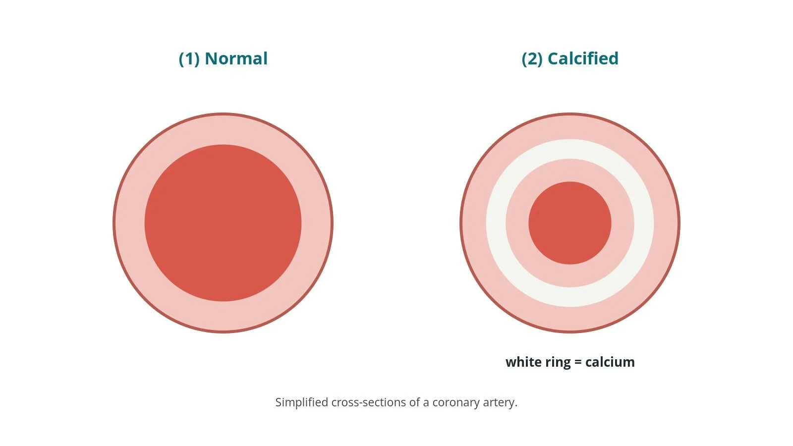
 Rotational and orbital atherectomy: Using a spinning tip to sand down hard, calcified plaque before ballooning or stenting. See also: IVL guide.
 Cutting balloon angioplasty: A balloon with small blades that scores tough plaque so the artery can be opened. See also: IVL guide.
 Coronary thrombectomy: Removing blood clot from a heart artery, usually during a heart attack. Covered in the heart attack (primary PCI) guide.
 Distal protection: Using a filter or similar device to catch debris that could travel downstream during treatment.
 Graft stenting: Placing a stent in a narrowed bypass graft.
Structural Heart
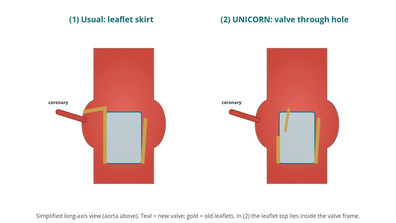
 Transcatheter valve implantation (aortic, mitral, pulmonic): Replacing a diseased heart valve with a new valve delivered by catheter. Guides: aortic (TAVI) · mitral (TMVI) · pulmonary (PPVI).
 Balloon valvuloplasty: Stretching open a narrowed heart valve with a balloon (the linked guide covers the mitral valve).
 Transcatheter mitral valve repair (TEER): Clipping the edges of the mitral valve leaflets together by catheter to reduce a leak.
 Left atrial appendage closure: Sealing off a small pouch of the left atrium where clots can form in atrial fibrillation.
 Paravalvular leak closure: Plugging a leak around the edge of a replacement valve.
 Alcohol septal ablation: Treating a thickened heart wall in HOCM by creating a small, controlled area of scar.
 ASD and PFO closure: Closing a hole or flap-like opening between the upper chambers of the heart with a closure device.
 PDA closure: Closing a blood vessel between the aorta and pulmonary artery that should have closed after birth.
 Coarctation angioplasty: Widening a narrowed section of the aorta with a balloon, often with a stent.
 Coil embolisation: Blocking an unwanted blood vessel or connection with small metal coils. See also: PDA closure · BT shunt closure.
 Septostomy: Making or enlarging an opening between the upper chambers, mainly in babies with some congenital heart conditions.
 Pulmonary angioplasty: Widening narrowed arteries in the lungs with a balloon, sometimes with a stent.
Peripheral Vascular
 Peripheral angiography: Contrast pictures of arteries outside the heart, such as those in the legs.
 Carotid stenting: Placing a stent in a narrowed neck artery that supplies the brain.
 Renal stenting: Placing a stent in a narrowed artery to the kidney.
 Renal denervation: Treating nerves around the kidney arteries by catheter, as a treatment for high blood pressure.
Support and Rescue
 Pericardiocentesis: Draining fluid from the sac around the heart with a needle and small drain.
 Percutaneous pericardial window: Making an opening in the sac around the heart with a balloon so fluid can keep draining.
 Temporary pacemaker placement: Passing a temporary pacing wire through a vein to keep the heart beating at a safe rate. See also: permanent pacemaker guide.
 Intra-aortic balloon pump: A balloon in the aorta that inflates and deflates with each heartbeat to support the heart.
 Percutaneous LV support devices: Small pumps placed by catheter to help the left ventricle pump blood during high-risk procedures or shock.
 ECMO: A machine that takes over the work of the heart and lungs for a time, connected through large tubes.
 Foreign body removal: Retrieving lost or broken equipment from the heart or blood vessels using snares or similar tools.
Electrophysiology (often a separate lab)
 EP studies: Recording the heart's electrical signals from inside the heart to find the cause of a rhythm problem.
 Radiofrequency ablation: Using heat at the catheter tip to treat small areas of tissue causing an abnormal rhythm. See also: AF ablation guide.
 Cryoablation: Using freezing to treat tissue causing an abnormal rhythm, for example in atrial fibrillation.
 Pulsed field ablation: Using short electrical pulses rather than heat or cold to treat rhythm-causing tissue. See also: AF ablation guide.
 Laser ablation (AF, SVT, VT): Using laser energy to treat tissue causing abnormal heart rhythms. See also: AF ablation guide.
 Pacemaker and defibrillator implants: Fitting a device under the skin to keep the heart rate safe or treat dangerous rhythms. See also the S-ICD guide.
Imaging Adjuncts
 IVUS: Intravascular ultrasound: a tiny ultrasound probe inside the artery to see the vessel wall and plaque. See also: PCI guide.
 OCT: Optical coherence tomography: light-based imaging inside the artery that shows very fine detail. See also: PCI guide.
 FFR: Fractional flow reserve: a pressure wire test showing whether a narrowing is limiting blood flow. See also: coronary angiogram guide.
 Electromechanical mapping: Building a map of the heart's electrical activity and wall movement from inside the heart. See also: AF ablation guide.
Disclaimer
Education only. This page is for general education for healthcare students, cath lab staff and curious readers. It isn't medical advice and doesn't replace advice from your own doctor, heart team or local unit protocols. Equipment and medicines are described generically. No product or manufacturer is recommended. If you think someone is having a heart emergency, call 999.
Disclosure: I work for a company that makes devices used in some of these procedures. This page is independent education, isn't sponsored or reviewed by any company, and doesn't recommend any product.
The diagrams on this page are original illustrations created for rodnieoro.com.