Pericardiocentesis: a Cath Lab 101 guide
Reviewed by Rodnie Oro, cath lab charge nurse · Last reviewed: October 2026 · Education only, not medical advice
Pericardiocentesis in one minute
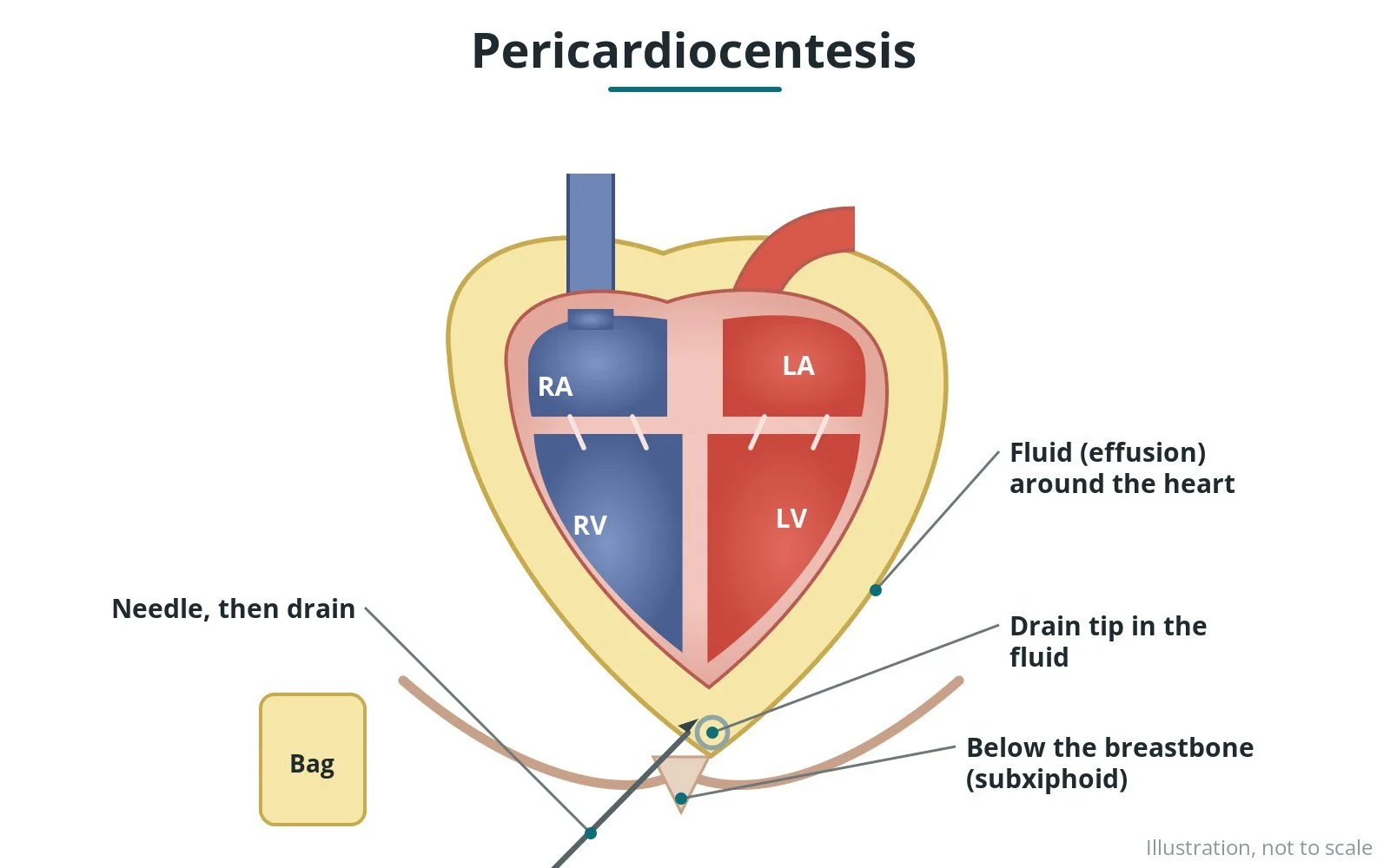
What it is: draining fluid from the sac around the heart (pericardium) with a needle, then leaving a small drain in place for a short time.
Who it is for: people with a large pericardial effusion, especially if it is pressing on the heart (tamponade), or to test the fluid.
Main risks: injury to the heart or a blood vessel, rhythm problems, a punctured lung, infection and bleeding.
Usually: under local anaesthetic, guided by echo and X-ray, 30–60 minutes; the drain usually stays in for 1–3 days.
On this page: What is pericardiocentesis? · Who is it for? · When is it done? · Step by step · Recovery · Risks · Kit list · First time in the lab? · FAQ
What is pericardiocentesis?
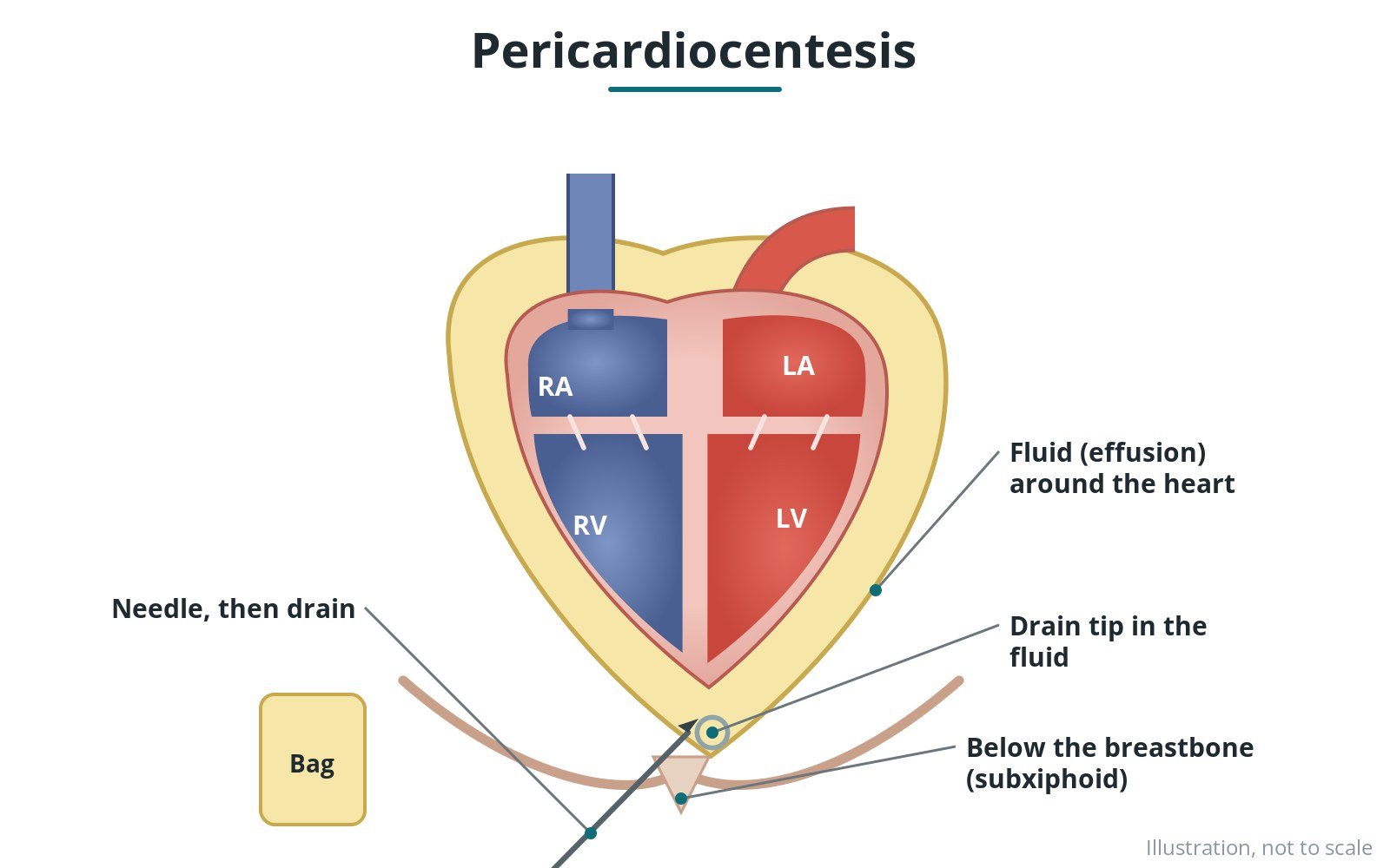
The heart sits in a thin sac, the pericardium. Fluid can build up between the sac and the heart (a pericardial effusion). If there is a lot, it can squeeze the heart so it can't fill properly: cardiac tamponade, an emergency.
In pericardiocentesis:
- echo finds the best spot, most often just under the breastbone
- a needle is passed into the fluid under local anaesthetic
- a guidewire is passed through it, and a soft drain is placed
- fluid is drained and can be sent for tests
The procedure may be done in the cath lab, at the bedside, or as an emergency.
New to the acronyms? See the Cath Lab Abbreviations A–Z.
Who is it for?
It may be considered for people with:
- tamponade (fluid pressing on the heart)
- a large or growing effusion
- fluid that needs testing to find the cause
- fluid after heart surgery or a procedure
The decision is made by your heart team, who will talk through the reasons, the alternatives and the risks with you.
When is it done?
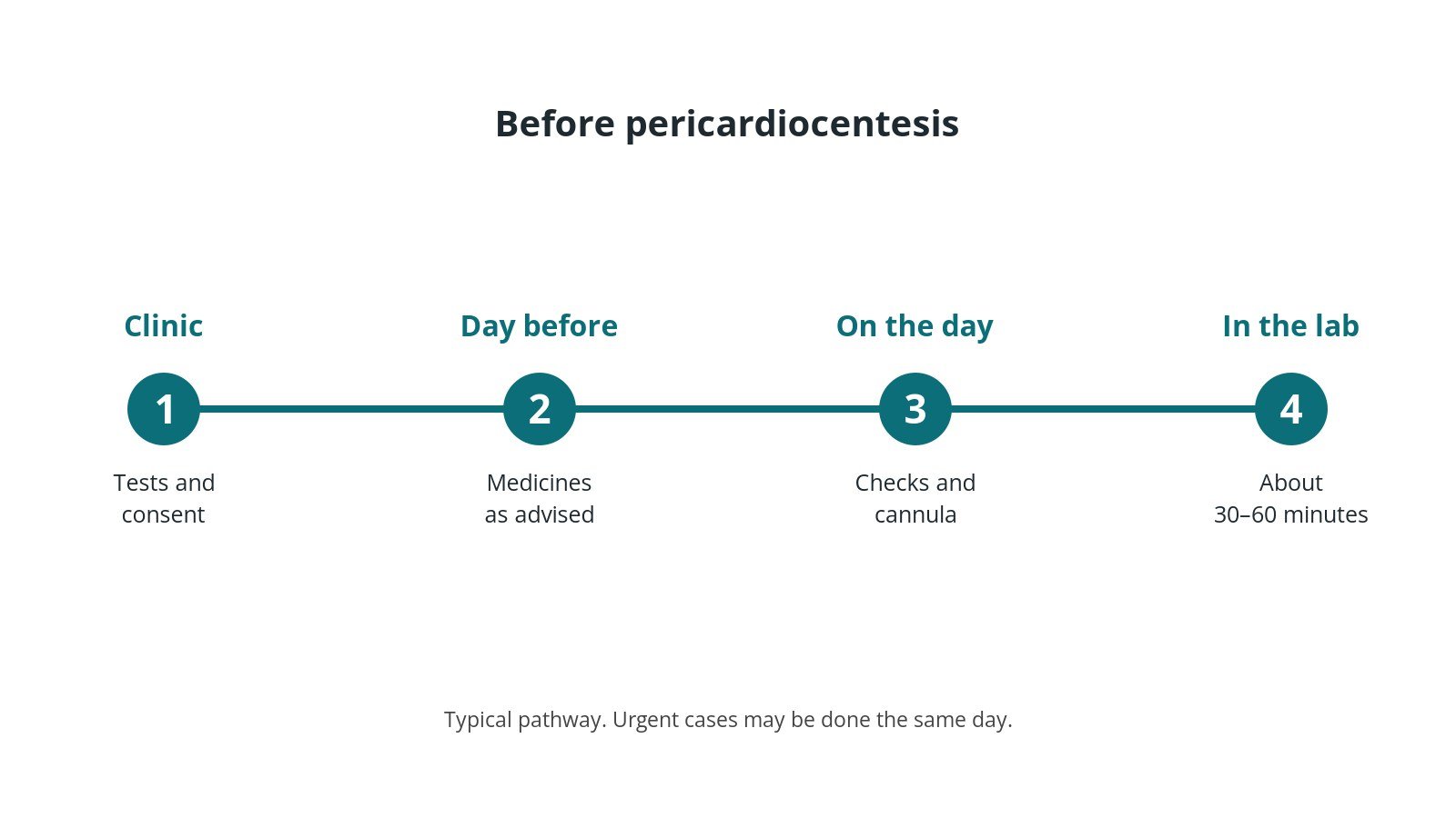
It may be planned or an emergency. If time allows:
- echo to measure the fluid and plan the approach
- blood tests, including clotting
- consent
Step by step
1. Getting ready
You lie on the table, usually propped up slightly. ECG, blood pressure and oxygen are monitored. The skin below the breastbone (or on the chest) is cleaned and numbed.
2. Access
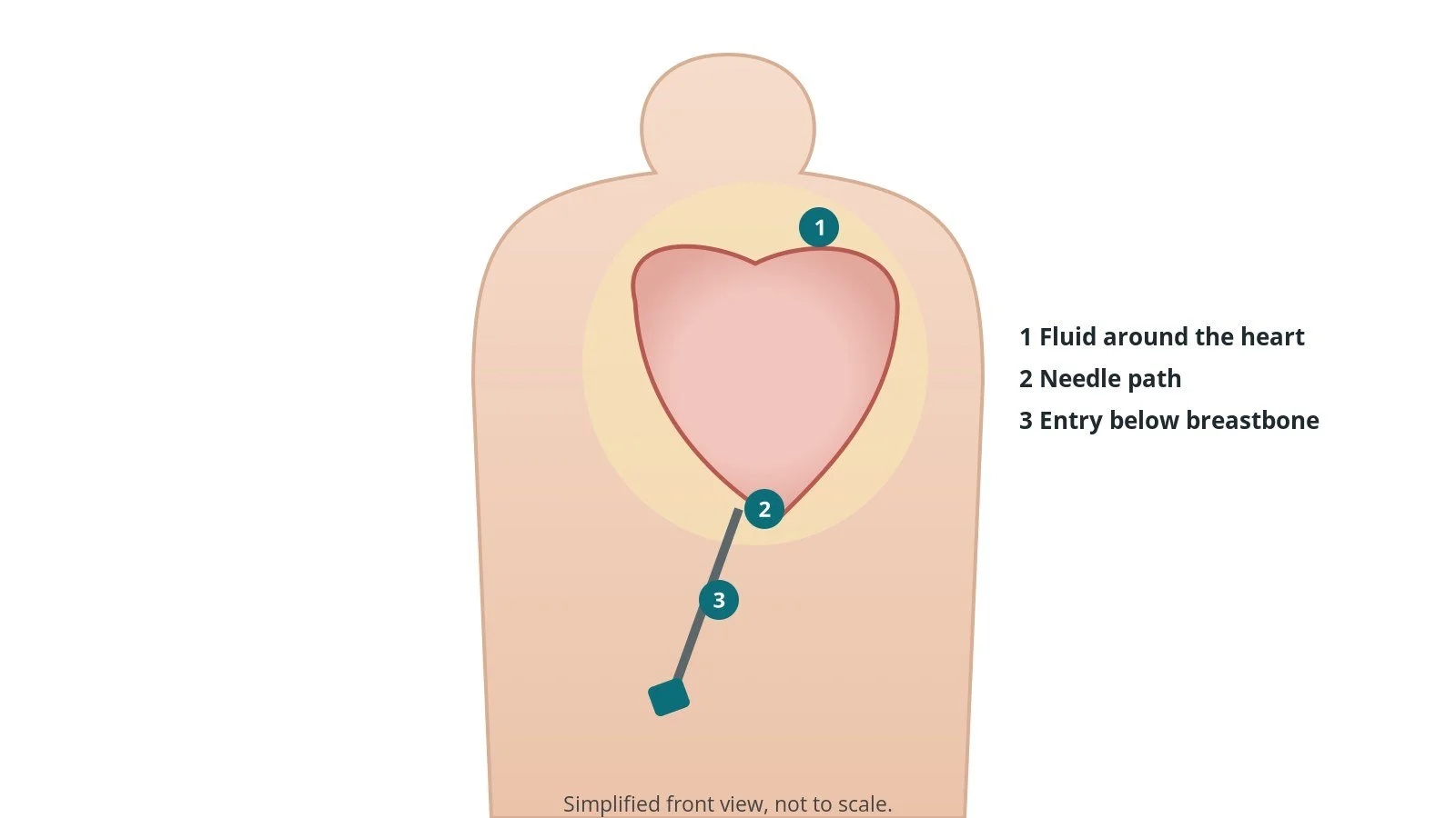
Using echo and X-ray, a needle is passed into the fluid, usually from just below the breastbone. A small amount of contrast or agitated saline may confirm the position.
3. Drainage
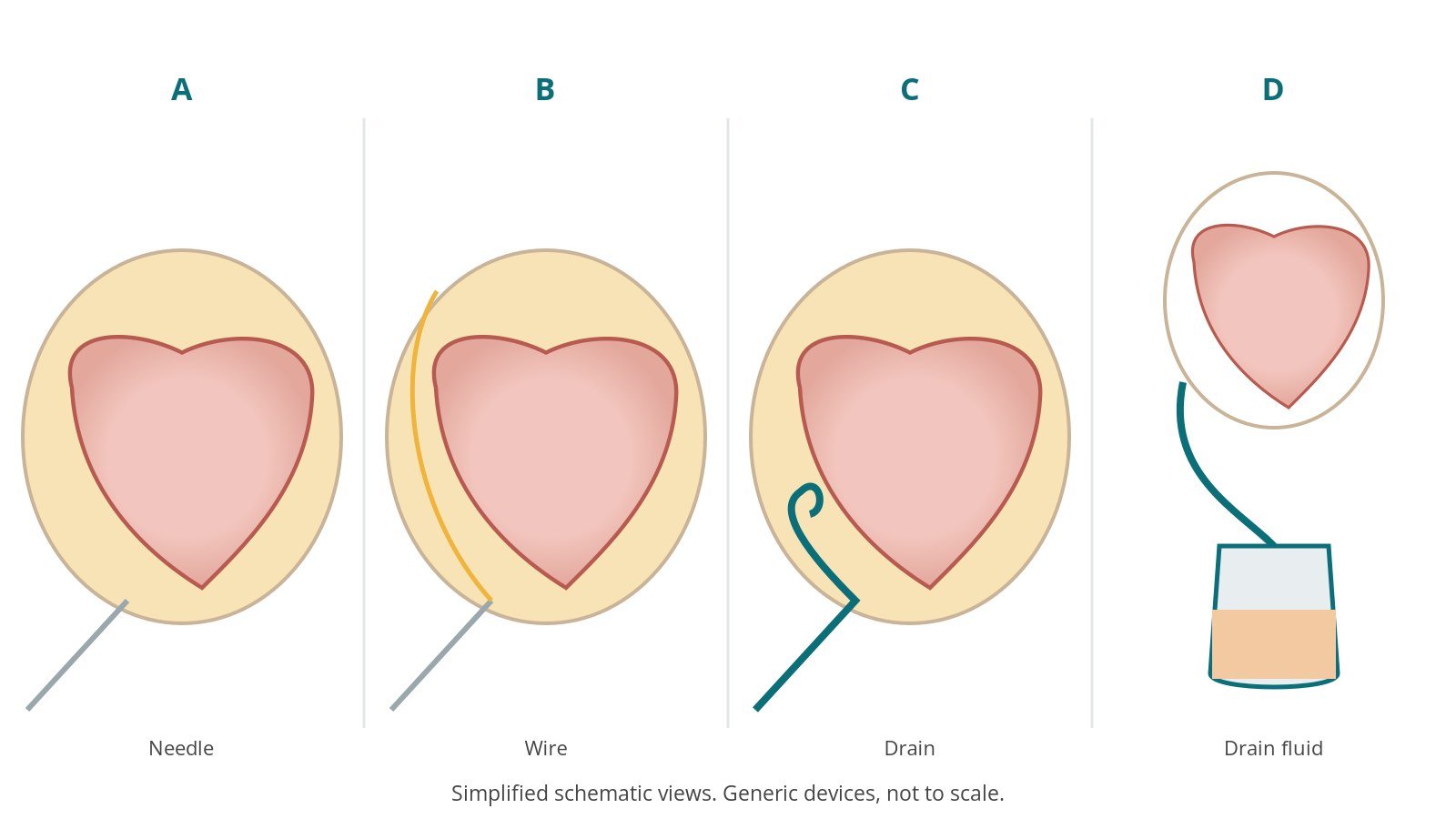
- A. Needle. The needle enters the fluid under echo guidance.
- B. Wire. A guidewire is passed and its position checked on X-ray.
- C. Drain. A soft pigtail drain is placed over the wire.
- D. Drain fluid. Fluid is drained and samples sent.
You may feel pressure under the breastbone. Many people breathe more easily once fluid is drained.
4. Finishing
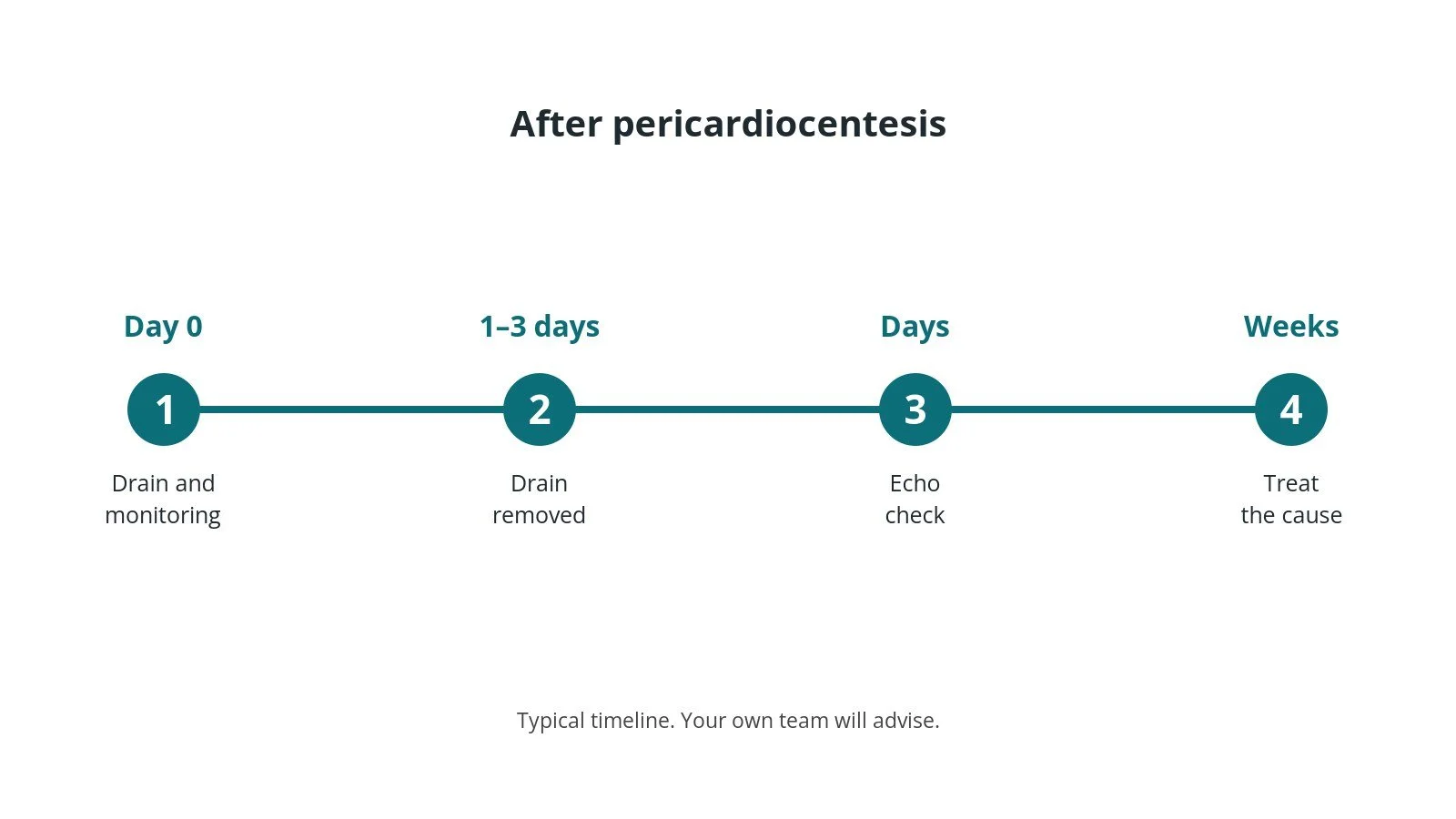
The drain is stitched or secured and connected to a bag. It is usually removed after 1–3 days when drainage slows.
Recovery and aftercare
- Monitoring: checks of rhythm, blood pressure and drain output, as per centre protocol.
- Drain: usually stays 1–3 days; repeat echo checks the fluid.
- Removal: quick and done on the ward.
- Follow-up: depends on the cause of the fluid.
Call 999 if you have chest pain, breathlessness, fainting, signs of a stroke, or heavy bleeding or swelling at the access site.
Risks and complications
Your team will explain the risks that apply to you. Possible problems include:
- injury to the heart muscle or a coronary artery
- heart rhythm problems
- punctured lung (pneumothorax)
- bleeding
- infection
- fluid returning
Cath lab kit list (printable)
Barts example kit list
An example from one UK centre, written generically. Every lab has its own protocol, and sizes and doses vary.
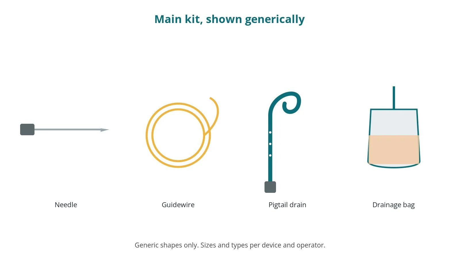
- pericardiocentesis set: needle, guidewire, dilator and pigtail drain, per device and operator
- local anaesthetic, as per centre protocol
- echo machine with sterile probe cover
- drainage bag and three-way tap
- sample pots
Procedural steps (Barts example)
- Echo to plan; mark the site
- Local anaesthetic
- Needle under echo; confirm position
- Wire; check on X-ray
- Dilator; pigtail drain; drain fluid
- Samples; secure drain
First time in the cath lab?
💡 First-time tip
- Echo first. Have the echo ready before starting.
- Label samples. Know which tests are requested.
- Record volume. Measure fluid drained.
- Watch BP. It often improves once fluid is out.
FAQ
Will it hurt?
Local anaesthetic is used; you may feel pressure.
How long does the drain stay?
Usually 1–3 days.
Will the fluid come back?
It can; the team will treat the cause.
Related Cath Lab 101 guides
Sources
All checked on 10 October 2026. Each page's own date is shown where it gives one.
- Adler Y, et al. 2015 ESC Guidelines for the diagnosis and management of pericardial diseases. Eur Heart J 2015;36(42):2921–2964. PubMed 26320112
- StatPearls (NCBI Bookshelf), search: pericardiocentesis. ncbi.nlm.nih.gov/books
Disclaimer
Education only. This page is for general education for healthcare students, cath lab staff and curious readers. It isn't medical advice and doesn't replace advice from your own doctor, heart team or local unit protocols. Equipment and medicines are described generically. No product or manufacturer is recommended. If you think someone is having a heart emergency, call 999.
Disclosure: I work in the medical device industry. This page is independent education and isn't sponsored or reviewed by any company.
All diagrams on this page are original illustrations created for rodnieoro.com.