Renal artery stenting: a Cath Lab 101 guide
Reviewed by Rodnie Oro, cath lab charge nurse · Last reviewed: October 2026 · Education only, not medical advice
Renal artery stenting in one minute
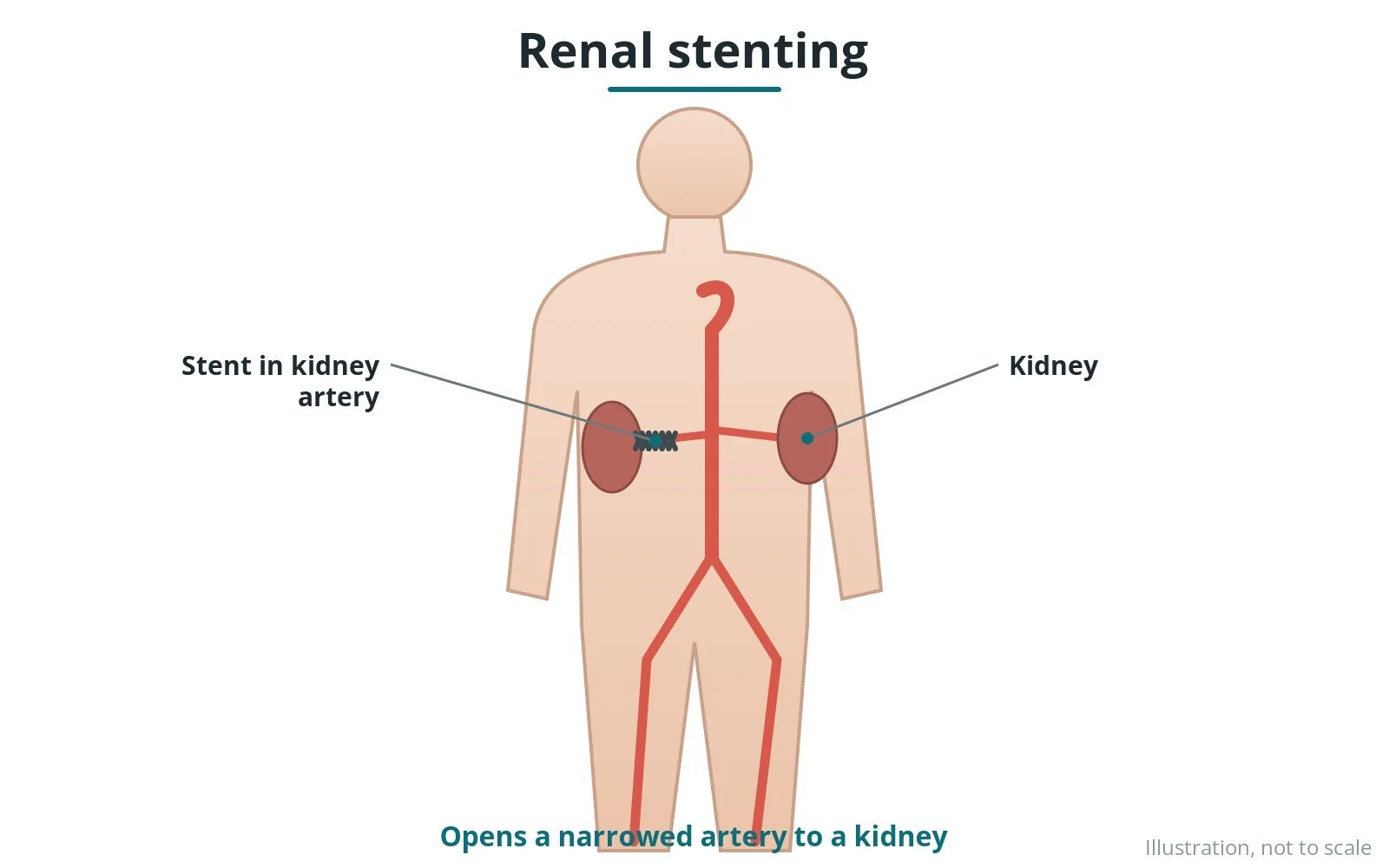
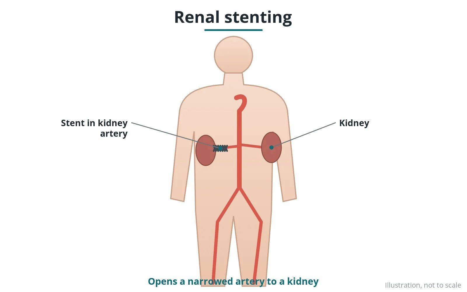
What it is: placing a stent in a narrowed artery to the kidney (renal artery) through a catheter.
Who it is for: selected people with renal artery narrowing causing problems such as sudden fluid on the lungs, rapidly worsening kidney function or very hard-to-control blood pressure. Most people are treated with medicines alone.
Main risks: bruising at the access site, kidney effects from dye or debris, artery injury and, rarely, loss of kidney function.
Usually: under local anaesthetic, about an hour, often one night in hospital.
On this page: What is renal artery stenting? · Who is it for? · When is it done? · Step by step · Recovery · Risks · Kit list · First time in the lab? · FAQ
What is renal artery stenting?
The renal arteries supply the kidneys. Narrowing, usually from atherosclerosis, can reduce blood flow, raise blood pressure and affect kidney function.
- a catheter is passed from the groin or wrist to the opening of the renal artery
- a guidewire crosses the narrowing
- a balloon-expandable stent is placed, usually right at the opening
For most people, medicines to control blood pressure and cholesterol are the main treatment. Stenting is reserved for specific situations. A different condition, fibromuscular dysplasia, is usually treated with a balloon alone.
New to the acronyms? See the Cath Lab Abbreviations A–Z.
Who is it for?
It may be considered for people with:
- sudden fluid on the lungs (flash pulmonary oedema) with renal artery narrowing
- rapidly worsening kidney function with a severe narrowing
- very hard-to-control blood pressure despite several medicines, after specialist review
The decision is made by your heart team, who will talk through the reasons, the alternatives and the risks with you.
When is it done?
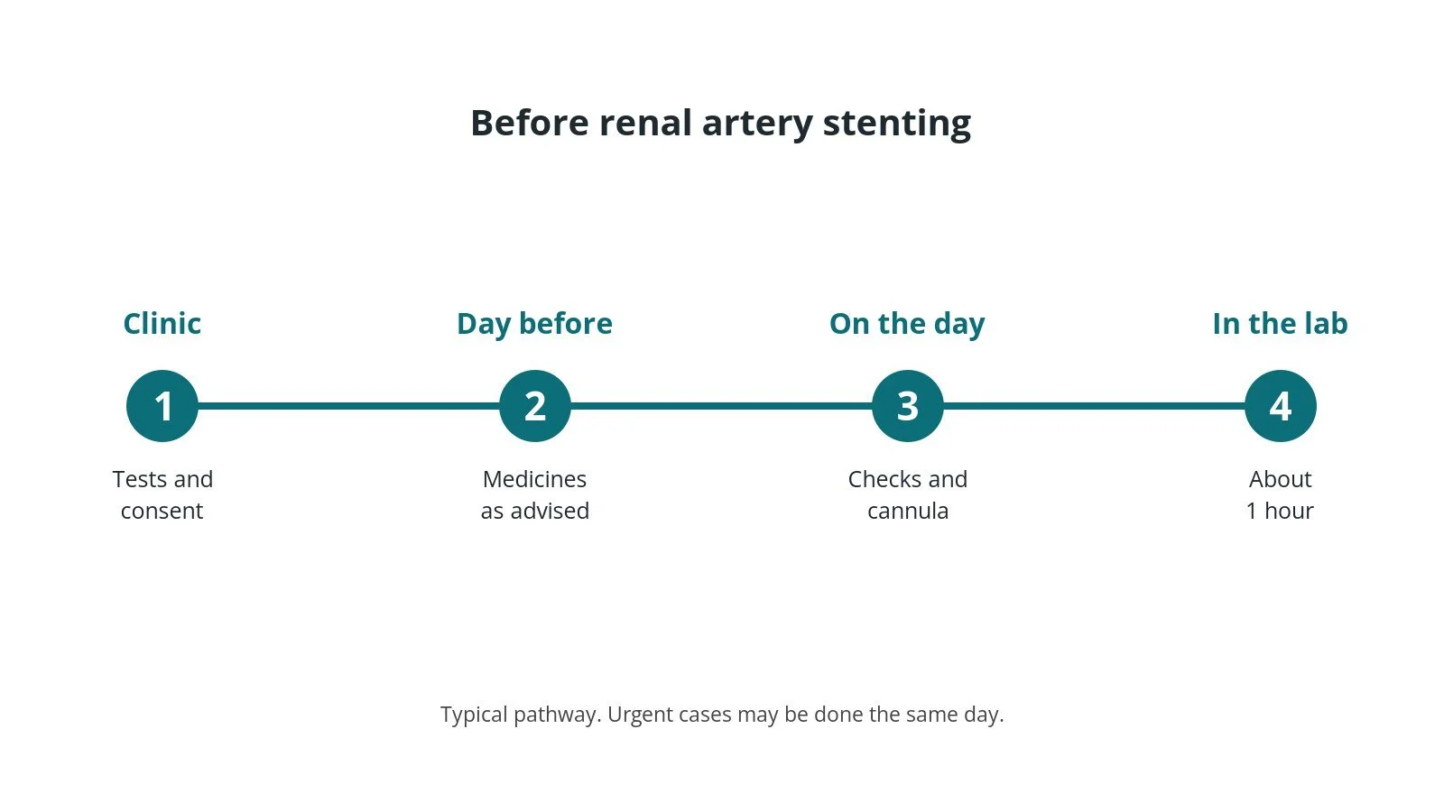
It can be planned in advance or done urgently. Before it, you usually have:
- blood tests, including kidney function and blood count
- an ECG, and often an echo or other scan
- a review of your medicines, especially blood thinners and diabetes medicines, as per centre protocol
- a chance to ask questions and give consent
Step by step
1. Getting ready
You lie on the X-ray table. ECG, blood pressure and oxygen levels are monitored. The skin over the access site is cleaned and numbed with local anaesthetic; some people also have light sedation, as per centre protocol.
2. Access
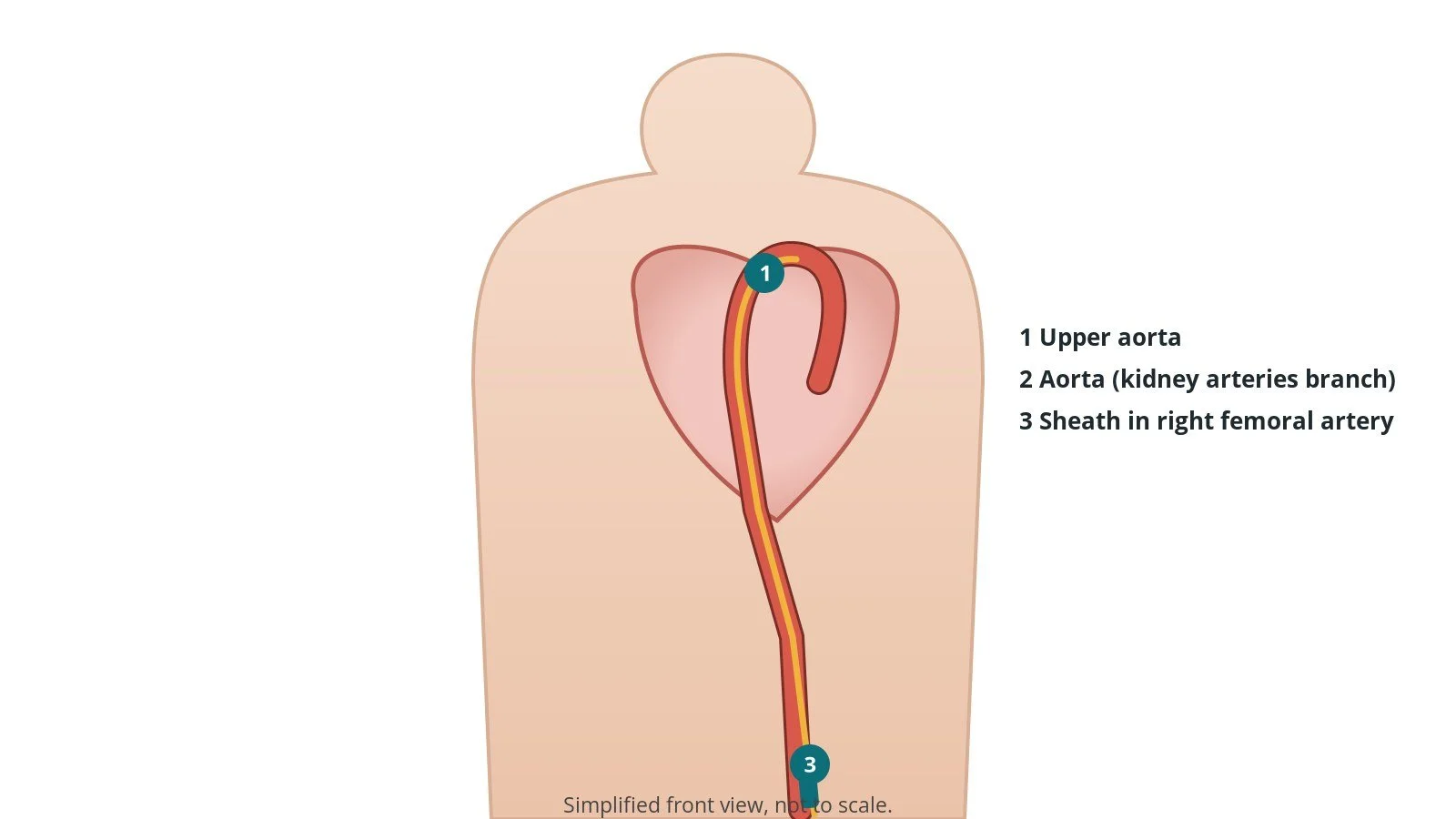
A sheath is placed in the femoral or radial artery, sizes per device and operator. Heparin is given, as per centre protocol.
3. Stenting
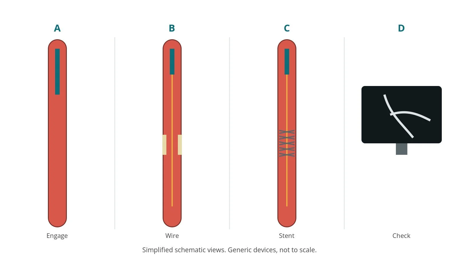
- A. Engage. A guide catheter engages the renal artery.
- B. Wire. A guidewire crosses the narrowing.
- C. Stent. A balloon-expandable stent is placed at the opening.
- D. Check. An angiogram checks the result.
4. Finishing
A final angiogram checks the result. The sheath is removed and the wrist closed with a pressure band, or the groin with pressure or a closure device, per device and operator.
Recovery and aftercare
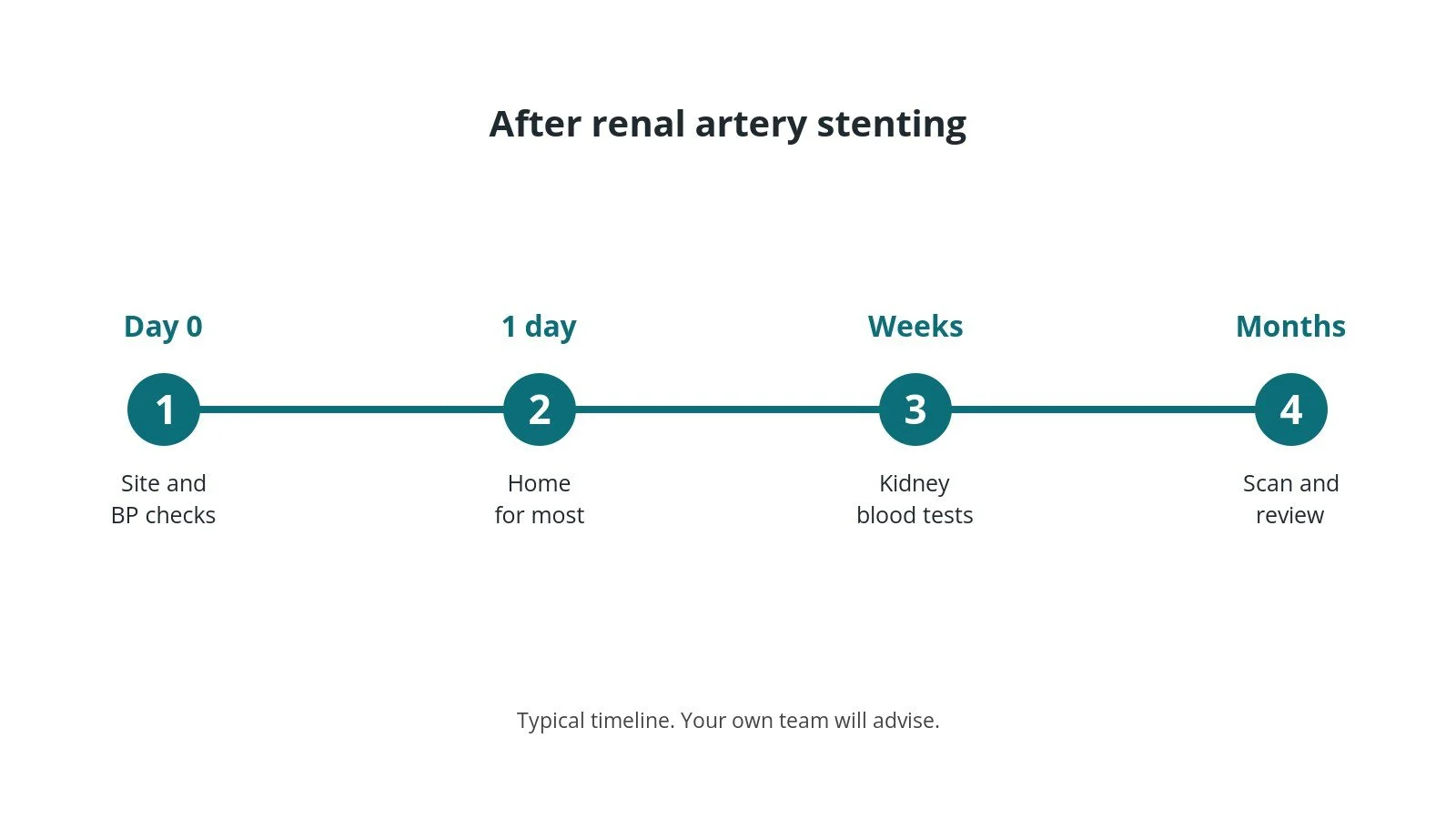
- First hours: checks of the access site, blood pressure and kidney function, as per centre protocol.
- Hospital stay: often one night.
- Medicines: blood pressure medicines may be adjusted; blood thinners as prescribed.
- Follow-up: kidney blood tests and an ultrasound scan.
Call 999 if you have chest pain, breathlessness, fainting, signs of a stroke, or heavy bleeding or swelling at the access site.
Risks and complications
Your team will explain the risks that apply to you. Possible problems include:
- bruising or bleeding at the access site
- kidney effects from contrast dye or debris
- artery tear or blockage
- narrowing again later
- loss of kidney function (rare)
Cath lab kit list (printable)
Barts example kit list
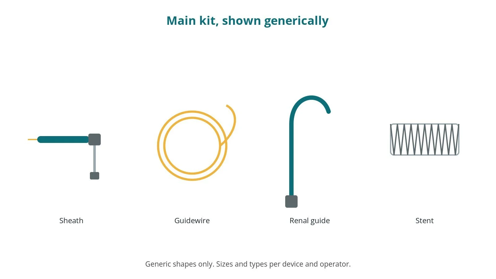
An example from one UK centre, written generically. Every lab has its own protocol, and sizes and doses vary.
- arterial sheath, size per device and operator
- renal guide catheter, per operator
- guidewires, per operator
- balloon-expandable stent, size per device and operator
- heparin, as per centre protocol
Procedural steps (Barts example)
- Access; heparin
- Aortogram to locate renal arteries
- Guide engaged; wire across
- Stent at the opening; flare if needed
- Final angiogram; sheath out
First time in the cath lab?
💡 First-time tip
- Kidney bloods. Note baseline kidney function.
- Minimise dye. Keep contrast totals down.
- Blood pressure. Watch for drops after stenting.
- Flank pain. Report back or side pain straight away.
FAQ
Will it cure my blood pressure?
Not usually; most people still need medicines.
Is it common?
No. It is used only in selected cases.
Is this renal denervation?
No. That treats nerves, not narrowings.
Related Cath Lab 101 guides
Sources
All checked on 10 October 2026. Each page's own date is shown where it gives one.
- McEvoy JW, et al. 2024 ESC Guidelines for the management of elevated blood pressure and hypertension. Eur Heart J 2024;45(38):3912–4018. PubMed 39210715
- StatPearls (NCBI Bookshelf), search: renal artery stenosis stenting. ncbi.nlm.nih.gov/books
Disclaimer
Education only. This page is for general education for healthcare students, cath lab staff and curious readers. It isn't medical advice and doesn't replace advice from your own doctor, heart team or local unit protocols. Equipment and medicines are described generically. No product or manufacturer is recommended. If you think someone is having a heart emergency, call 999.
Disclosure: I work for a company that makes a device used in this procedure. This page is independent education, isn't sponsored or reviewed by any company, and doesn't recommend any product.
All diagrams on this page are original illustrations created for rodnieoro.com.