Electroanatomical and electromechanical mapping: a Cath Lab 101 guide
Reviewed by Rodnie Oro, cath lab charge nurse · Last reviewed: October 2026 · Education only, not medical advice
Electroanatomical mapping in one minute
What it is: building a 3D map of the heart from inside, showing its electrical activity and, in electromechanical mapping, how the wall moves.
Who it is for: mainly people having ablation for complex rhythms; electromechanical mapping has also been used in research, for example to guide injections into heart muscle.
Main risks: the risks of the EP procedure it is part of: bruising in the groin, fluid around the heart, and rarely stroke.
Usually: part of an EP study or ablation.
On this page: What is electroanatomical mapping? · Who is it for? · When is it done? · Step by step · Recovery · Risks · Kit list · First time in the lab? · FAQ
What is electroanatomical mapping?
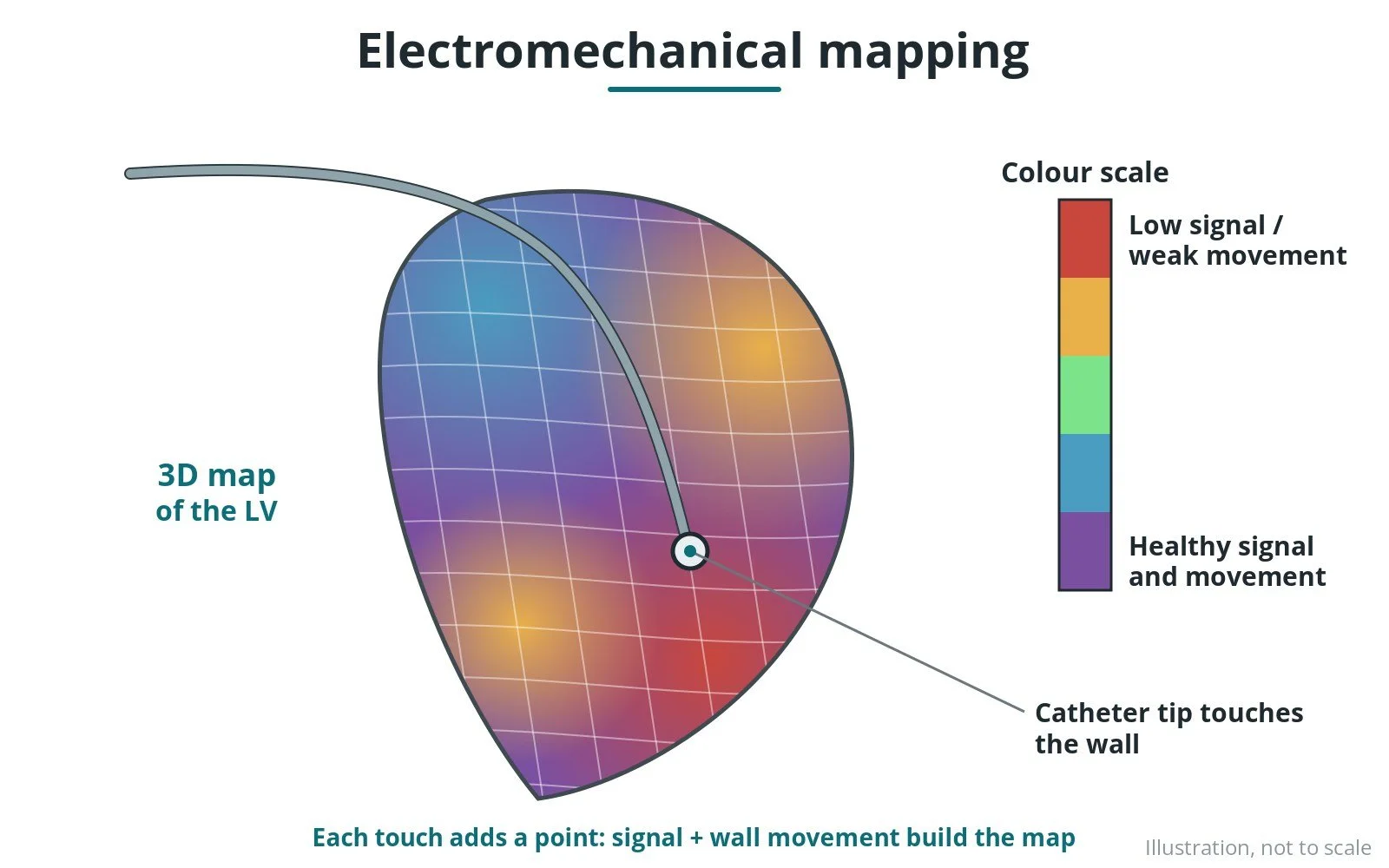
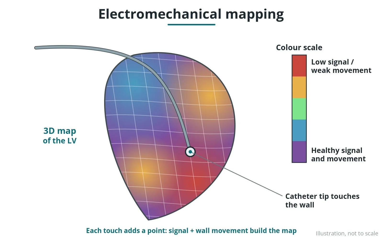
A mapping system tracks the position of a catheter inside the heart in 3D.
- as the catheter touches many points, the system builds a shape of the chamber
- it colours the map by timing of electrical activity or voltage (healthy versus scarred tissue)
- electromechanical mapping also measures how much each area moves, to show weak but living muscle
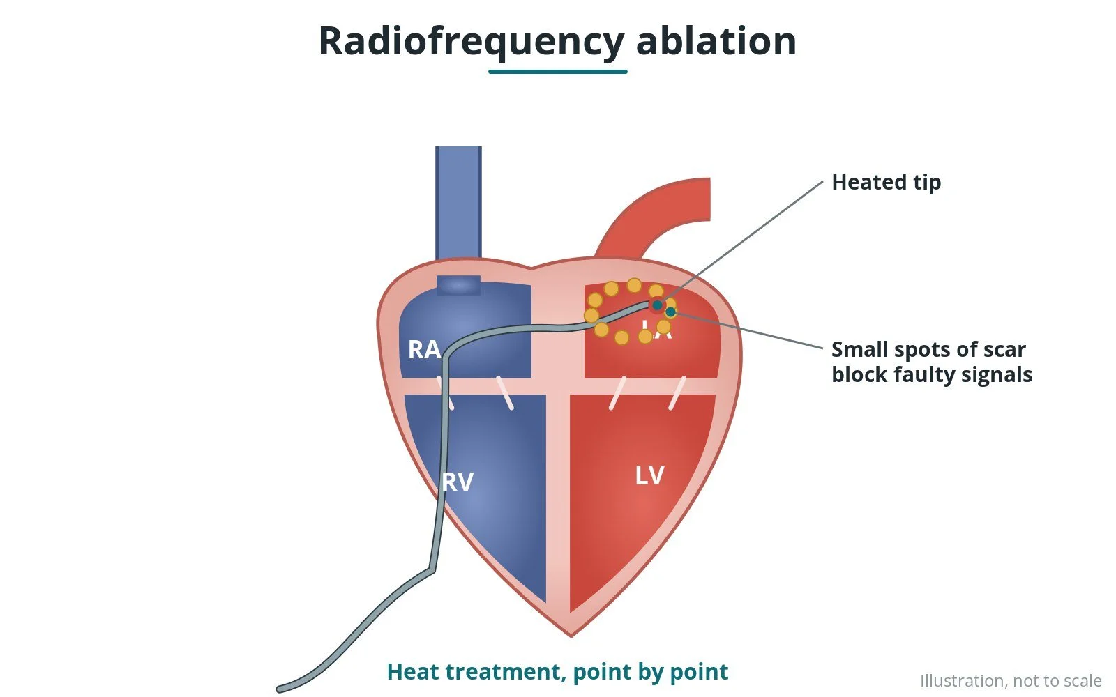
The map guides where to ablate, and reduces X-ray use.
New to the acronyms? See the Cath Lab Abbreviations A–Z.
Who is it for?
It may be considered for people with:
- ablation for AF, flutter or VT
- complex congenital heart rhythms
- research on heart muscle viability
The decision is made by your heart team, who will talk through the reasons, the alternatives and the risks with you.
When is it done?
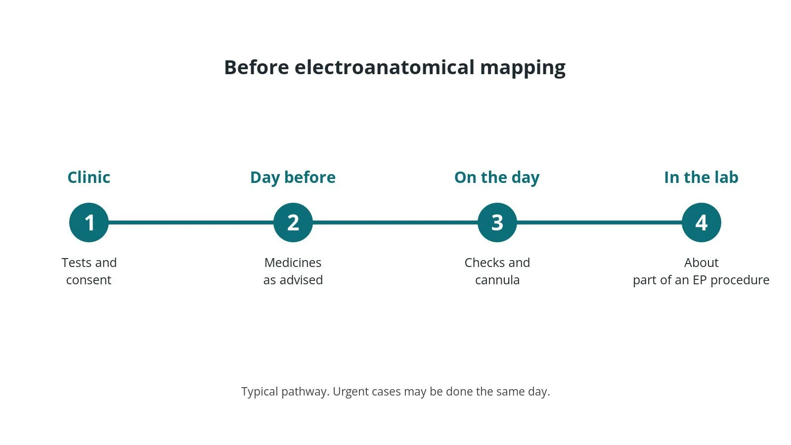
It can be planned in advance or done urgently. Before it, you usually have:
- blood tests, including kidney function and blood count
- an ECG, and often an echo or other scan
- a review of your medicines, especially blood thinners and diabetes medicines, as per centre protocol
- a chance to ask questions and give consent
Step by step
1. Getting ready
You lie on the table. Sticky pads go on your chest and back, and ECG, blood pressure and oxygen are monitored. You usually have sedation or a general anaesthetic, as per centre protocol. The groin is cleaned and numbed.
2. Access
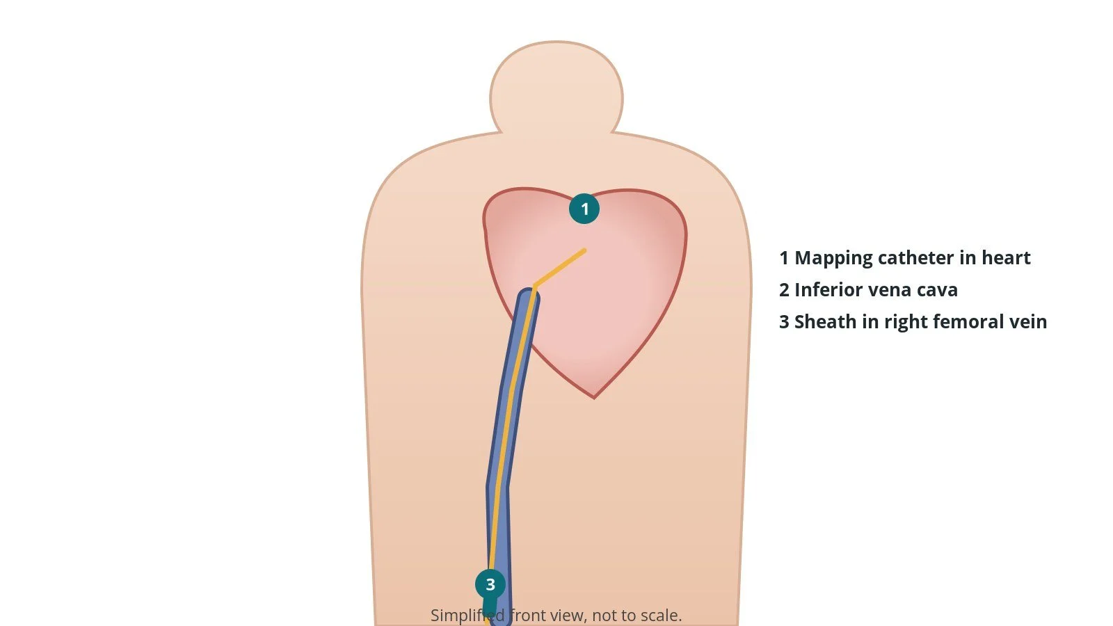
Using ultrasound, sheaths are placed in the femoral vein in the groin, sizes per device and operator. Catheters are passed up to the heart under X-ray and a 3D mapping system.
3. Mapping
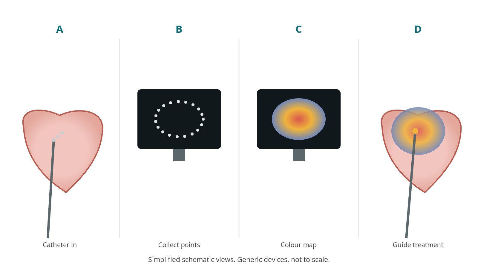
- A. Catheter in. A mapping catheter is placed in the chamber.
- B. Collect points. It touches many points to build a 3D shape.
- C. Colour map. Timing and voltage colour the map.
- D. Guide treatment. The map guides ablation.
4. Finishing
The catheters and sheaths are removed. The groin is closed with pressure or a stitch, per device and operator.
Recovery and aftercare
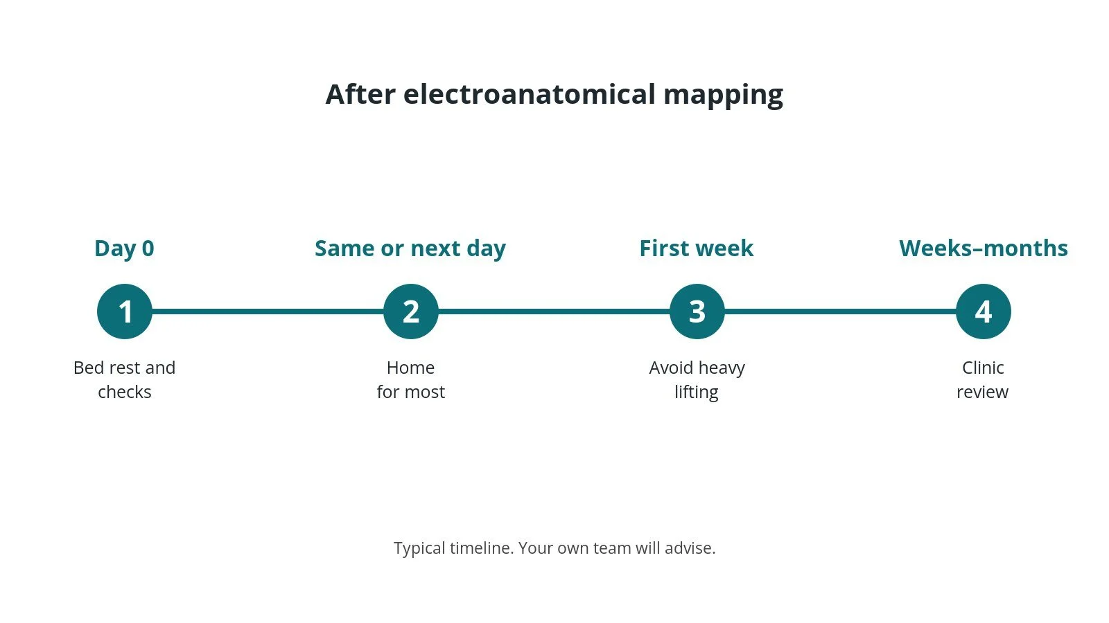
- First hours: bed rest while the groin settles; checks of the groin, rhythm and blood pressure, as per centre protocol.
- Going home: often the same day or the next morning.
- Groin: avoid heavy lifting and strenuous exercise for about a week.
- Medicines: take blood thinners and rhythm medicines exactly as advised.
- Driving: DVLA rules depend on the rhythm problem; ask your team.
- Follow-up: clinic review, sometimes with an ECG or heart monitor.
Call 999 if you have chest pain, breathlessness, fainting, signs of a stroke, or heavy bleeding or swelling at the access site.
Risks and complications
Your team will explain the risks that apply to you. Possible problems include:
- bruising or bleeding in the groin
- damage to a vein or artery in the groin
- new rhythm problems, sometimes needing a pacemaker (uncommon)
- fluid or bleeding around the heart (tamponade) (uncommon)
- stroke or blood clots (rare)
- the original rhythm problem coming back
Cath lab kit list (printable)
Barts example kit list
An example from one UK centre, written generically. Every lab has its own protocol, and sizes and doses vary.
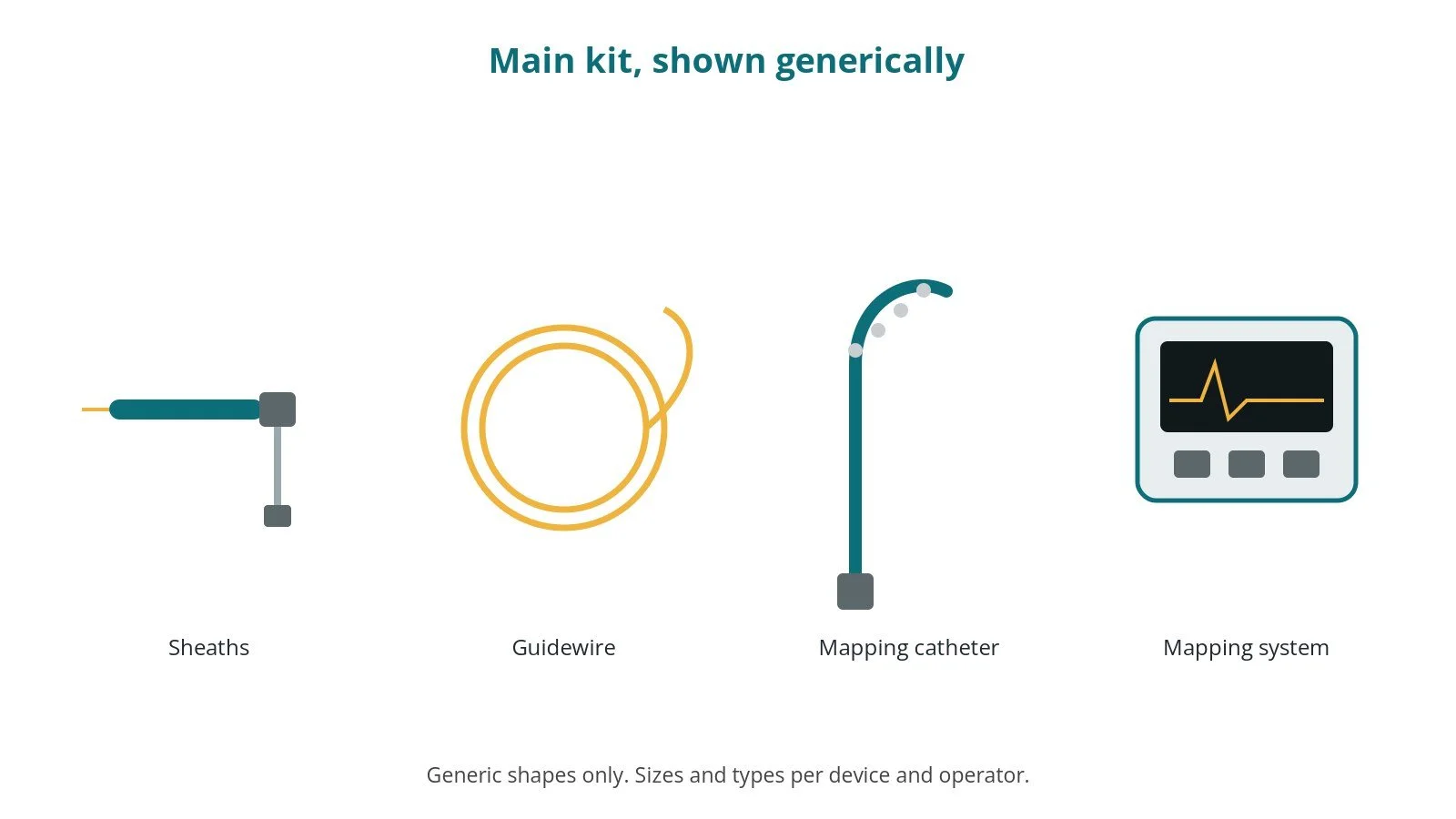
- venous sheaths in the groin, sizes per device and operator
- diagnostic EP catheters, per operator
- recording system and stimulator
- ultrasound for vein access
- heparin, as per centre protocol
- sedation or anaesthetic, as per centre protocol
- 3D mapping system and patches, per device and operator
- mapping catheter, per device and operator
Procedural steps (Barts example)
- Mapping patches on before draping
- Access; catheters in
- Geometry built
- Activation and voltage maps
- Ablation guided by map
First time in the cath lab?
💡 First-time tip
- Patches first. Place before draping.
- Keep still. Movement distorts the map.
- Breathing steady. Respiration gating helps.
- Save maps. Store them as per centre protocol.
FAQ
Is mapping a treatment?
No; it guides treatment.
Does it use more X-ray?
Usually less.
Will I feel it?
No.
Related Cath Lab 101 guides
Sources
All checked on 10 October 2026. Each page's own date is shown where it gives one.
- Van Gelder IC, et al. 2024 ESC Guidelines for the management of atrial fibrillation. Eur Heart J 2024;45(36):3314–3414. PubMed 39210723
- Zeppenfeld K, et al. 2022 ESC Guidelines for the management of patients with ventricular arrhythmias and the prevention of sudden cardiac death. Eur Heart J 2022;43(40):3997–4126. PubMed 36017572
- StatPearls (NCBI Bookshelf), search: electroanatomic mapping. ncbi.nlm.nih.gov/books
Disclaimer
Education only. This page is for general education for healthcare students, cath lab staff and curious readers. It isn't medical advice and doesn't replace advice from your own doctor, heart team or local unit protocols. Equipment and medicines are described generically. No product or manufacturer is recommended. If you think someone is having a heart emergency, call 999.
Disclosure: I work for a company that makes a device used in this procedure. This page is independent education, isn't sponsored or reviewed by any company, and doesn't recommend any product.
All diagrams on this page are original illustrations created for rodnieoro.com.