Permanent pacemaker (PPM), ICD and CRT: a Cath Lab 101 guide
Reviewed by Rodnie Oro, cath lab charge nurse · Last reviewed: October 2026 · Education only, not medical advice
PPM in one minute
What it is: PPM stands for permanent pacemaker. It is a small battery-powered device placed under the skin below the collarbone, with one or more thin wires (leads) that run through a vein into the heart. It watches the heartbeat and sends a tiny electrical pulse whenever the heart is too slow or pauses.
Who needs one: mostly people with a slow or unreliable heartbeat, such as heart block or sick sinus syndrome, that causes dizziness, blackouts, breathlessness or tiredness. Related devices: an ICD can also shock a dangerously fast rhythm, and CRT (a biventricular device) helps both pumping chambers beat together in some types of heart failure.
Main risks: bruising or a collection of blood at the wound is the most common. Uncommon but important problems include infection, a lead moving out of place and a collapsed lung. Your heart team will explain your own risk.
Usually: local anaesthetic with or without sedation, about an hour, home the same day or the next day.
On this page: What is a PPM? · Who is it for? · When is it done? · Step by step · Recovery · Risks · Kit list · First time in the lab? · FAQ
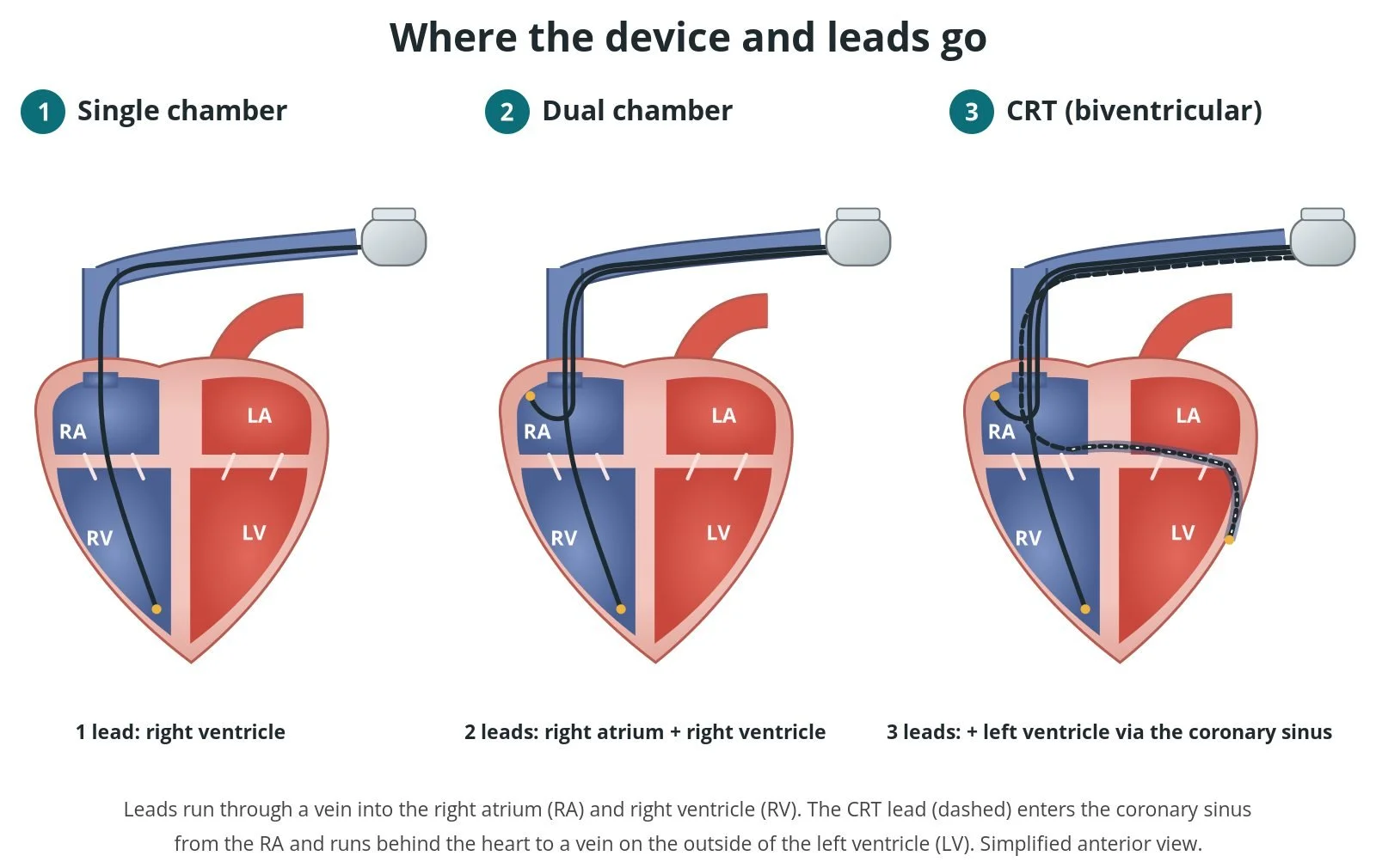
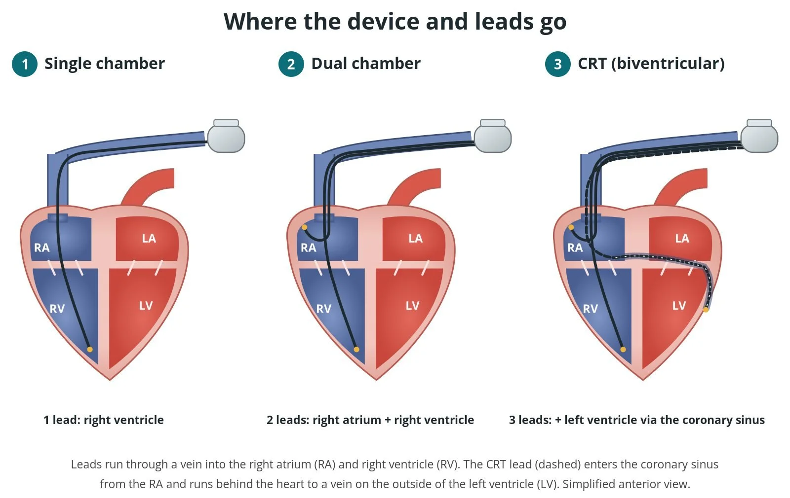
Where the leads go. (1) Single chamber: one lead in the right ventricle (RV). (2) Dual chamber: leads in the right atrium (RA) and RV. (3) CRT: a third lead runs through the coronary sinus to a vein on the left ventricle (LV).
What is a PPM?
Every heartbeat starts with an electrical signal from the sinoatrial (SA) node, the heart's natural pacemaker at the top of the right atrium. The signal spreads across the upper chambers (atria), passes through the atrioventricular (AV) node and then runs down the His bundle and bundle branches so the lower chambers (ventricles) contract. If the SA node fires too slowly, or the signal is delayed or blocked on the way down (heart block), the heart rate can drop too low to keep you well.
A permanent pacemaker has two parts:
the generator (the "box"): a sealed battery and small computer, about the size of a matchbox
the leads: insulated wires that sense the heart's own activity and deliver pacing pulses
Pacemakers only pace when needed. Most of the time they simply watch. The main types are:
Single chamber: one lead, usually in the right ventricle
Dual chamber: two leads, one in the right atrium and one in the right ventricle, so the upper and lower chambers stay in step
CRT (cardiac resynchronisation therapy): a third lead goes through the coronary sinus (a vein on the back of the heart) to the outside of the left ventricle, so both ventricles beat together. A CRT device can be a pacemaker (CRT-P) or a defibrillator (CRT-D)
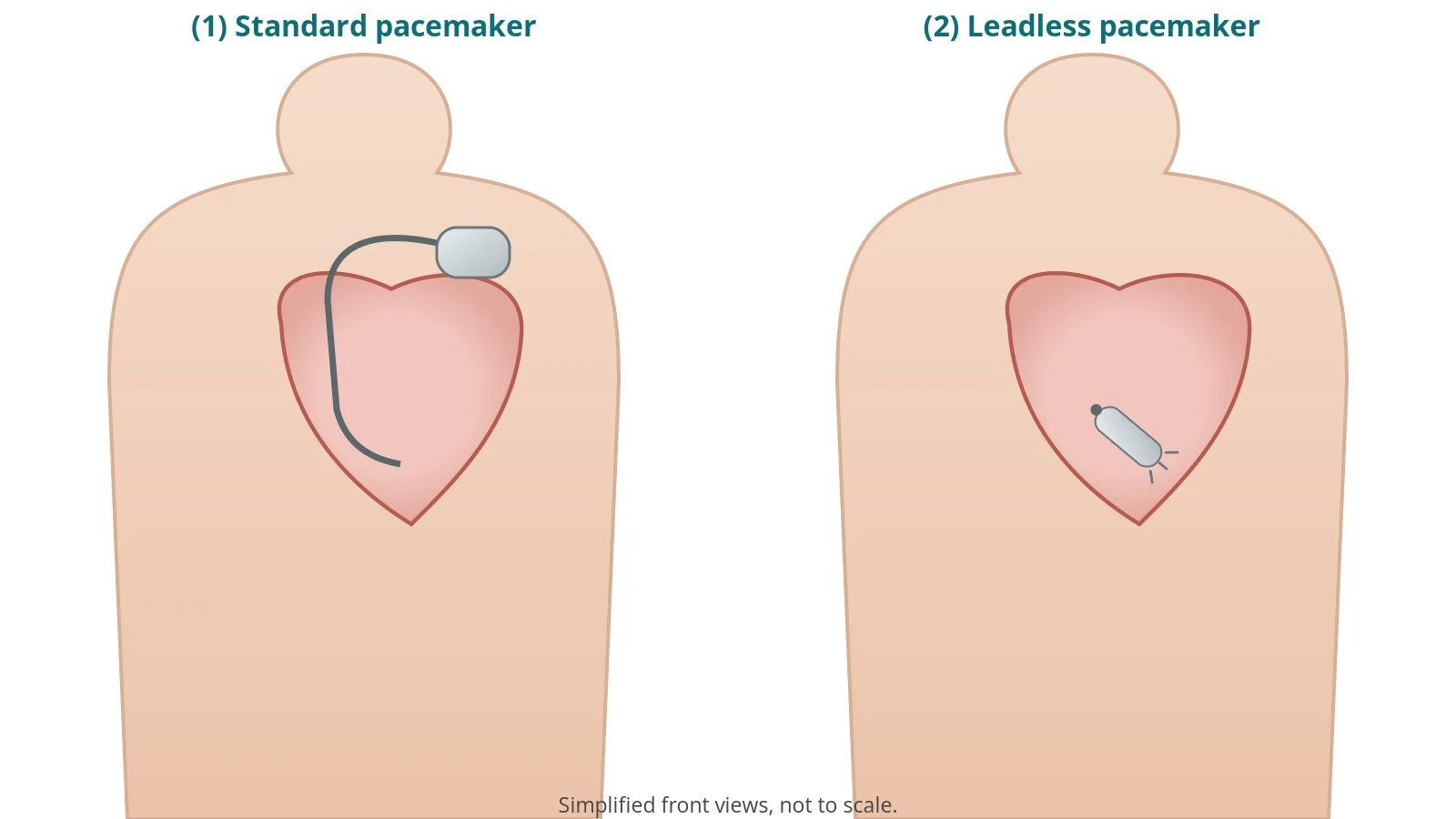
Leadless pacemaker: a small self-contained capsule placed inside the right ventricle through a vein in the groin, with no leads or chest wound. It suits some people with a slow heartbeat
Conduction system pacing: some centres place the ventricular lead on the His bundle or the left bundle branch area, to use the heart's own wiring
An ICD (implantable cardioverter defibrillator) looks and is fitted much like a pacemaker. It can pace, and it can also treat a dangerously fast ventricular rhythm with rapid pacing or a shock. A subcutaneous ICD sits entirely under the skin with no lead in the heart; see the separate S-ICD guide.
New to the acronyms? See the Cath Lab Abbreviations A–Z.
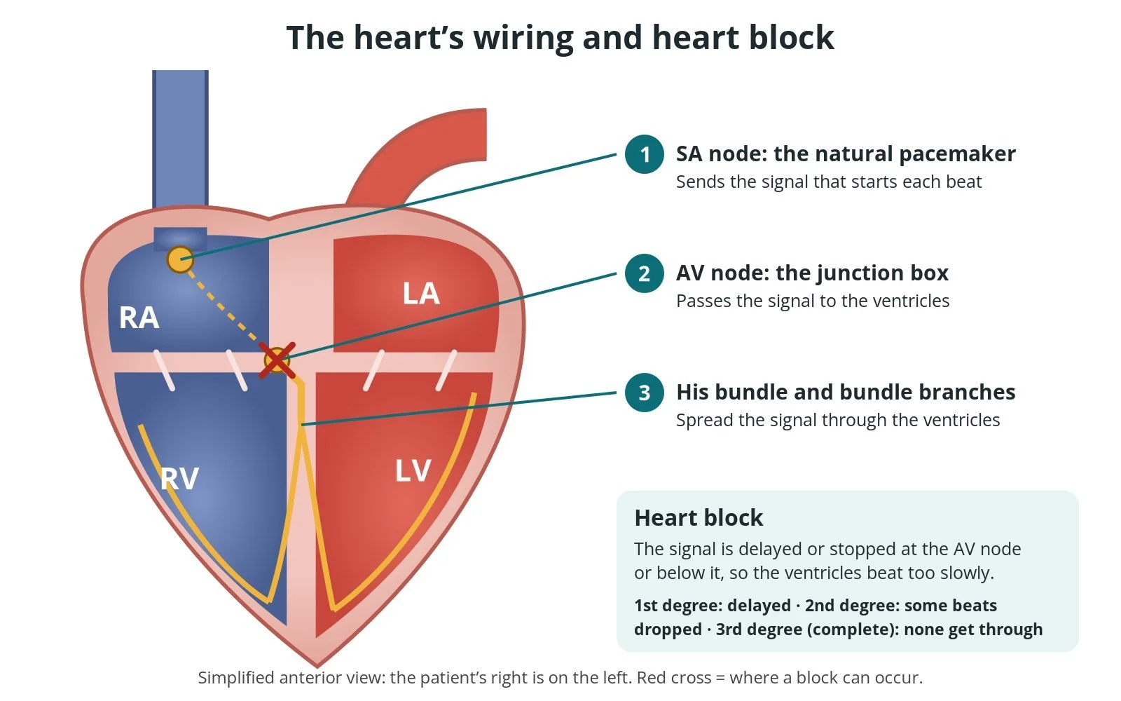
The heart’s wiring: (1) the SA node starts each beat; (2) the AV node passes it to the ventricles; (3) the His bundle and bundle branches spread it. In heart block the signal is delayed or stopped at or below the AV node.
Pacing modes in brief
A pacemaker's mode is written as three to five letters (the NBG code):
Chamber paced: O (none), A (atrium), V (ventricle) or D (dual, both)
Chamber sensed: O, A, V or D
Response to sensing: O (none), I (inhibited), T (triggered) or D (both)
Rate response: R if the rate rises with activity; left off if not
Multisite pacing: O, A, V or D; usually left off
So DDDR paces and senses both chambers, can inhibit or trigger, and speeds up with activity. VVI paces and senses the ventricle only and holds back when the heart beats by itself. DDD(R), VVI(R) and AAI(R) are the modes you will see most.
Who is it for?
The decision is made by a cardiologist, often after an ECG, heart monitor or echo. The commonest reasons in UK and European guidelines are:
heart block, especially second-degree (Mobitz II) or complete (third-degree) heart block, even without symptoms in some cases
sick sinus syndrome: a slow or pausing SA node that causes symptoms
slow atrial fibrillation that causes symptoms
some types of fainting (reflex syncope) with long pauses, in carefully selected people
heart failure with a weak left ventricle and a wide QRS on the ECG (usually left bundle branch block): a CRT device may be offered
a high risk of a life-threatening fast rhythm, or survival of one: an ICD may be offered
NICE recommends dual-chamber pacing for symptomatic slow heart rates from sick sinus syndrome and/or AV block with some exceptions (TA88). NICE TA314 sets out who should be offered an ICD or CRT. Temporary causes, such as medicines that slow the heart or a reversible problem, are treated first.
When is it done?
Most pacemakers are planned procedures, done as a day case or with one night in hospital. Someone with complete heart block may have it done urgently, sometimes after a temporary pacing wire through a vein in the neck or groin. Before the procedure, the team usually:
records ECGs and reviews any monitor or loop recorder results
checks bloods, including kidney function, clotting and potassium
plans blood thinners: whether to continue, pause or bridge anticoagulants or antiplatelets, as per centre protocol
checks for infection and agrees which side to implant (usually the left, away from the dominant arm)
gives antibiotics before the procedure, as per centre protocol
Step by step
1. Preparation
The patient lies flat on the X-ray table with a cannula in place and ECG, blood pressure and oxygen monitoring. The skin is cleaned and draped. Antibiotics are given before the skin incision. Local anaesthetic numbs the area, with sedation if needed. All doses are as per centre protocol.
2. Access
A cut of a few centimetres is made below the collarbone. The operator reaches a vein in one of three ways: a cut-down onto the cephalic vein in the groove between the chest and shoulder muscles; a needle into the axillary vein, often with X-ray or ultrasound guidance; or a needle into the subclavian vein under the collarbone. A guidewire and a peel-away sheath follow. Sheath size is per device and operator.
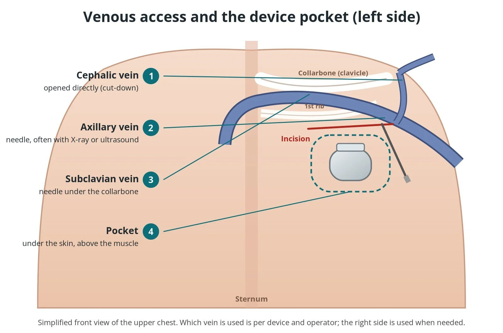
Ways into the vein: (1) the cephalic vein, opened directly; (2) the axillary vein, by needle; (3) the subclavian vein, by needle under the collarbone. (4) The device sits in a pocket under the skin, above the muscle.
3. Placing the leads
Under X-ray, each lead is passed through the vein into the superior vena cava and the right atrium. The ventricular lead crosses the tricuspid valve into the right ventricle, at the apex or on the septum. The atrial lead is usually placed in the right atrial appendage. Most leads are held in place by a small screw at the tip (active fixation); some use soft tines (passive fixation).
For CRT, the coronary sinus is entered from the right atrium, a small dye injection maps its branches, and the left ventricular lead is advanced over a fine wire into a vein on the outer wall of the left ventricle.
4. Testing
Each lead is tested with a pacing analyser, usually by a cardiac physiologist:
sensing: how well the lead "hears" the heart's own signal
pacing threshold: the smallest pulse that makes the heart beat
impedance: a check of the lead and its contact with the heart
The operator may also test at high output to check the lead does not stimulate the diaphragm, especially with CRT. Once the numbers are acceptable, each lead is tied to the muscle with a non-absorbable suture on its protective sleeve.
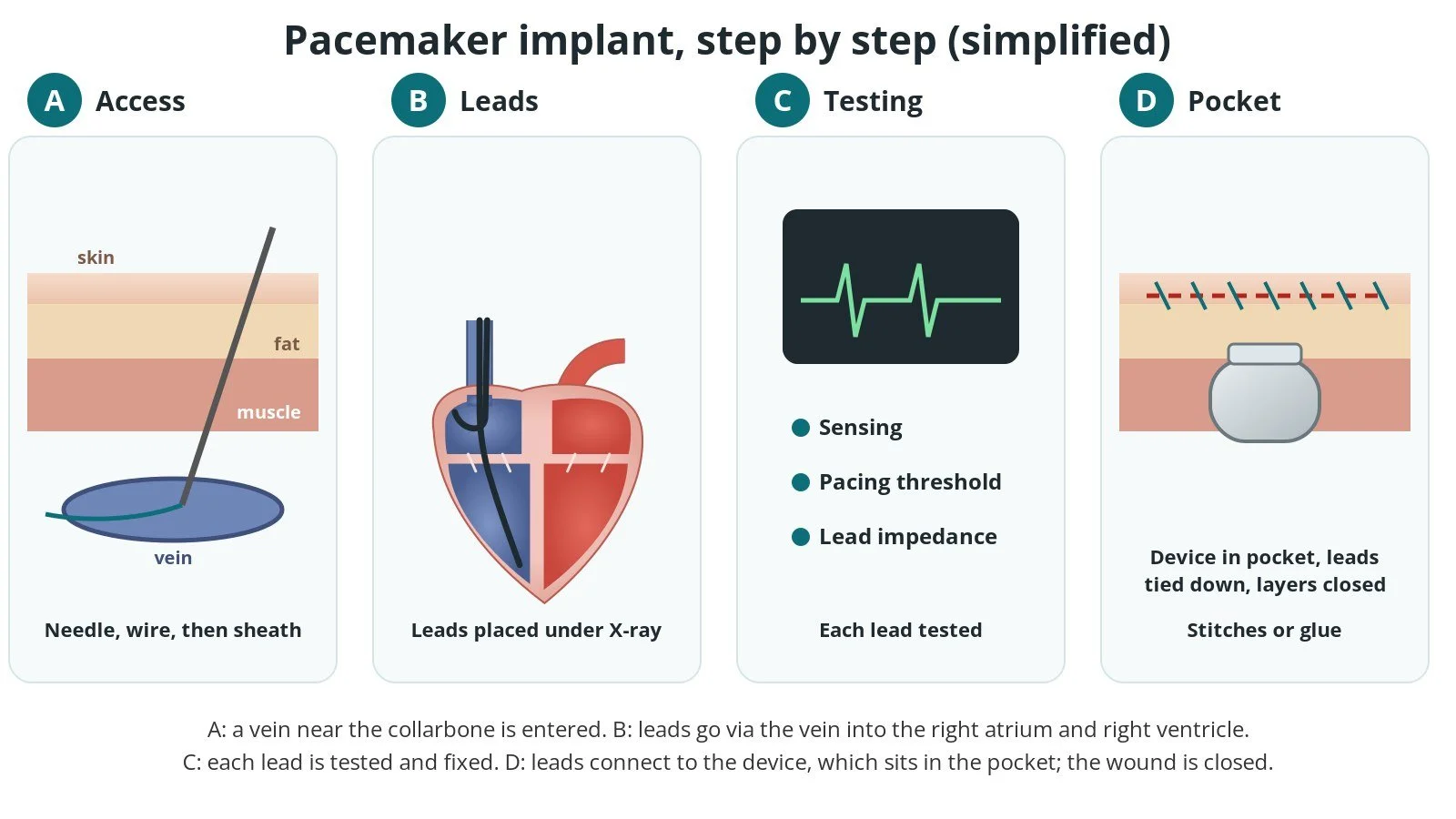
A: a vein near the collarbone is entered with a needle, wire and sheath. B: the leads go via the vein into the right atrium and right ventricle under X-ray. C: each lead is tested. D: the device goes in the pocket and the wound is closed.
5. Pocket and closure
A pocket is made under the skin, above the chest muscle (sometimes under it). The leads are screwed into the generator, the excess lead is coiled behind it, and the generator is placed in the pocket. The wound is closed in layers with dissolvable stitches or skin glue, and a dressing is applied. A final check confirms the device is pacing and sensing correctly.
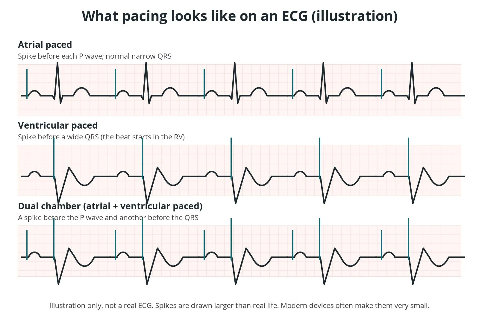
Illustrated ECG: atrial pacing puts a spike before each P wave; ventricular pacing puts a spike before a wide QRS; dual-chamber pacing shows both. Not a real ECG.
Recovery and aftercare
Same day or next morning. A chest X-ray checks lead positions and rules out a collapsed lung, and a device check confirms it is working. You'll receive a device ID card.
Wound care. Keep the wound clean and dry. Follow your team's advice on showering and dressings. Expect soreness and tiredness for a few days.
Arm movement. Gentle arm movements are fine within the first few days. For 4 to 6 weeks, don't lift the elbow on the device side above shoulder height, and avoid heavy lifting and vigorous exercise, so the leads can settle. Do the shoulder exercises your team gives you.
Driving (UK). After a pacemaker, you must not drive a car or motorcycle for at least one week, and you must tell the DVLA. Bus and lorry drivers must wait at least 6 weeks. ICD rules are stricter: after an ICD for a dangerous rhythm, car driving usually stops for 6 months, and an ICD bars bus and lorry licences. Check the current GOV.UK guidance.
Follow-up. A pacemaker clinic check at about 6 weeks, a review at 3 to 6 months, then at least yearly, often with home remote monitoring too. The battery usually lasts several years, and a "box change" replaces the generator but usually keeps the leads.
Contact your clinic or NHS 111 if the wound becomes red, swollen, hot or leaks, if you have a fever, or if your original symptoms come back. Call 999 for chest pain, severe breathlessness, fainting or collapse.
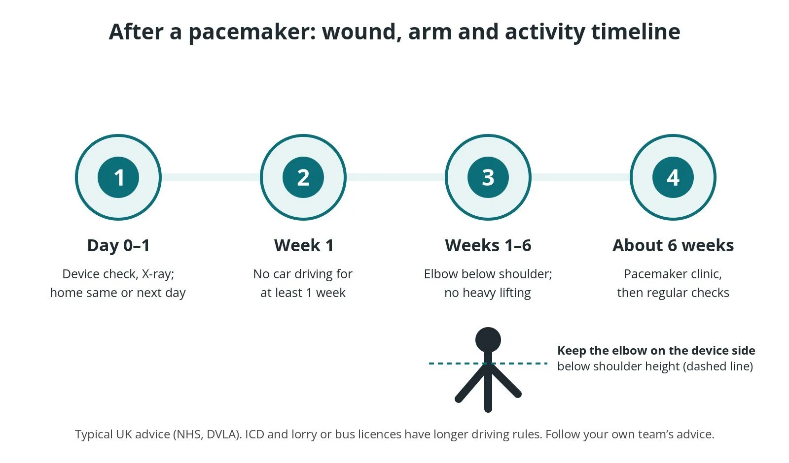
After a pacemaker: (1) device check and X-ray, home the same or next day; (2) no car driving for at least a week; (3) elbow below shoulder height and no heavy lifting for 4 to 6 weeks; (4) clinic check at about 6 weeks, then regular checks.
Risks and complications
Pacemaker implantation is common and most procedures go smoothly. Possible problems include:
Bruising or a haematoma (collection of blood) in the pocket, the most common problem, more likely on blood thinners
Lead displacement: a lead moves and needs repositioning, usually in the first weeks
Pneumothorax: a collapsed lung from the needle, uncommon, and less likely with cephalic or axillary access
Infection of the pocket or leads. This is uncommon but serious, and often means removing the whole system
Cardiac perforation by a lead, with fluid around the heart (rare)
Diaphragm stimulation (hiccups), mainly with CRT leads
Pacemaker syndrome or pacing-related weakening of the heart with long-term right ventricular pacing, in some people
Your heart team will explain how these apply to you.
Cath lab kit list (printable)
Drugs, doses and dilutions: always as per centre protocol. Equipment, sizes and suture choices also vary by unit, operator and device. Device and product names below are generic. Always check the sheath, lead and wire required in the chosen device's instructions for use.
Example: Barts Heart Centre kit list — always follow your own centre protocol
Typical setup for a transvenous pacemaker or ICD implant under local anaesthetic, with or without mild to moderate sedation, shared as one real-world example. It is not a protocol for your unit.
Basic kit (every implant)
device implant pack
chlorhexidine skin prep applicator × 2
antimicrobial incise drape
medium image intensifier cover
diathermy pen and scratch pad
500 mL sterile saline bag
Sutures (operator preference)
non-absorbable braided suture (securing the leads)
absorbable tie (cephalic cut-down)
absorbable suture for deep tissue
absorbable suture for skin (curved or straight)
adhesive skin closure strips and a dressing
Lead revision or box change: an electrosurgical cutting device may be used instead of standard diathermy, as per operator.
Extra for CRT or conduction system pacing
shoulder radiation shield pad
20 mL Luer-lock syringe
small trolley cover
hydrophilic guidewire, length per device and operator
0.014" coronary guidewire (floppy or support), per operator
small bowl of contrast for coronary sinus injections
Extra for device or lead extraction
shoulder radiation shield pad
small trolley cover
hydrophilic guidewire, length per device and operator
large swabs
extra non-absorbable sutures
extraction is usually done with surgical back-up, as per centre protocol
Extra for a subcutaneous ICD
usually under general anaesthetic; if a regional nerve block is used: ultrasound, probe cover, large chlorhexidine applicators × 2 and a nerve block needle
local anaesthetic agents as per centre protocol
arm board to rest the arm out to the side, with padding to secure it
multi-piece drape set (arm, two sides, leg, head)
extra non-absorbable and deep-tissue sutures, and two dressings
Medications (scrub nurse): local anaesthetic, antibiotics, sedation and any heparin as per centre protocol.
Imaging: fluoroscopy, with contrast for venography or the coronary sinus as needed, as per centre protocol.
Procedural steps (Barts example)
Check side, antibiotics given, skin prep and drape
Local anaesthetic; incision below the left collarbone
Cephalic cut-down, or axillary or subclavian puncture; wire and peel-away sheath (size per device and operator)
Leads to the right ventricle (and right atrium, and coronary sinus for CRT) under fluoroscopy
Test sensing, threshold and impedance; secure each lead with non-absorbable suture
Connect the leads, form the pocket, place the generator
Close in layers; dressing; final device check
First time in the cath lab?
💡 First-time tip
Watch the ECG the whole time. Wires in the right ventricle can trigger extra beats, and someone with heart block can become very slow. Know where the external pacing pads and temporary wire are. Check antibiotics are in before the knife. Infection is the complication everyone works hardest to prevent. Keep the field clean. Minimise traffic and door opening, and keep the pocket covered. Count and keep the lead stylets and packaging. The lead and device serial numbers go in the record and the patient's ID card. Have the physiologist and analyser ready before the leads go in, and know the plan if numbers are poor.
FAQ
What does PPM stand for?
PPM is the medical abbreviation for permanent pacemaker: an implanted device that stops the heart from beating too slowly.
Is a pacemaker the same as an ICD?
No. Both can pace a slow heart, but an ICD can also treat a dangerously fast rhythm with rapid pacing or a shock. A CRT device helps both ventricles beat together and can be either type.
Am I awake for it?
Usually yes, under local anaesthetic, often with sedation to help you relax. Some procedures, such as a subcutaneous ICD, may use a general anaesthetic.
Can I have an MRI scan with a pacemaker?
Many modern systems are MRI-conditional, so a scan can be done safely under set conditions if the whole system (generator and leads) is approved. The device is checked and reprogrammed before and after. Tell the scanning team and bring your device card.
Will phones and airport security affect it?
Everyday use is fine. Don't keep a phone, or headphones with magnets, in a pocket right over the device. Show your ID card at airport security and ask for a hand search rather than a handheld metal detector held over the device.
How long does the battery last?
Usually several years, depending on how much pacing you need and the type of device. The clinic watches the battery, and a box change is a smaller procedure than the first implant.
Related Cath Lab 101 guides
Sources
All checked on 10 October 2026. Each page's own date is shown where it gives one.
NHS. Pacemakers. nhs.uk/tests-and-treatments/pacemaker-implantation (page last reviewed 30 Mar 2026)
British Heart Foundation. What is a pacemaker and what does it do? bhf.org.uk (published 19 Jun 2024)
NICE. Dual-chamber pacemakers for symptomatic bradycardia due to sick sinus syndrome and/or atrioventricular block (TA88). nice.org.uk/guidance/ta88 (published 23 Feb 2005, last updated 1 Nov 2014)
NICE. Implantable cardioverter defibrillators and cardiac resynchronisation therapy for arrhythmias and heart failure (TA314, which replaced TA120). nice.org.uk/guidance/ta314 (published 25 Jun 2014)
Glikson M, et al. 2021 ESC Guidelines on cardiac pacing and cardiac resynchronization therapy. Eur Heart J 2021;42(35):3427–3520. PubMed 34455430
DVLA. Pacemakers and driving. gov.uk/pacemakers-and-driving
DVLA. Cardiovascular disorders: assessing fitness to drive (pacemaker, ICD and CRT standards). gov.uk (last updated 7 Nov 2025)
MHRA. Safety guidelines for magnetic resonance imaging equipment in clinical use. gov.uk (last updated 30 Sep 2026)
Disclaimer
Education only. This page is for general education for healthcare students, cath lab staff and curious readers. It isn't medical advice and doesn't replace advice from your own doctor, heart team or local unit protocols. Equipment and medicines are described generically. No product or manufacturer is recommended. If you think someone is having a heart emergency, call 999.
Disclosure: I work in the medical device industry. This page is independent education and isn't sponsored or reviewed by any company.
All diagrams on this page are original illustrations created for rodnieoro.com.