Subcutaneous ICD (S-ICD): a Cath Lab 101 guide
Reviewed by Rodnie Oro, cath lab charge nurse · Last reviewed: October 2026 · Education only, not medical advice
S-ICD in one minute
What it is: an implantable cardioverter defibrillator (ICD) watches the heart rhythm and gives a shock if a dangerous fast rhythm starts. A subcutaneous ICD (S-ICD) does this with the generator under the skin on the left side of the chest and a lead under the skin beside the breastbone, so nothing goes inside the heart or veins.
Who it is for: people who need an ICD but do not need a pacemaker for a slow heart rate or CRT, and where avoiding wires in the heart is helpful, for example younger people or those with difficult veins or a higher infection risk.
Main risks: infection, bleeding or bruising, shocks that are not needed, and the device or lead moving. Your team will explain your own risk.
Usually: under general anaesthetic or sedation with a nerve block, about 1 to 1.5 hours, often home the same or next day.
On this page: What is an S-ICD? · Who is it for? · When is it done? · Step by step · Recovery · Risks · Kit list · First time in the lab? · FAQ
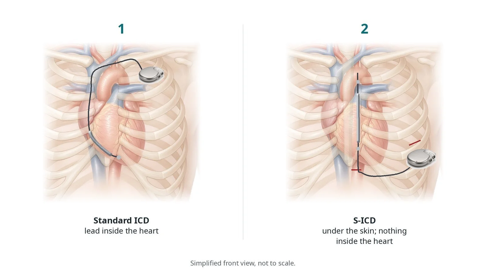
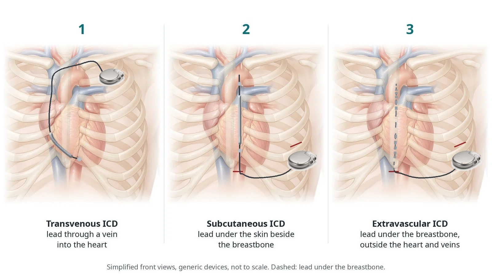
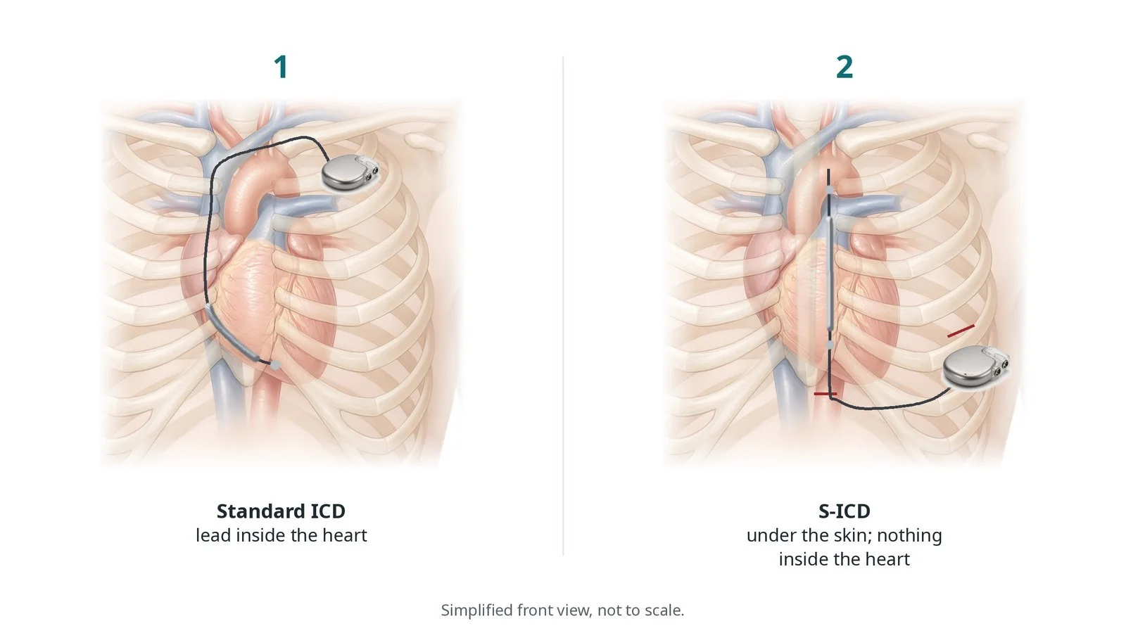
(1) Standard ICD: lead inside the heart. (2) S-ICD: generator under the skin on the left side and a lead beside the breastbone; nothing inside the heart.
What is an S-ICD?
Some people are at risk of ventricular tachycardia (VT) or ventricular fibrillation (VF), fast rhythms from the lower chambers that can cause sudden cardiac arrest. An ICD constantly monitors the heart rhythm and, if it detects one of these rhythms, delivers a shock to restore a normal rhythm.
A standard (transvenous) ICD has leads passed through a vein into the heart. A subcutaneous ICD (S-ICD) works differently:
the generator (the battery and computer) sits under the skin on the left side of the chest, near the armpit, often under or beside a muscle
a single lead runs under the skin from the generator to the lower end of the breastbone, then up beside it
the shock passes from the generator, through the heart, to the lead
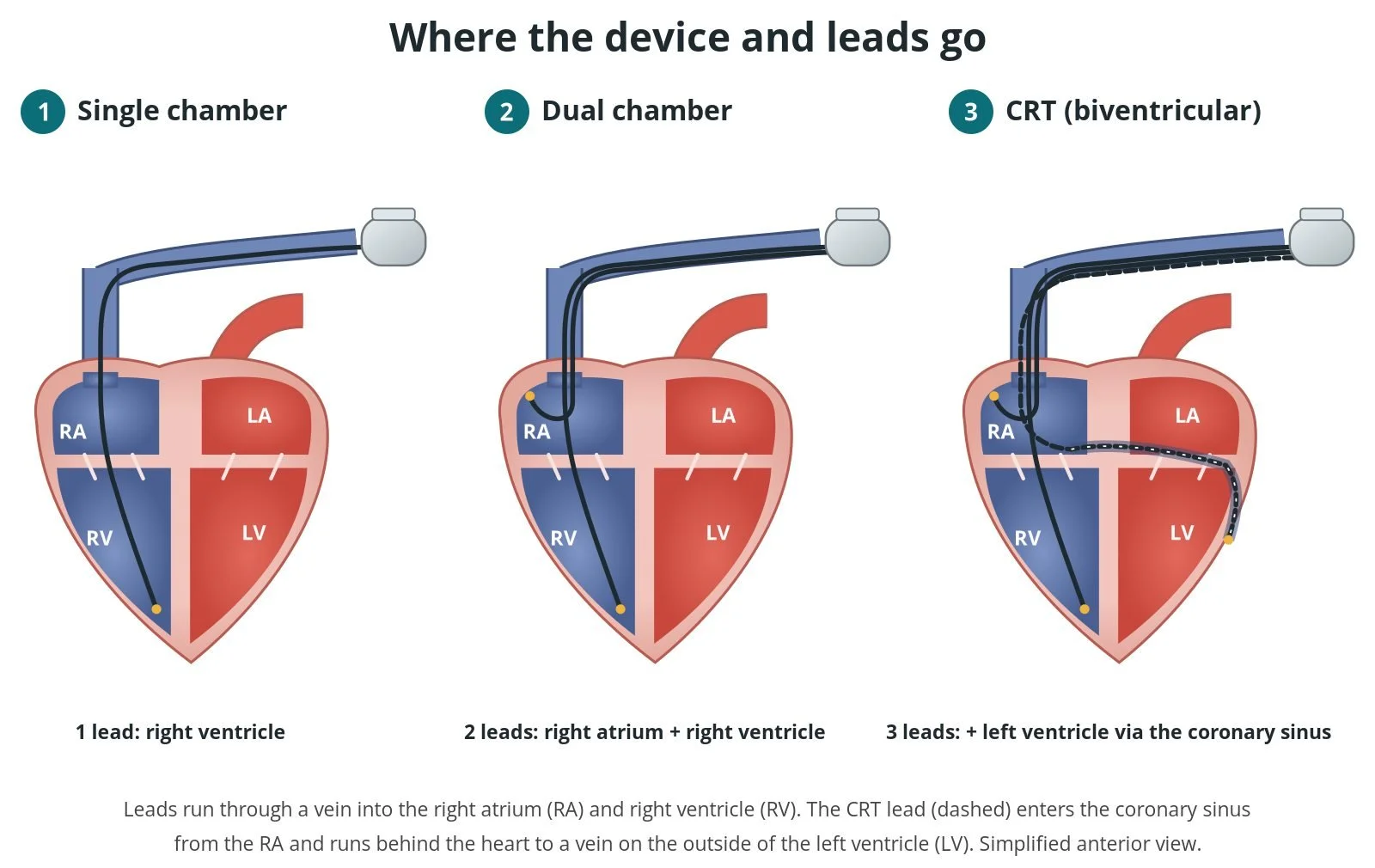
Because nothing enters the veins or heart, there is no risk of lead problems inside the heart or blood infections from leads. But an S-ICD cannot pace the heart for a slow rhythm, give anti-tachycardia pacing, or provide CRT, so it does not suit everyone. See the pacemaker, ICD and CRT guide for standard devices.
New to the acronyms? See the Cath Lab Abbreviations A–Z.
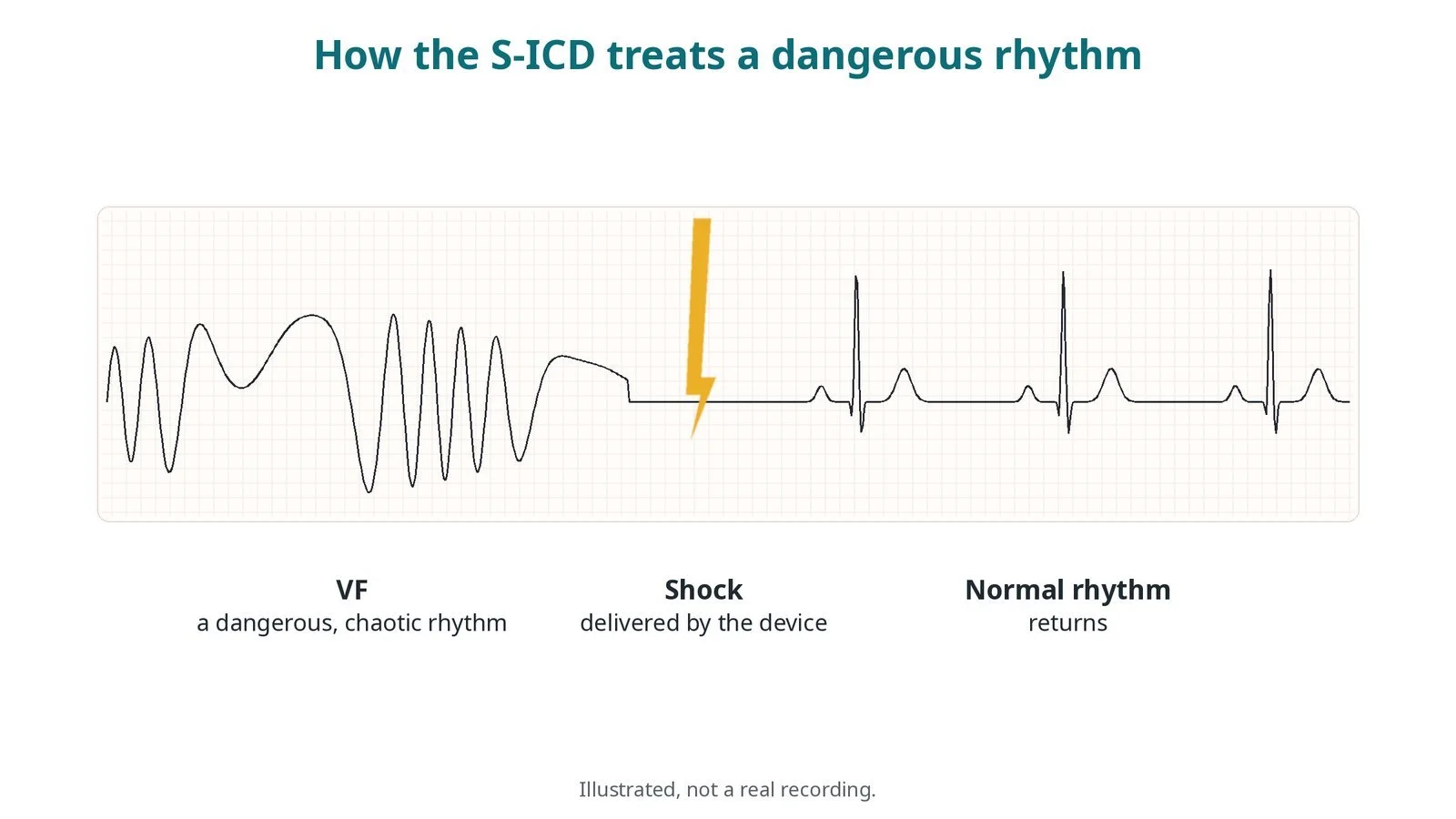
Illustrated, not a real recording. The S-ICD detects a dangerous rhythm (VF), delivers a shock, and a normal rhythm returns.
Who is it for?
NICE (TA314) recommends ICDs for people who have survived a cardiac arrest or dangerous ventricular rhythm, and for selected people at high risk of one, such as some people with heart failure and a weak heart muscle, or inherited heart conditions. The ESC 2022 ventricular arrhythmia guidelines say an S-ICD should be considered as an alternative to a transvenous ICD for people who need an ICD when pacing for a slow heart rate, CRT or anti-tachycardia pacing is not needed.
An S-ICD is often chosen for:
younger people, who may need devices for decades
people with no suitable vein access or with congenital heart disease
people at higher risk of infection, or who have had a previous device infection
Before implant, an ECG screening test checks that the device will be able to sense the heart's signals reliably from under the skin.
When is it done?
It is usually planned. Before it, the team usually:
carries out the ECG screening, lying and standing
checks blood tests and blood thinners, which may need adjusting, as per centre protocol
checks allergies and gives skin-cleaning instructions
explains driving rules, shocks and living with a device
Step by step
1. Anaesthetic and set-up
The implant is usually done under general anaesthetic or deep sedation, sometimes with an ultrasound-guided nerve block of the chest wall. The left arm is supported on an arm board away from the body. Antibiotics are given as per centre protocol. X-ray is used to check positions.
2. Planning the incisions
Using surface landmarks and often X-ray, the team marks where the generator and lead will go. Most implants use two small incisions: one on the left side of the chest for the generator, and one near the lower end of the breastbone. A third incision higher up is used in some techniques.
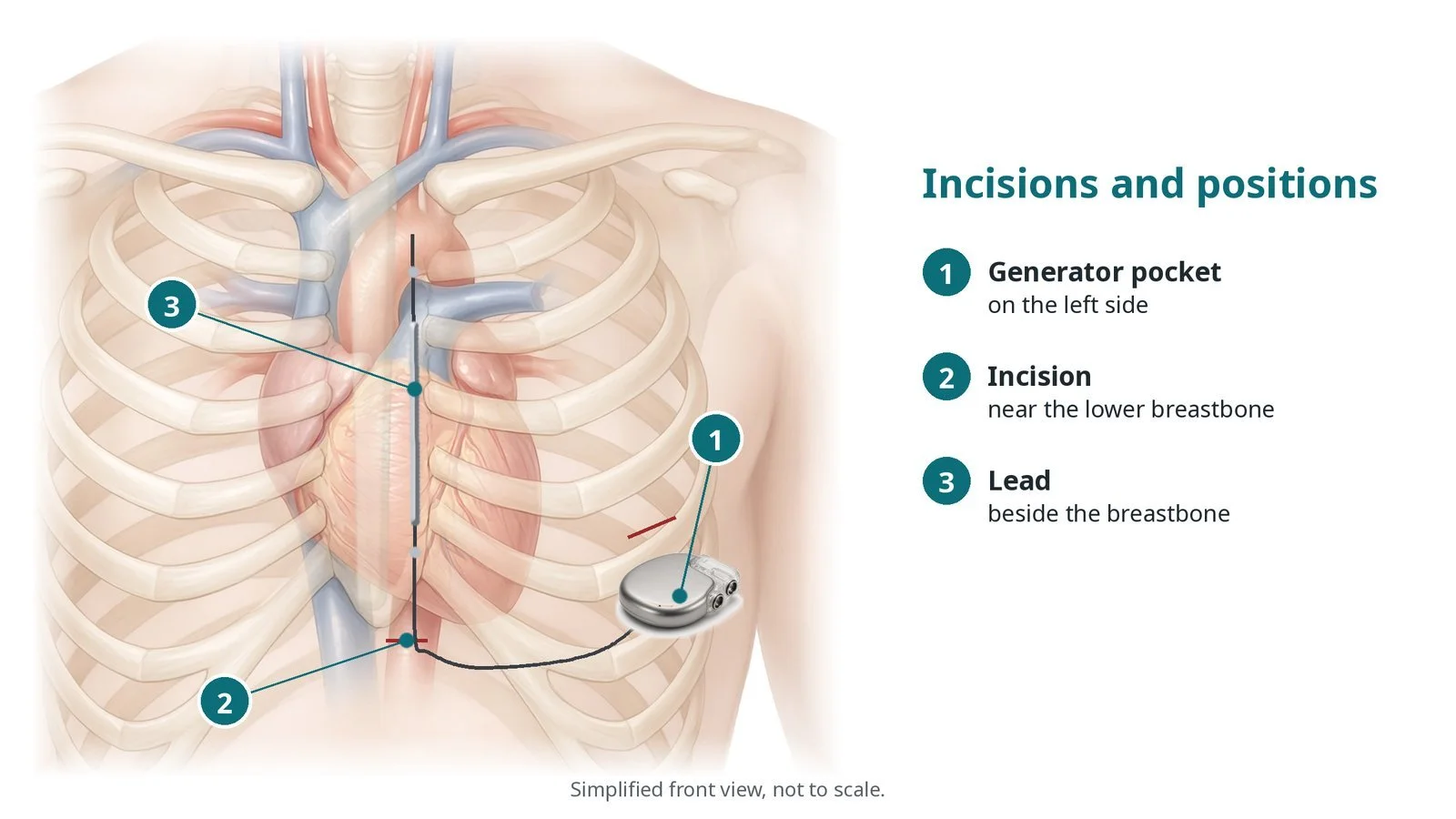
Incisions and positions: (1) generator pocket on the left side; (2) incision near the lower breastbone; (3) lead beside the breastbone.
3. Implanting the device
A. Pocket. A pocket is made on the left side of the chest for the generator, often between muscle layers.
B. Tunnel across. A tunnelling tool is passed under the skin from the pocket to the incision near the lower breastbone, and the lead is pulled through.
C. Tunnel up. The lead is tunnelled up beside the breastbone and secured with sutures, per operator.
D. Connect and close. The lead is connected to the generator, which is placed in the pocket. Some teams test the device by briefly starting VF under anaesthetic and checking it shocks correctly (defibrillation testing); others do not, per operator. The incisions are closed in layers.
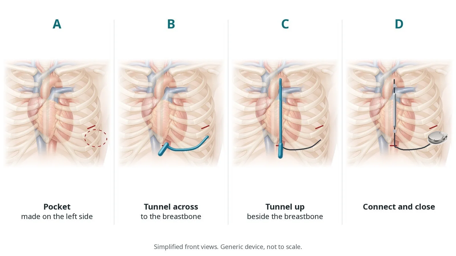
A: make the pocket. B: tunnel across. C: tunnel up beside the breastbone. D: connect and close.
4. Finishing
The device is programmed and checked. A chest X-ray may be taken to confirm positions. Dressings are applied.
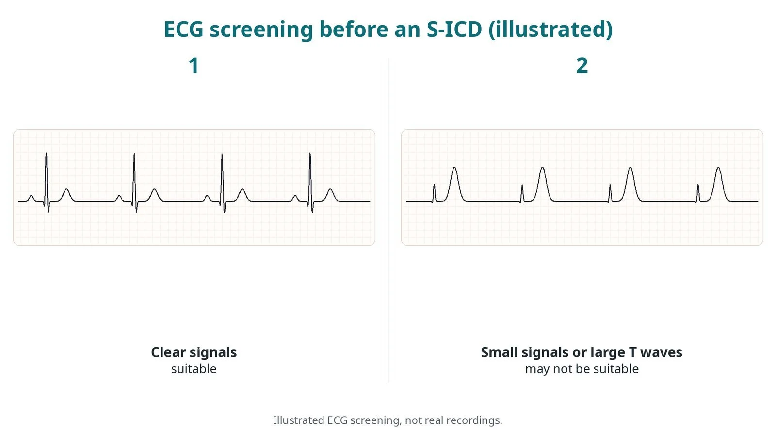
Illustrated ECG screening, not real recordings. (1) Clear signals: suitable. (2) Small signals or large T waves: may not be suitable.
Recovery and aftercare
First hours: checks of the wounds, blood pressure and heart rhythm, as per centre protocol. The wounds can be sore for a few days.
Going home: often the same or next day after a device check.
Wounds: keep them clean and dry, and follow the dressing advice. Watch for redness, swelling, discharge or fever.
Arm and activity: avoid heavy lifting and vigorous arm movement for a few weeks, or as advised.
Follow-up: device clinic checks, often with remote monitoring.
If you get a shock: follow the plan your team gives you. A single shock with no symptoms usually means contacting the device clinic; repeated shocks or feeling unwell means calling 999.
Driving: DVLA rules after an ICD depend on why it was fitted. You must not drive for a set period, which is longer if the ICD was fitted after a dangerous rhythm, and further limits apply after shocks. An ICD permanently bars a bus or lorry licence. Check the DVLA guidance and ask your team.
Call 999 if you have more than one shock, chest pain, fainting or feel very unwell.
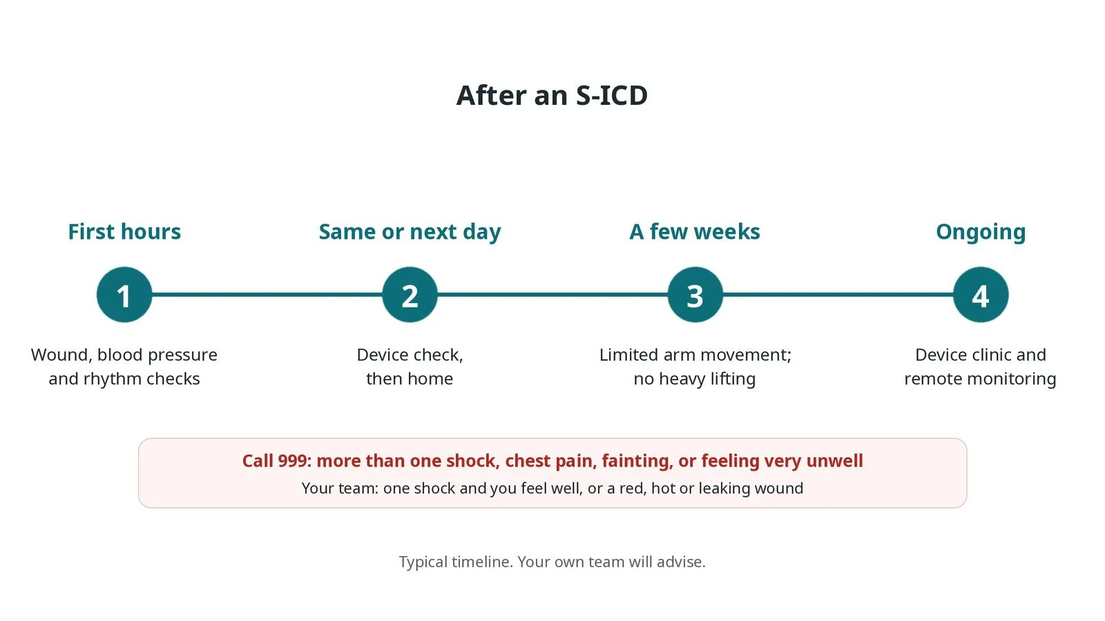
After an S-ICD: (1) checks; (2) device check, home; (3) limited arm movement for a few weeks; (4) device clinic and remote monitoring.
Risks and complications
Your team will discuss your own risks. Possible problems include:
wound infection or pocket infection, which may need the device removed
bleeding or a collection of blood in the pocket
shocks that are not needed (inappropriate shocks), for example from muscle noise or a fast normal rhythm
the lead or generator moving
pain or skin wearing over the device
risks of the anaesthetic and of defibrillation testing, if done
Cath lab kit list (printable)
Barts example kit list
An example from one UK centre, written generically. Every lab has its own protocol, and sizes and doses vary.
Basic device kit
device procedure pack, skin antiseptic applicators, adhesive incise drape, image intensifier cover
diathermy pen and scratch pad
sterile saline bag
sutures (operator preference): non-absorbable sutures to secure the lead; absorbable sutures for deep layers; skin closure suture or adhesive strips
wound dressings
S-ICD extras
S-ICD generator, lead and tunnelling tool, per device and operator
arm board and padding to hold the left arm out; drapes for the side approach
extra sutures for the additional incisions
extra dressings
If a nerve block is used
ultrasound machine and sterile probe cover
nerve block needle
local anaesthetics, as per centre protocol
Always
external defibrillator with pads applied, and programmer
Procedural steps (Barts example)
General anaesthetic or sedation (with or without nerve block); left arm on arm board
External defibrillator pads applied; skin prepared and draped
Landmarks and incisions marked
Generator pocket made on the left side of the chest
Lead tunnelled to the lower sternum, then up beside the sternum; secured
Lead connected; generator placed in the pocket
Defibrillation testing if the operator uses it
Layered closure; device programmed; dressings
First time in the cath lab?
💡 First-time tip
Pads on first. External defibrillator pads go on before draping, placed away from where the device will go. Know the arm set-up. The arm board and padding must hold the left arm safely away from the body for the side approach. Defibrillation testing. If the team induces VF, know your role and keep everyone clear. Count everything. Tunnelling tools and sutures need counting like any surgical case. Infection matters. Strict sterile technique protects the device; a device infection usually means removing it.
FAQ
Can an S-ICD pace my heart?
Only briefly after a shock. It cannot pace for a slow heart rate long term, give anti-tachycardia pacing or provide CRT.
Will I feel a shock?
Yes. A shock is like a sudden thump in the chest. It is over in a moment.
Can I have an MRI scan?
Some S-ICD systems are MR conditional. Always tell the scanning team and carry your device card.
How long does the battery last?
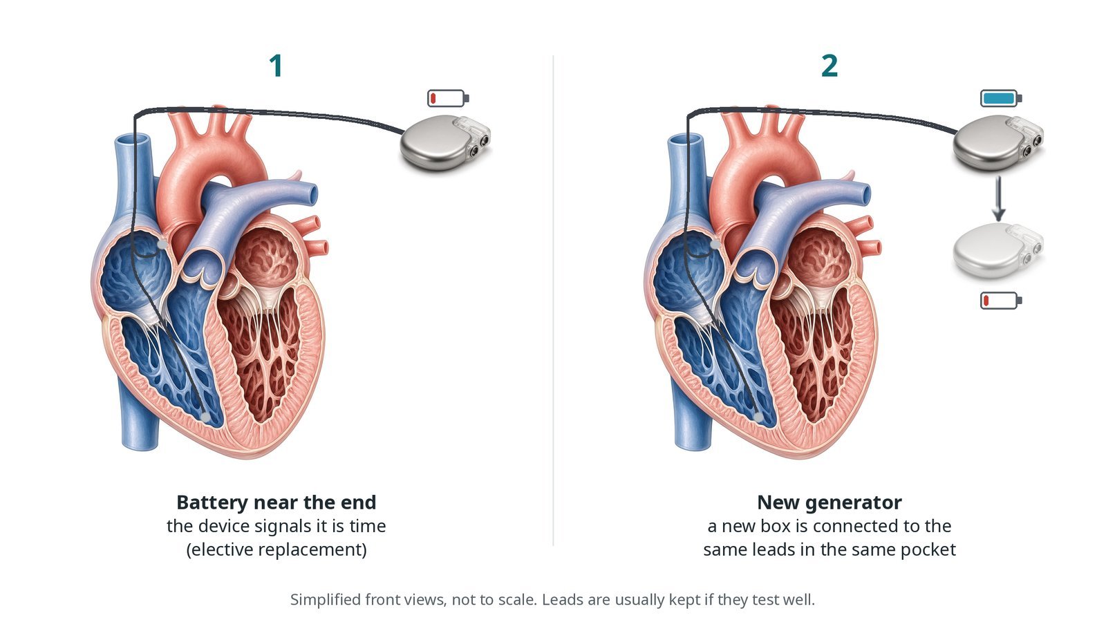
Several years. The generator is replaced in a smaller procedure when the battery runs low; the lead usually stays.
Related Cath Lab 101 guides
Sources
All checked on 10 October 2026. Each page's own date is shown where it gives one.
NICE. Implantable cardioverter defibrillators and cardiac resynchronisation therapy for arrhythmias and heart failure (TA314). nice.org.uk/guidance/ta314
Zeppenfeld K, et al. 2022 ESC Guidelines for the management of patients with ventricular arrhythmias and the prevention of sudden cardiac death. Eur Heart J 2022;43(40):3997–4126. PubMed 36017572
British Heart Foundation. Implantable cardioverter defibrillator (ICD). bhf.org.uk
DVLA / GOV.UK. Cardiovascular disorders: assessing fitness to drive (ICDs). gov.uk
Disclaimer
Education only. This page is for general education for healthcare students, cath lab staff and curious readers. It isn't medical advice and doesn't replace advice from your own doctor, heart team or local unit protocols. Equipment and medicines are described generically. No product or manufacturer is recommended. If you think someone is having a heart emergency, call 999.
Disclosure: I work for a company that makes a device used in this procedure. This page is independent education, isn't sponsored or reviewed by any company, and doesn't recommend any product.
All diagrams on this page are original illustrations created for rodnieoro.com.