Coarctation of the aorta stenting: a Cath Lab 101 guide
Reviewed by Rodnie Oro, cath lab charge nurse · Last reviewed: October 2026 · Education only, not medical advice
Coarctation stenting in one minute
What it is: coarctation is a narrowing of the aorta, the main artery from the heart, usually just beyond the arteries to the head and left arm. It is present from birth. In older children and adults it can often be treated by stretching the narrowing with a balloon and holding it open with a stent, passed up the artery from the groin.
Who it is for: people with a significant narrowing, or a re-narrowing after earlier surgery, causing high blood pressure or a large pressure difference, as decided by a congenital heart team. Babies are usually treated with surgery.
Main risks: bleeding or damage at the groin artery, a tear or bulge in the aorta, the stent moving and, rarely, stroke. Your team will explain your own risk.
Usually: under general anaesthetic, about 1 to 2 hours, usually 1 to 2 nights in hospital.
On this page: What is coarctation of the aorta? · Who is it for? · When is it done? · Step by step · Recovery · Risks · Kit list · First time in the lab? · FAQ
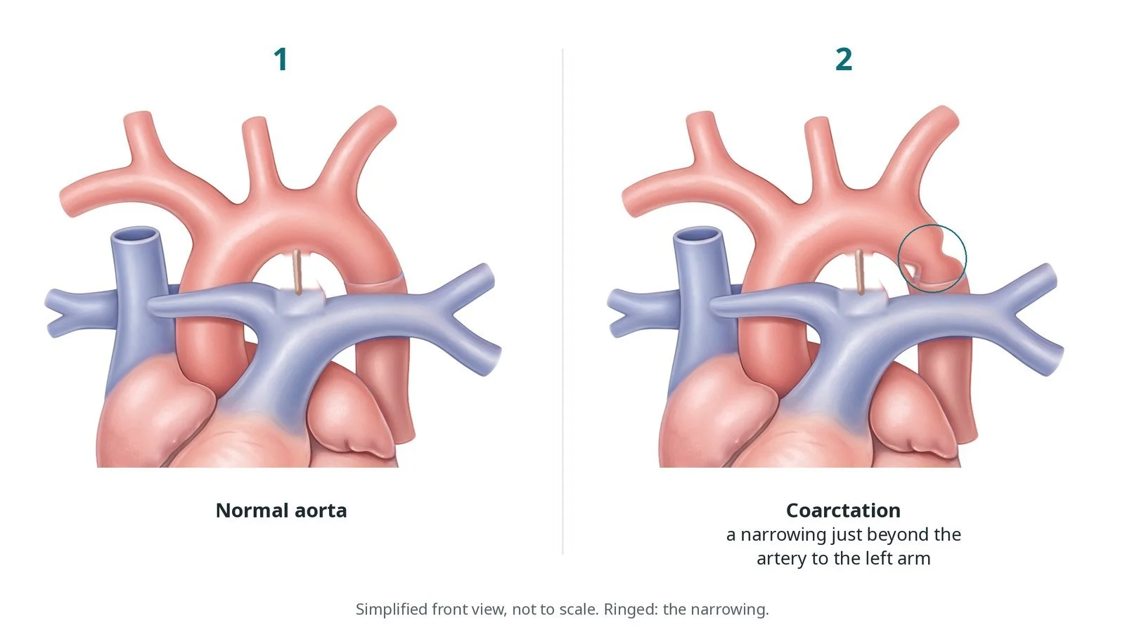
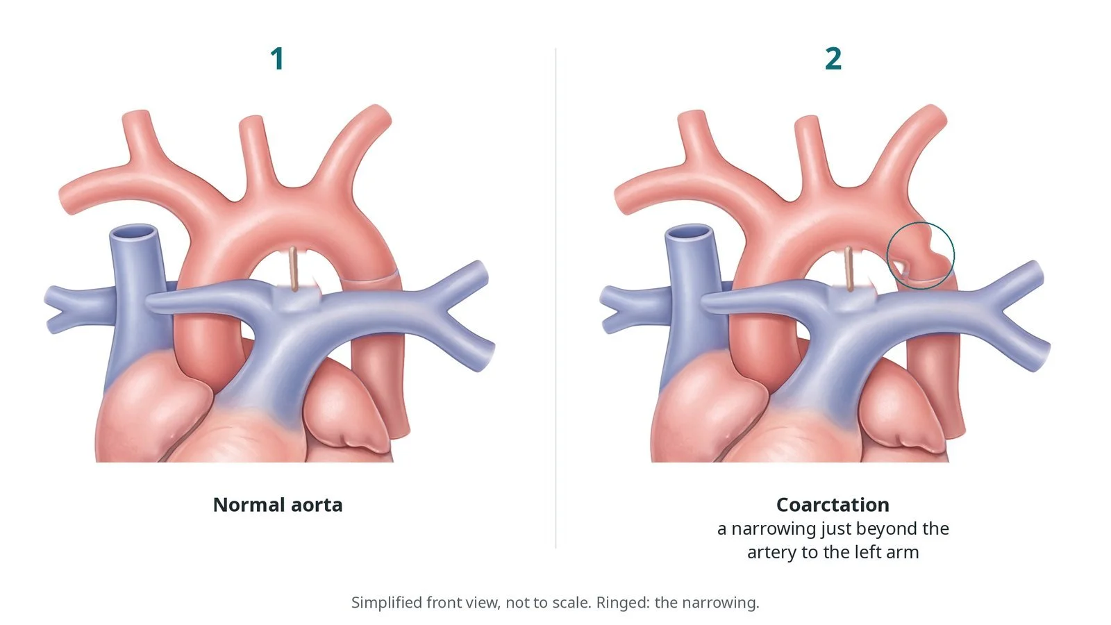
(1) Normal aorta. (2) Coarctation: a narrowing just beyond the artery to the left arm.
What is coarctation of the aorta?
The aorta leaves the heart, arches over and gives off the arteries to the head and arms, then runs down through the chest and abdomen as the descending aorta. In coarctation, there is a narrowing, usually just after the artery to the left arm (the left subclavian artery), near where the ductus arteriosus joined the aorta before birth.
The narrowing makes the heart work harder and causes:
high blood pressure in the arms and head, with lower pressure and weaker pulses in the legs
extra blood vessels (collaterals) that grow around the narrowing
over time, thickening of the left ventricle, and a higher risk of heart attack, stroke and aortic problems if untreated
Coarctation is often found in babies or children, but sometimes not until adulthood, for example when a young person is found to have high blood pressure. It is linked with a bicuspid aortic valve and with Turner syndrome. People treated as children, by surgery or a catheter, can develop a re-coarctation later and need lifelong follow-up.
New to the acronyms? See the Cath Lab Abbreviations A–Z.
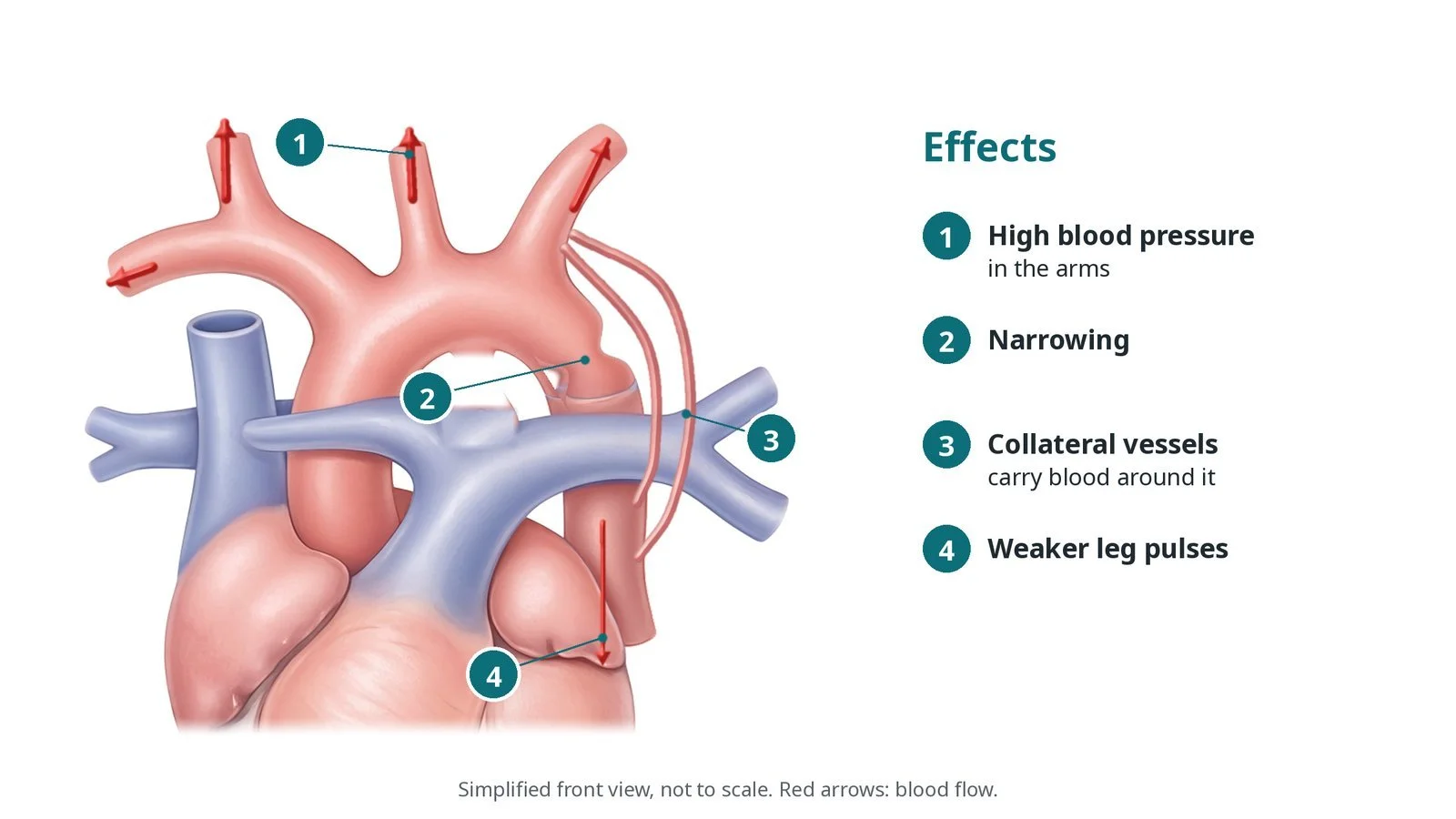
Effects of coarctation: (1) high blood pressure in the arms; (2) narrowing; (3) collateral vessels; (4) weaker leg pulses.
Who is it for?
The decision is made by a congenital heart team. In line with the ESC 2020 guidelines for adult congenital heart disease, treatment is recommended for people with high blood pressure and a significant pressure difference across the narrowing, or a significant narrowing on imaging, and considered in some others. In adults and older children, stenting is usually preferred when it is technically suitable. In babies and young children, surgery is usually the first treatment, and balloon stretching alone may be used in some cases.
When is it done?
It is usually a planned procedure. Before it, the team usually arranges:
MRI or CT of the aorta to measure the narrowing and plan the stent, and an echo
blood pressure measurements in the arms and legs, and often 24-hour monitoring
blood tests, including a group and save
a discussion of risks and alternatives, including surgery
Step by step
1. Anaesthetic and set-up
The procedure is usually done under general anaesthetic, with biplane X-ray. Antibiotics are given as per centre protocol. A surgical team is available in case of complications.
2. Access and measuring
The main access is the femoral artery in the groin, under ultrasound, for the large long sheath. Sizes are per device and operator. Many operators pre-place a suture-based closure device.
A femoral vein sheath is often placed as well, for example for a temporary pacing wire used to briefly lower the blood pressure during stent expansion, per operator.
Heparin is given, dose as per centre protocol. A pigtail catheter measures the pressure above and below the narrowing (the gradient), and an aortogram shows its shape and size.
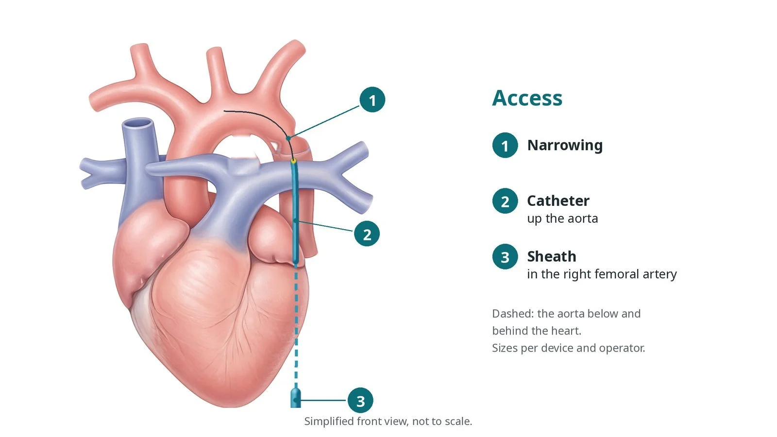
Access: (1) narrowing; (2) catheter up the aorta; (3) sheath in the right femoral artery.
3. Stenting
A. Wire across. A catheter and wire cross the narrowing and a stiff exchange wire is parked in a safe position high in the arch or in the right subclavian artery, per operator.
B. Long sheath. The long sheath is advanced across the narrowing. The stent is mounted on its balloon outside the body.
C. Expand. The stent, often a covered stent to seal any tear, is positioned using angiograms through the sheath and expanded on a two-layer (inner and outer) balloon: the inner balloon first, to check the position, then the outer. Inflation volumes are per device and operator. The blood pressure may be briefly lowered with rapid pacing.
D. Check. Pressures and an aortogram are repeated to confirm the gradient has gone and there is no tear or leak. A further balloon may be used to shape the stent.
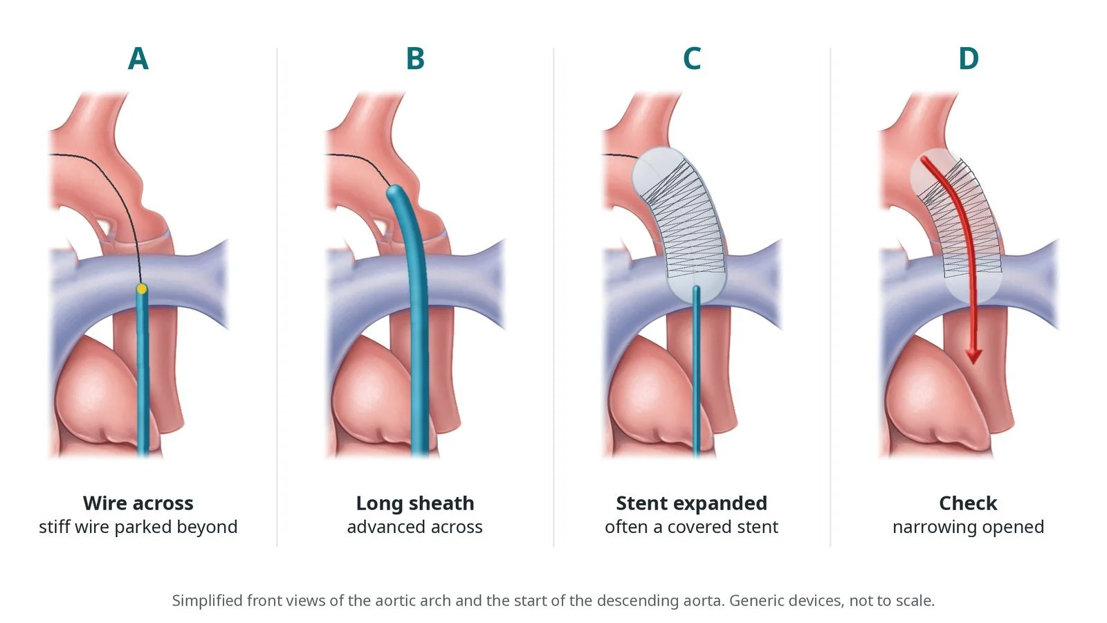
A: wire across. B: long sheath across. C: stent expanded. D: check: narrowing opened.
4. Finishing
The sheaths are removed and the artery is closed with the closure device sutures or pressure, per operator.
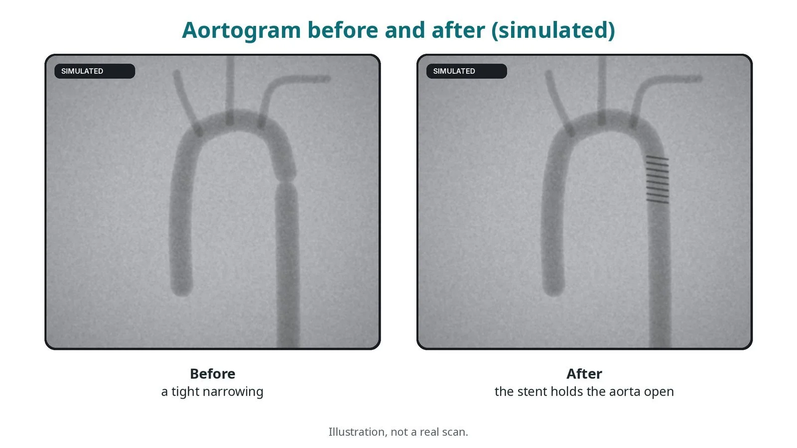
Illustration, not a real scan. Before: a tight narrowing. After: the stent holds the aorta open.
Recovery and aftercare
First hours: bed rest with checks of the groin, leg pulses and blood pressure, as per centre protocol.
Going home: usually after 1 to 2 nights.
Medicines: antiplatelet medicine for a period, as advised. Blood pressure medicines are reviewed; some people still need them.
Activity: avoid heavy lifting and strenuous exercise for a few weeks, or as advised.
Follow-up: lifelong with the congenital heart team, with imaging (CT or MRI) of the stent.
Driving: follow the DVLA guidance for congenital heart disease and aortic conditions, and ask your team.
Call 999 if you have sudden severe chest, back or abdominal pain, heavy bleeding from the groin, or a cold, pale or painful leg.
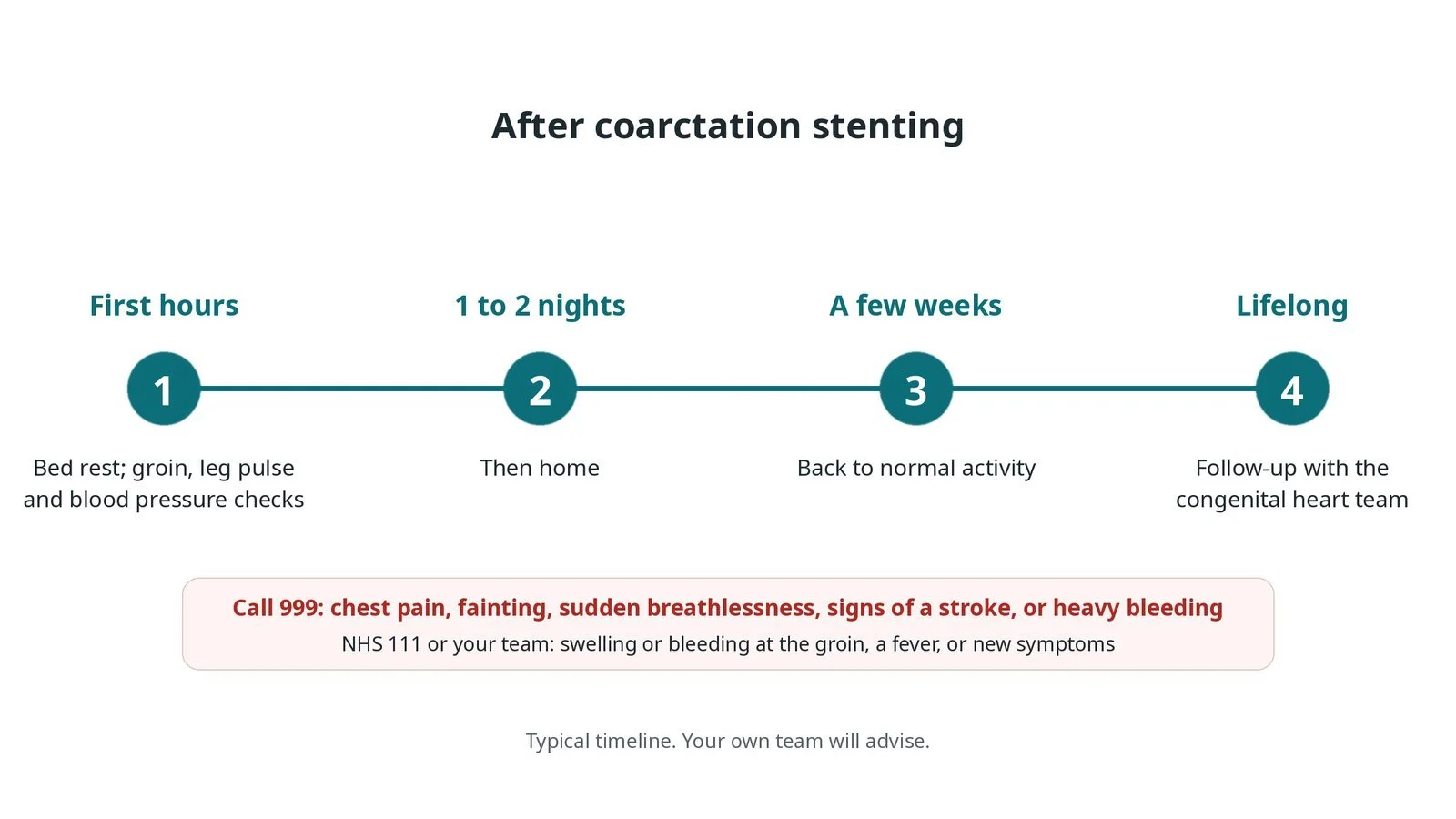
After coarctation stenting: (1) bed rest and checks; (2) home after 1 to 2 nights; (3) normal activity after a few weeks; (4) lifelong follow-up.
Risks and complications
Your team will discuss your own risks. Possible problems include:
bleeding, a false aneurysm or a blocked artery at the groin
a tear (dissection) or rupture of the aorta, or a later bulge (aneurysm) at the stent
the stent moving or the balloon bursting
stroke or blood clots
re-narrowing over time, which may need further ballooning
Cath lab kit list (printable)
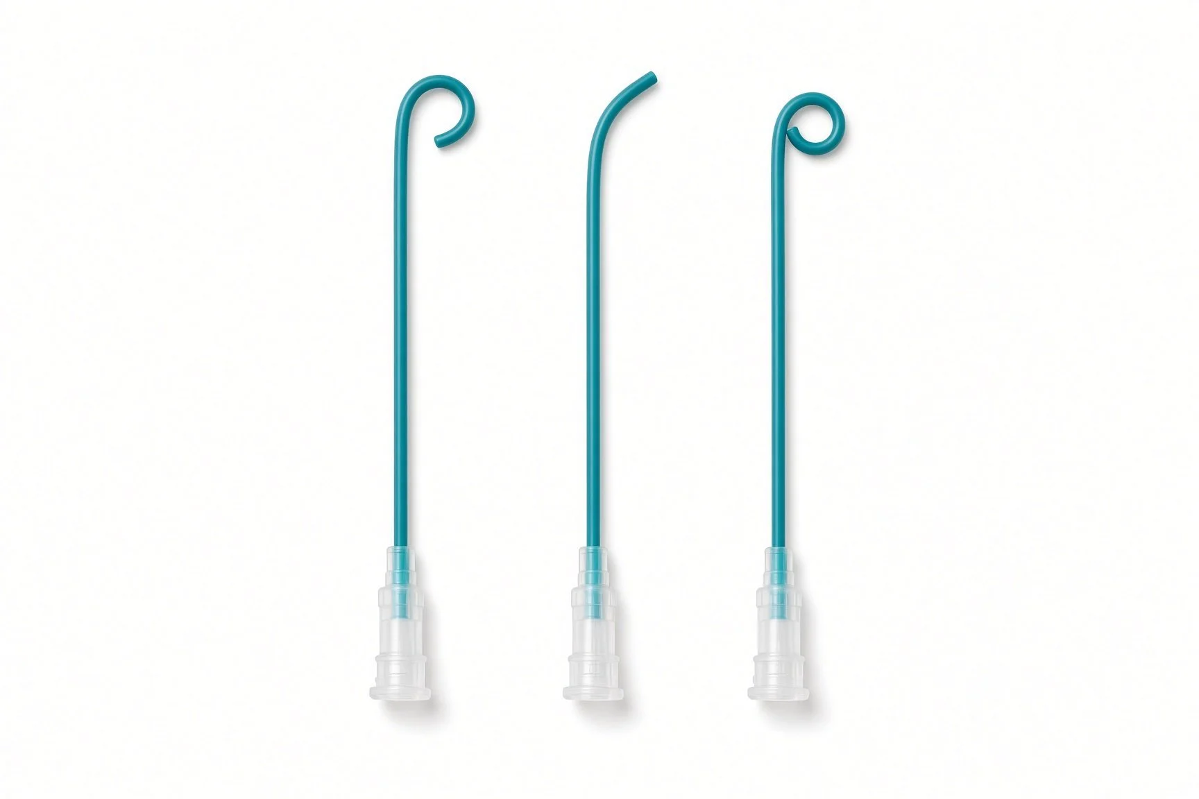
Pigtail diagnostic catheter and power injector (from the guide kit list; no published kit-page picture)
Barts example kit list
An example from one UK centre, written generically. Every lab has its own protocol, and sizes and doses vary.
general anaesthetic set-up; biplane X-ray
femoral arterial access; femoral venous sheath if used; sizes per device and operator
suture-based vascular closure devices, per operator
long sheath, size and length per device and operator
stiff exchange guidewire, per operator
pigtail diagnostic catheter and power injector
covered stent (size per device and operator), mounted on a two-layer (inner and outer) stent-delivery balloon
contrast–saline mix for balloon inflation, as per centre protocol
labelled syringes for the inner balloon, outer balloon and flush; 3-way taps
temporary pacing wire and box, if rapid pacing is used
heparin and local anaesthetic, as per centre protocol
Procedural steps (Barts example)
General anaesthetic; femoral arterial (and venous) access; closure sutures pre-placed if used
Heparin; pressures above and below the narrowing; aortogram
Narrowing crossed; stiff wire parked
Long sheath across the narrowing; stent mounted and positioned
Inner then outer balloon inflation (rapid pacing if used)
Repeat pressures and aortogram
Sheaths removed; closure; leg pulses checked
First time in the cath lab?
💡 First-time tip
Label the balloon syringes. The inner and outer balloon need separate, clearly labelled syringes. Know where the covered stents are. A covered stent is the first rescue for a tear. Watch the blood pressure. Sudden severe pain or a falling blood pressure can mean a tear; tell the team straight away. Pacing ready. If rapid pacing is planned, test the pacing wire before the stent goes in. Check the legs. Leg pulses before and after the case help spot access problems.
FAQ
Will I still need blood pressure tablets?
Some people do. Blood pressure often improves, but not always to normal, so it is checked regularly.
Is the stent permanent?
Yes. In growing children, a stent may be stretched further later.
Is surgery better?
It depends on age and anatomy. Your team will explain which is best for you.
Can I have an MRI scan afterwards?
Most stents are labelled MR conditional. Always tell the scanning team; follow-up imaging of the stent is often by CT or MRI.
Related Cath Lab 101 guides
Sources
All checked on 10 October 2026. Each page's own date is shown where it gives one.
Baumgartner H, et al. 2020 ESC Guidelines for the management of adult congenital heart disease. Eur Heart J 2021;42(6):563–645. PubMed 32860028
British Heart Foundation. Coarctation of the aorta. bhf.org.uk
NHS. Congenital heart disease: types. nhs.uk
DVLA / GOV.UK. Cardiovascular disorders: assessing fitness to drive (congenital heart disease; aortic disorders). gov.uk
Disclaimer
Education only. This page is for general education for healthcare students, cath lab staff and curious readers. It isn't medical advice and doesn't replace advice from your own doctor, heart team or local unit protocols. Equipment and medicines are described generically. No product or manufacturer is recommended. If you think someone is having a heart emergency, call 999.
Disclosure: I work in the medical device industry. This page is independent education and isn't sponsored or reviewed by any company.
All diagrams on this page are original illustrations created for rodnieoro.com.