Coronary angiogram: a Cath Lab 101 guide
Reviewed by Rodnie Oro, cath lab charge nurse · Last reviewed: October 2026 · Education only, not medical advice
Coronary angiogram in one minute
What it is: a test that takes X-ray pictures of the coronary arteries, the blood vessels that supply the heart muscle. A thin tube (a catheter) is passed from the wrist or groin to the heart, and a dye (contrast) is injected so the arteries show up.
Who it is for: people with chest pain or other tests that suggest coronary heart disease, people having a heart attack, and some people before heart valve surgery or other procedures.
Main risks: bruising or bleeding where the tube went in, and an allergic reaction to the dye. Serious problems, such as a stroke, heart attack or damage to an artery, are uncommon. Your team will explain your own risk.
Usually: under local anaesthetic, awake (sometimes with a mild sedative), about 30 minutes, home the same day for most.
On this page: What is a coronary angiogram? · Who is it for? · When is it done? · Step by step · Recovery · Risks · Kit list · First time in the lab? · FAQ
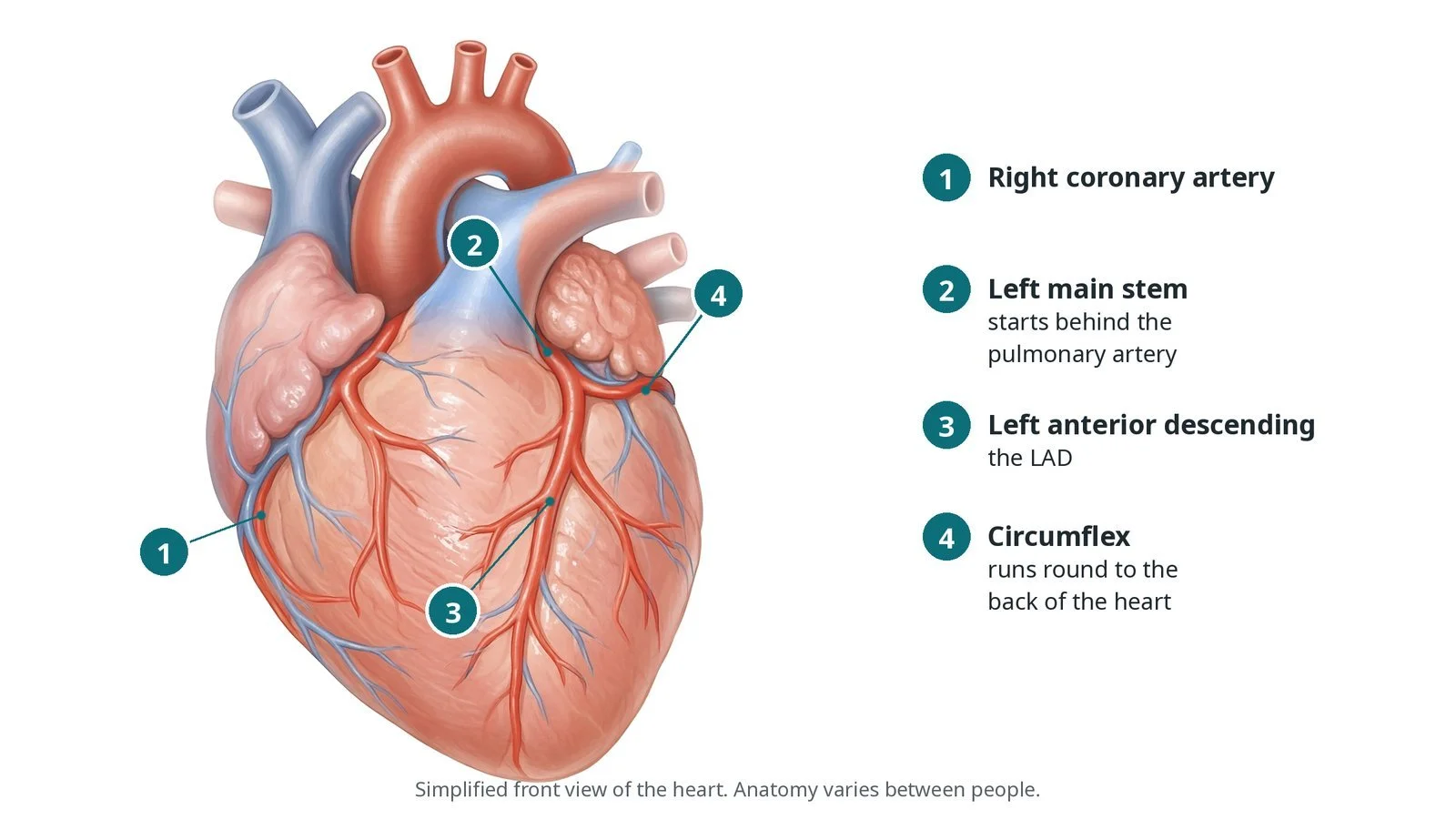
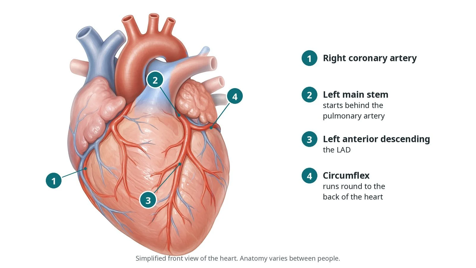
The coronary arteries: (1) right coronary; (2) left main stem; (3) left anterior descending; (4) circumflex, running behind the heart.
What is a coronary angiogram?
The heart muscle gets its blood from two main coronary arteries that branch off the aorta just above the aortic valve:
the left coronary artery, which starts as the left main stem (LMS) and splits into the left anterior descending (LAD) artery, running down the front of the heart, and the circumflex (LCx) artery, running around the back
the right coronary artery (RCA), which runs around the right side to the underside of the heart
Fatty plaque can build up in these arteries and narrow them (coronary heart disease). This can cause angina (chest pain on exertion) or, if a plaque breaks open and a clot forms, a heart attack.
A coronary angiogram (also called coronary angiography or a cardiac catheterisation) shows exactly where any narrowings are and how severe they look. Doctors use it to decide on treatment: medicines alone, PCI (stents) or bypass surgery (CABG). If a narrowing needs a stent, PCI can sometimes be done straight away in the same session.
New to the acronyms? See the Cath Lab Abbreviations A–Z.
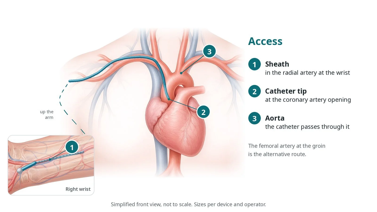
Access: (1) sheath in the radial artery at the wrist, the usual route; (2) catheter tip at the coronary artery opening; (3) catheter passing through the aorta. The femoral artery at the groin is the alternative.
Who is it for?
A coronary angiogram may be offered when:
you are having a heart attack or acute coronary syndrome. NICE guidance (NG185) covers urgent angiography and PCI for these
you have chest pain and other tests, such as a CT coronary angiogram, a stress test or an echo, suggest significant coronary heart disease. NICE (CG95) recommends CT coronary angiography as the first test for most people with suspected angina, with invasive angiography when the result is unclear or treatment is being planned
symptoms continue despite medicines and the team is considering PCI or bypass surgery
you need heart valve surgery or another procedure and the team needs to check the arteries first
there are signs of heart muscle weakness (heart failure) and the cause needs to be found
The ESC guidelines for chronic coronary syndromes describe similar uses. The decision is always made with you after a discussion of benefits and risks.
When is it done?
It can be emergency (during a heart attack), urgent (in hospital after chest pain) or planned (from a clinic). Before a planned test, the team usually:
checks blood tests, especially kidney function, as the dye is cleared by the kidneys
asks about allergies, especially to contrast or iodine
gives instructions about blood thinners and diabetes medicines, as per centre protocol
checks the pulses in the wrists and groins to choose the access site
explains the test and asks for your consent, including whether PCI could be done at the same time
Step by step
1. Getting ready
You lie on the X-ray table, awake. The team attaches ECG stickers and a blood pressure cuff and puts in a cannula (a small tube in a vein) for medicines. A mild sedative may be offered, as per centre protocol. The wrist or groin is cleaned and covered with sterile drapes.
2. Access: wrist or groin
Radial (wrist): the most common route in the UK. Local anaesthetic numbs the skin, the radial artery is punctured and a short tube called a sheath is inserted using the Seldinger technique (needle, then wire, then sheath over the wire). Medicines are given through the sheath to stop the artery going into spasm and to prevent clots, as per centre protocol.
Femoral (groin): used when the wrist arteries are small or blocked, or for larger equipment. The femoral artery is punctured, often under ultrasound.
Sheath and catheter sizes are per device and operator; smaller sizes may be used for small arteries.
3. Taking the pictures
A guidewire leads a pre-shaped diagnostic catheter up the arm or leg arteries to the aorta. The wire is removed and the catheter is flushed.
The catheter tip is gently "engaged" in the opening of the left coronary artery, and small puffs of contrast are injected while the X-ray camera records short films. The process is repeated for the right coronary artery, usually with a different catheter shape. Some operators use a single catheter shaped to reach both.
The X-ray camera (the C-arm) is moved around the chest to take pictures from several angles, so that every segment of each artery is seen clearly without overlap.
Sometimes a pigtail catheter is used to inject a larger amount of contrast into the left ventricle (a ventriculogram) or aorta (an aortogram), using a power injector.
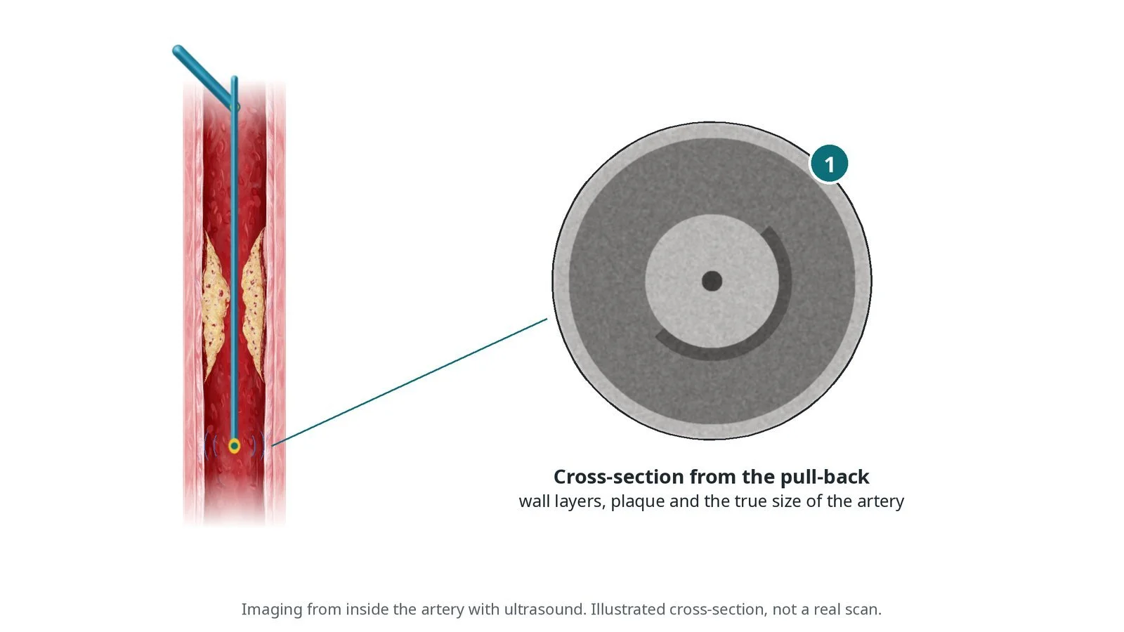
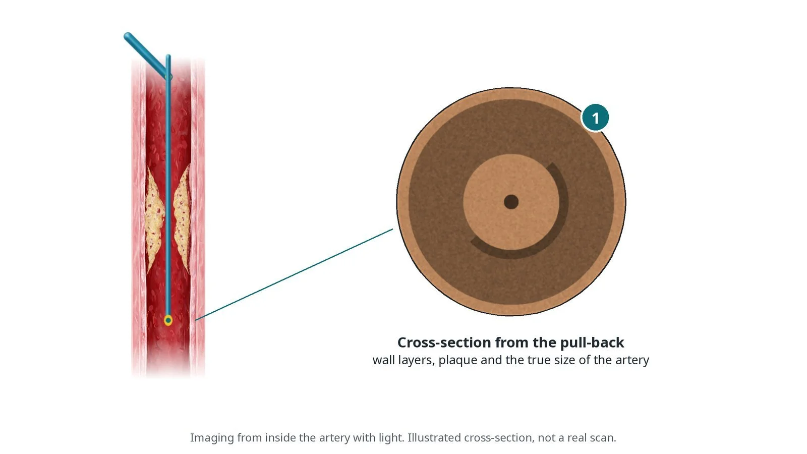
If a narrowing is borderline, the team may measure the pressure across it with a pressure wire, or look inside the artery with intravascular imaging, as per operator.
You may feel a warm flush when the dye is injected, and you may be asked to hold your breath for the pictures.
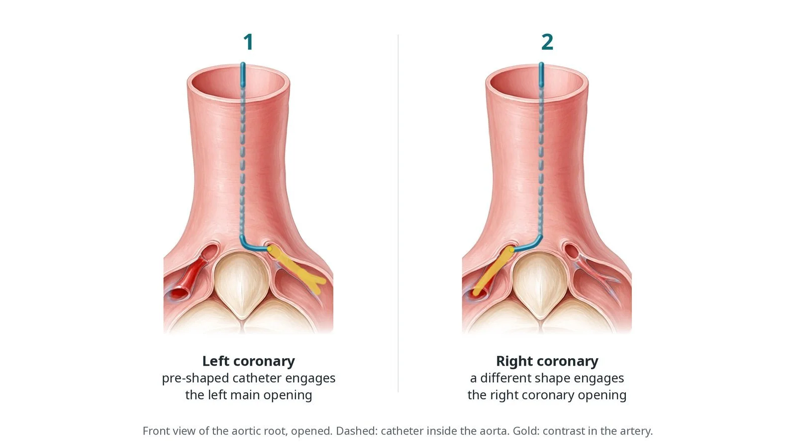
Pre-shaped catheters “engage” the opening of each coronary artery so contrast can be injected: (1) left coronary; (2) right coronary.
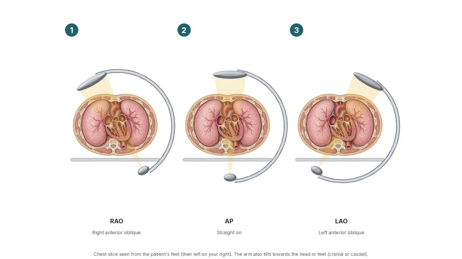
The C-arm moves around the patient: (1) RAO; (2) AP; (3) LAO, with cranial or caudal tilt, so each artery is seen without overlap.
4. Finishing
Radial: the sheath is removed and a compression band is put on the wrist and gradually loosened over a few hours, as per centre protocol.
Femoral: the artery is closed with firm pressure or a closure device, per operator.
The operator usually explains the results straight away.
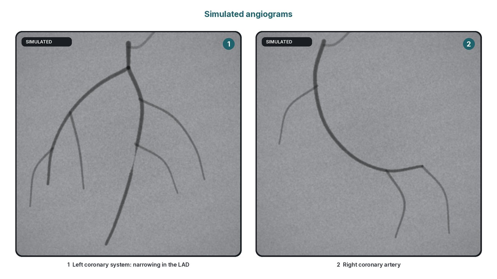
Illustration, not a real scan: contrast shows the artery as a dark line; (1) a narrowing appears as a thinner segment; (2) the right coronary artery.
Recovery and aftercare
First hours: checks of the wrist or groin, pulse and blood pressure. After the radial route you can usually sit up straight away. After the femoral route you lie flat for a while, as per centre protocol.
Going home: most people go home the same day. Someone should take you home and stay with you overnight, especially if you had a sedative.
Wrist or groin: avoid heavy lifting with that arm or straining for a few days, as advised. A small bruise is common.
Driving: the DVLA guidance has no separate rule for a diagnostic angiogram on its own; what applies depends on what was found and done (for example a stent or a heart attack) and on your licence type (car or bus/lorry). Most centres advise not driving for a short time while the wrist or groin heals. Check the DVLA guidance and ask your team.
Results and next steps: your team will explain the plan: medicines, PCI, surgery or no treatment needed.
Call 999 if you have chest pain that does not settle, or bleeding from the wrist or groin that does not stop with firm pressure for 10 minutes. Seek urgent help if your hand or leg becomes cold, pale or numb.
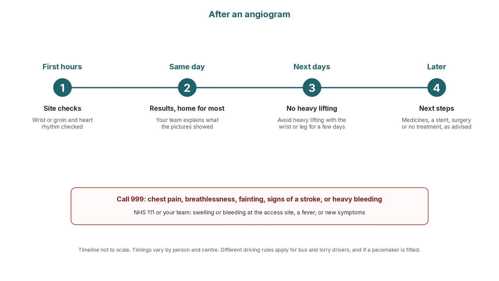
After an angiogram: (1) site checks; (2) results, home for most; (3) no heavy lifting for a few days; (4) next steps.
Risks and complications
Your team will discuss your own risks. Possible problems include:
bruising or bleeding at the wrist or groin, sometimes a swelling (haematoma)
the radial artery blocking afterwards, which usually causes no symptoms
an allergic reaction to the contrast dye
kidney strain from the dye, especially if kidney function is already reduced
irregular heart rhythms during the test
uncommon but serious: damage to an artery, stroke, heart attack or the need for emergency surgery
Cath lab kit list (printable)
Barts example kit list
An example from one UK centre, written generically. Every lab has its own protocol, and sizes and doses vary.
Set-up
angiography pack with drapes and gowns
skin antiseptic applicators
sterile saline bag and spike for flushing
transparent dressing to secure a radial sheath
radial or femoral arterial sheath, size per device and operator
IV drug line
saline bag, giving set, long extension line and 3-way tap
labelled syringes for drugs; blunt drawing-up needle
Diagnostic catheters (per operator)
left coronary: Judkins left-shaped catheter, curve size per operator and access route
right coronary: Judkins right-shaped catheter, or alternative shapes for unusual anatomy
single-catheter option shaped to reach both coronaries
pigtail catheter for a ventriculogram or aortogram
Wires
0.035" J-tip guidewire; hydrophilic wire and long exchange wire for tortuous arteries or spasm
0.014" coronary guidewire if needed
Medicines (all as per centre protocol)
local anaesthetic
heparin
nitrate and calcium-channel blocker for radial spasm, diluted as per centre protocol
sedative, if used
Ventriculogram / aortogram
high-pressure tubing, contrast syringe and power injector
forceps or clamps to tap out air and secure connections
Haemostasis
radial compression band (size to fit)
femoral closure device or manual compression, per operator
Procedural steps (Barts example)
Local anaesthetic to the wrist or groin
Sheath inserted using the Seldinger technique; blood aspirated to confirm position
Anti-spasm cocktail through a radial sheath, as per centre protocol
Catheter advanced over a guidewire; usually right coronary first from the wrist, left coronary first from the groin, per operator
Right coronary views, for example left anterior oblique (LAO) and cranial views, and right anterior oblique (RAO)
Left coronary views, for example RAO caudal, RAO cranial, LAO cranial and LAO caudal ("spider") views
Ventriculogram or aortogram if requested
Sheath removed; compression band or closure device; transfer to recovery
First time in the cath lab?
💡 First-time tip
Watch the pressure trace. When a catheter engages a coronary, a "damped" or "ventricularised" pressure trace means the tip may be blocking the artery. Tell the operator straight away. No bubbles. Flush and de-air the manifold and contrast lines carefully: air in a coronary can cause chest pain and rhythm problems. Label every syringe. Spasm cocktails, heparin and sedation all look the same in a syringe. Check the band. A radial band that is too tight or too loose causes problems. Check the hand colour and the site regularly. Know the views. Learn the standard LAO/RAO, cranial/caudal views so you can anticipate the C-arm moves.
FAQ
Is a coronary angiogram painful?
You should feel only a sting from the local anaesthetic. Some people feel a warm flush or a brief need to pass urine when the dye goes in.
How long does it take?
Usually about 30 minutes, longer if a stent is put in at the same time.
Am I awake?
Yes, usually. A mild sedative may be offered.
What is the difference between a CT coronary angiogram and a coronary angiogram?
A CT coronary angiogram uses a CT scanner and dye through an arm vein, with no catheter in the heart. An invasive coronary angiogram gives more detail and allows treatment at the same time.
Can a stent be put in during the same test?
Often yes, if you have agreed to it beforehand and the team thinks it is the right treatment. See the PCI guide.
Related Cath Lab 101 guides
Sources
All checked on 10 October 2026. Each page's own date is shown where it gives one.
NHS. Coronary angiography. nhs.uk/conditions/coronary-angiography (page last reviewed 30 Jan 2023)
NHS. Coronary heart disease. nhs.uk/conditions/coronary-heart-disease
NICE. Recent-onset chest pain of suspected cardiac origin: assessment and diagnosis (CG95). nice.org.uk/guidance/cg95
NICE. Acute coronary syndromes (NG185). nice.org.uk/guidance/ng185
Knuuti J, et al. 2019 ESC Guidelines for the diagnosis and management of chronic coronary syndromes. Eur Heart J 2020;41(3):407–477. PubMed 31504439
British Heart Foundation. Coronary angiogram. bhf.org.uk
DVLA / GOV.UK. Cardiovascular disorders: assessing fitness to drive. gov.uk
Disclaimer
Education only. This page is for general education for healthcare students, cath lab staff and curious readers. It isn't medical advice and doesn't replace advice from your own doctor, heart team or local unit protocols. Equipment and medicines are described generically. No product or manufacturer is recommended. If you think someone is having a heart attack, call 999.
Disclosure: I work for a company that makes a device used in this procedure. This page is independent education, isn't sponsored or reviewed by any company, and doesn't recommend any product.
All diagrams on this page are original illustrations created for rodnieoro.com.