Hi.
Welcome to a hub where you can watch and learn anything about Cath Lab procedures.

Welcome to a hub where you can watch and learn anything about Cath Lab procedures.
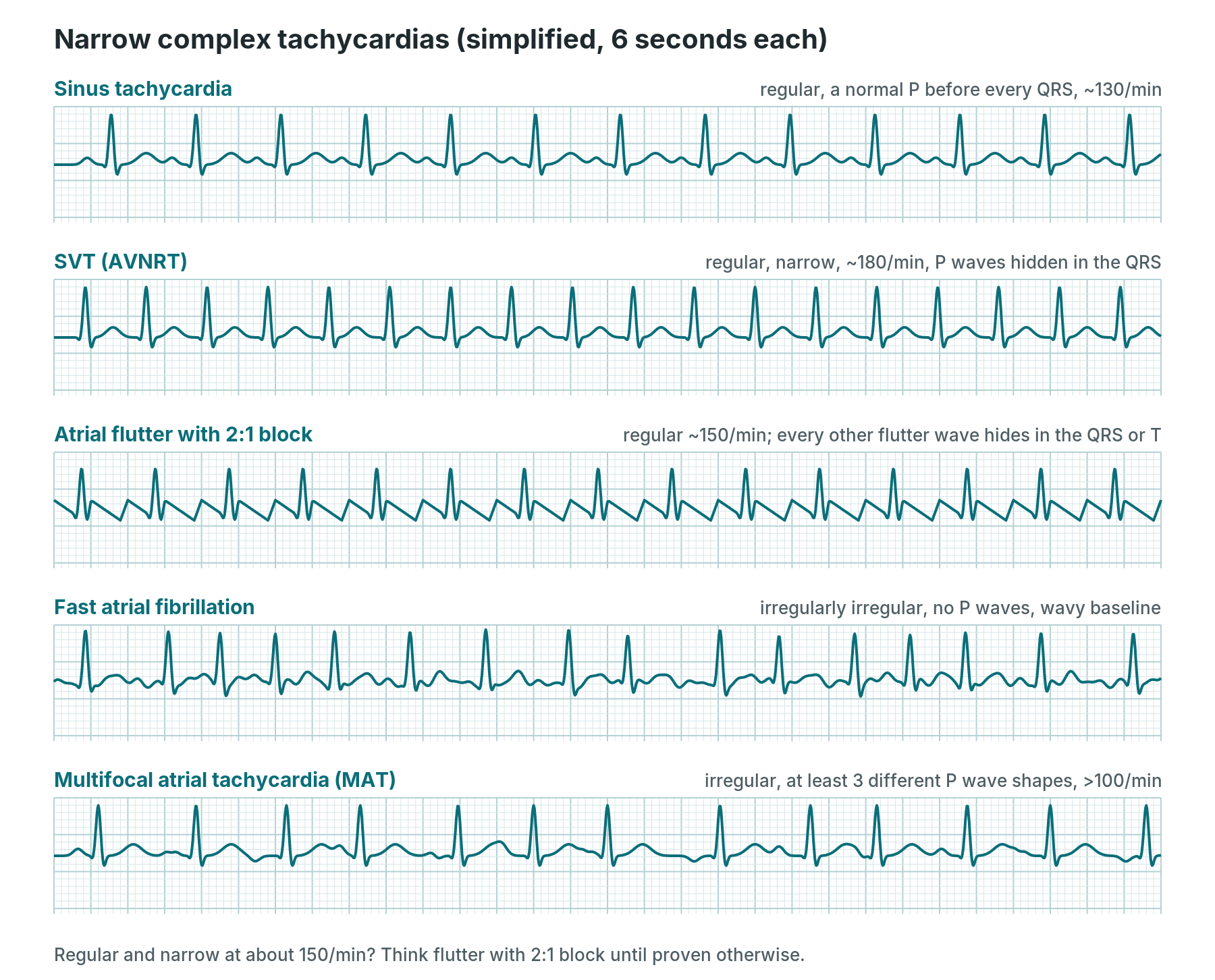
Advanced ECG guide for nurses and cath lab staff: abnormal ECGs explained, from SVT, AF, VT and heart block to bundle branch block, WPW, Brugada, STEMI equivalents, electrolytes, pacing problems and arrest rhythms, with causes, symptoms, treatment and what to call out in the lab.
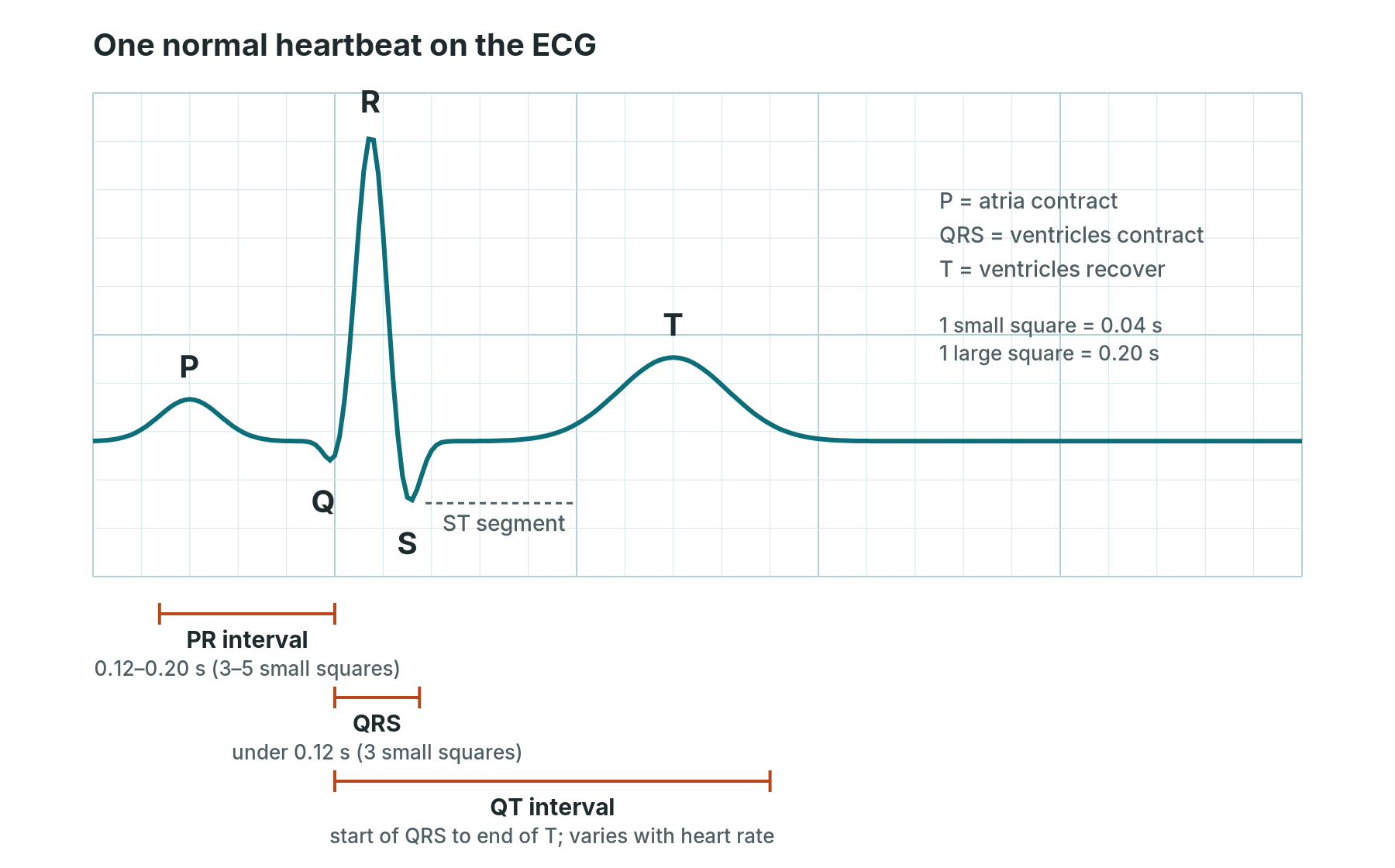
ECG basics for student nurses and new cath lab staff: 12-lead electrode placement, the normal ECG waveform, reading a rhythm strip step by step, AF, flutter, heart block, VT, VF, STEMI patterns and what to watch for during procedures.
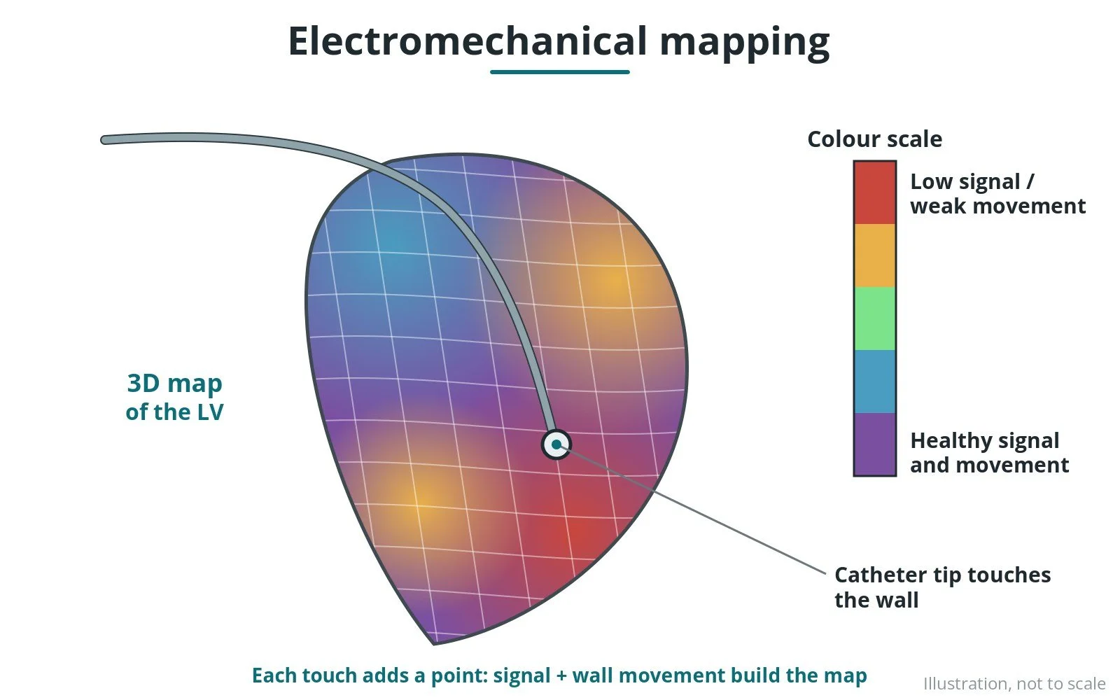
How a catheter and 3D system build a map of the heart's electrical activity and, in some systems, wall movement. Original diagrams; steps, kit, risks and recovery.
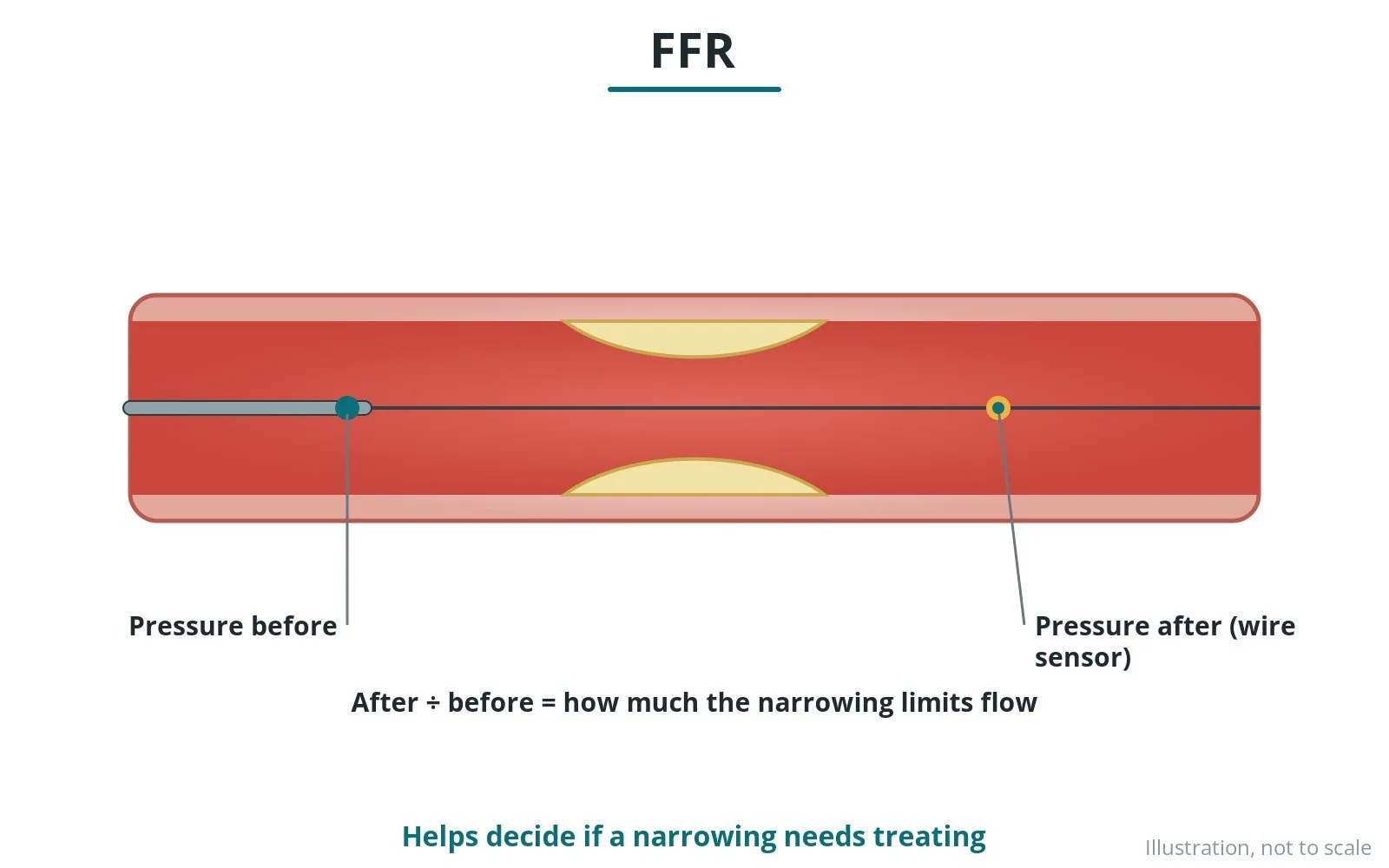
How a pressure wire measures the pressure drop across a narrowing to decide whether it needs treating. Original diagrams; steps, kit and risks.
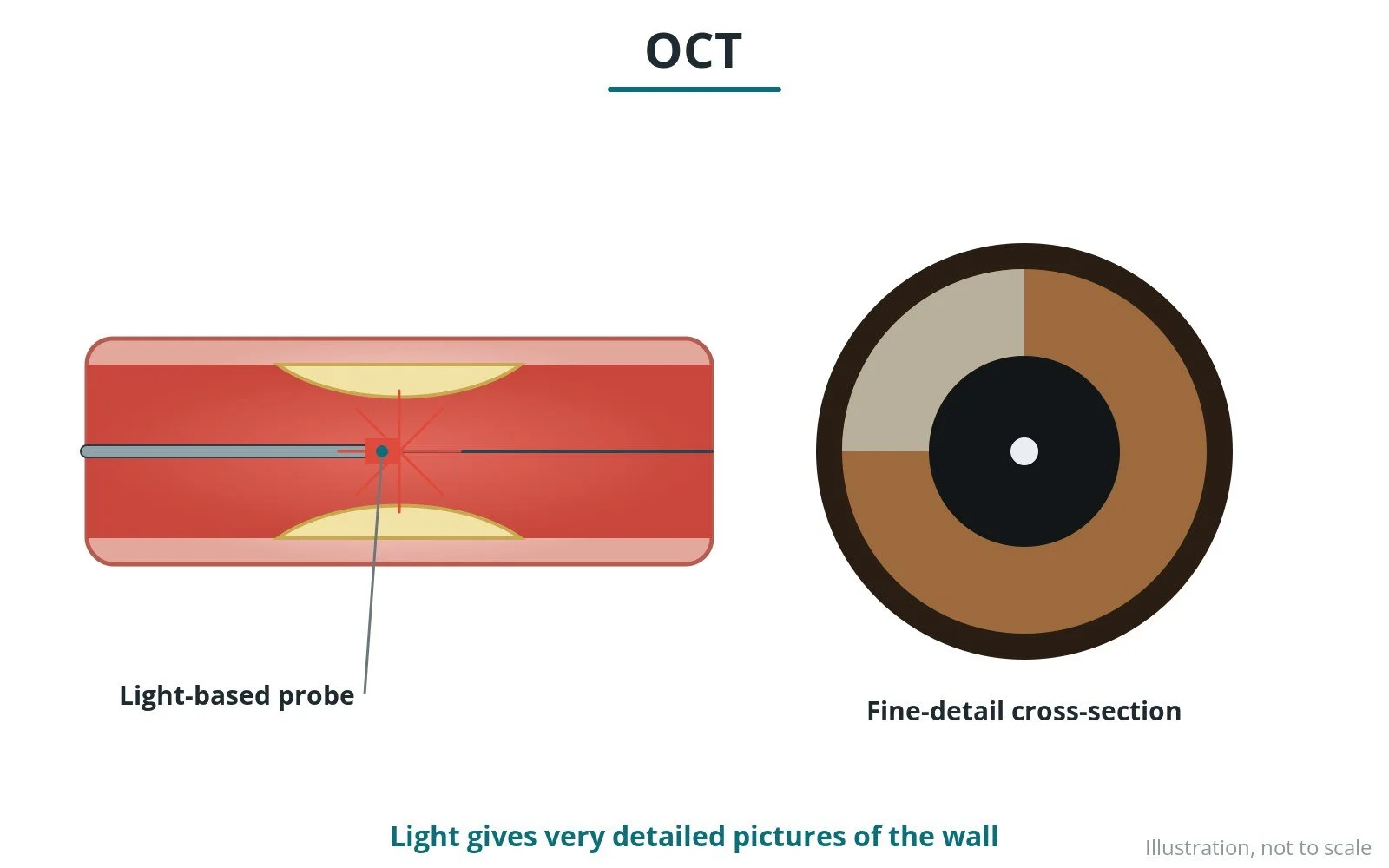
How light-based imaging on a catheter shows the inside of a heart artery in very fine detail during an angiogram or PCI. Original diagrams; steps, kit and risks.
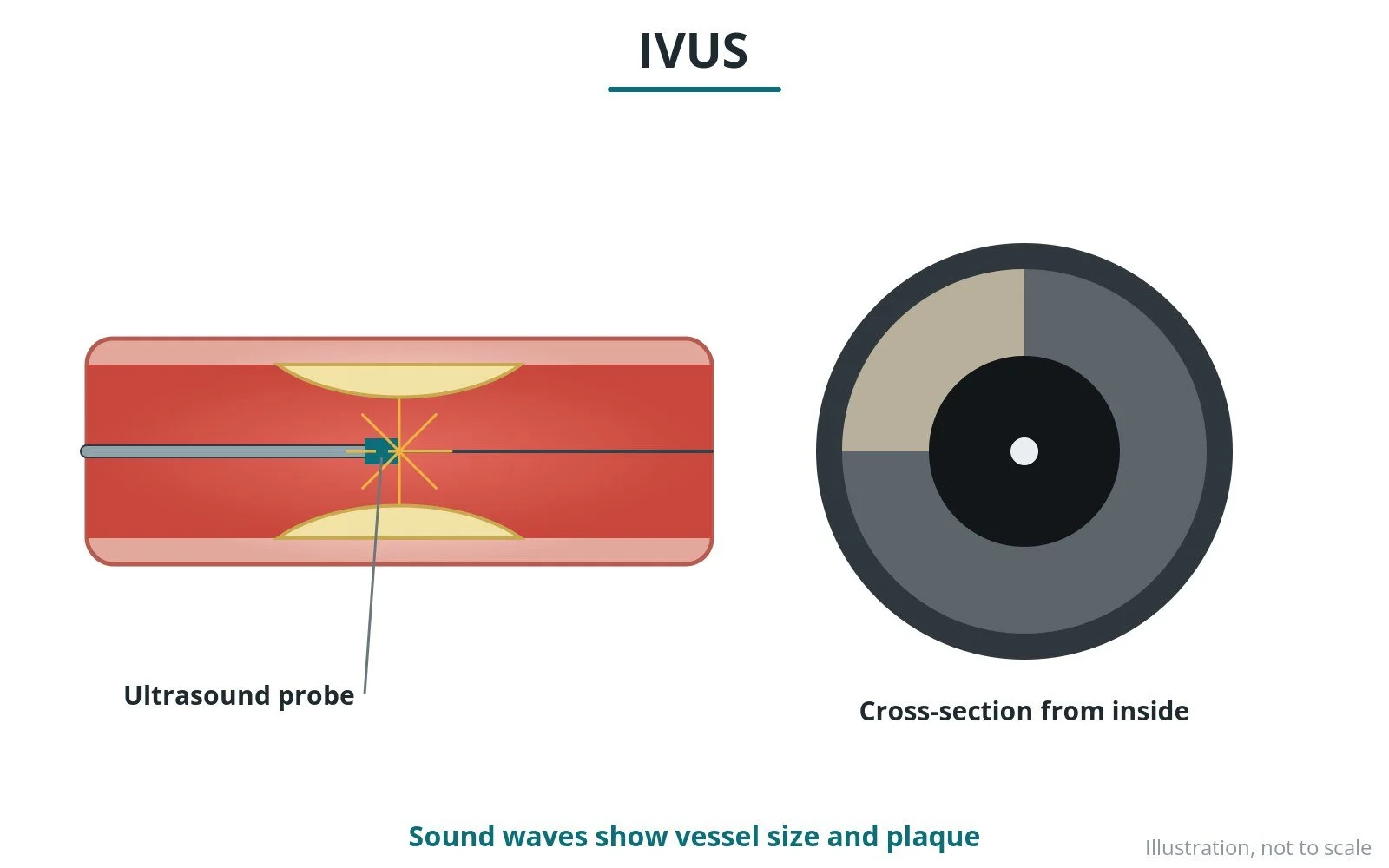
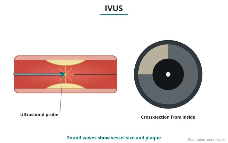
How a tiny ultrasound probe on a catheter shows the inside of a heart artery, the plaque and how well a stent has expanded. Original diagrams; steps, kit and risks.
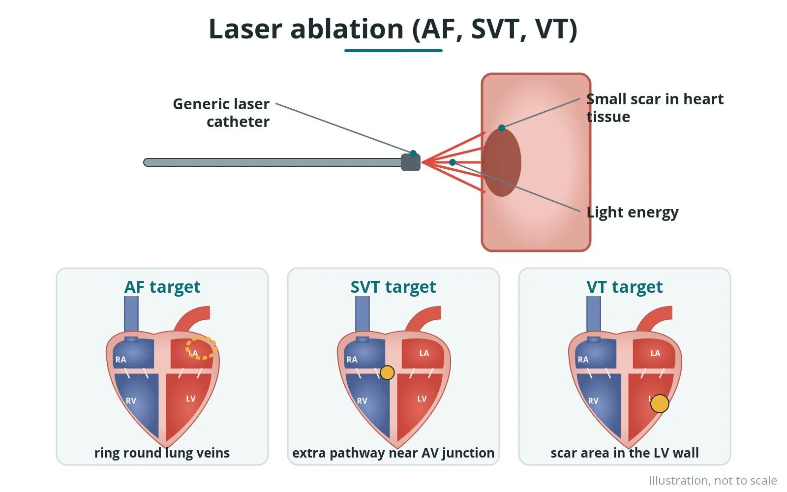
How laser energy, usually from a balloon catheter with a built-in camera, isolates the pulmonary veins in AF. Original diagrams; steps, kit, risks and recovery.
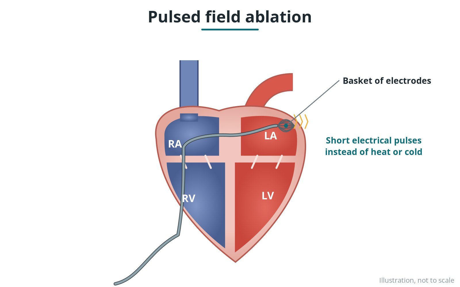
How very short, high-energy electrical pulses treat the heart tissue causing AF, rather than heat or cold. Original diagrams; steps, kit, risks and recovery.
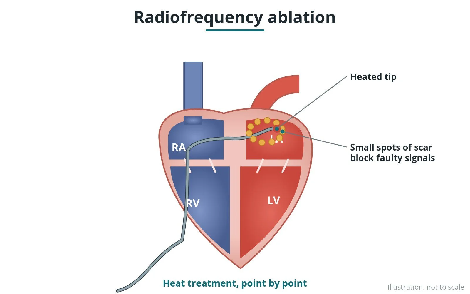
How heat from a catheter tip treats the small areas of heart tissue causing an abnormal rhythm. Original diagrams; steps, kit, risks and recovery.
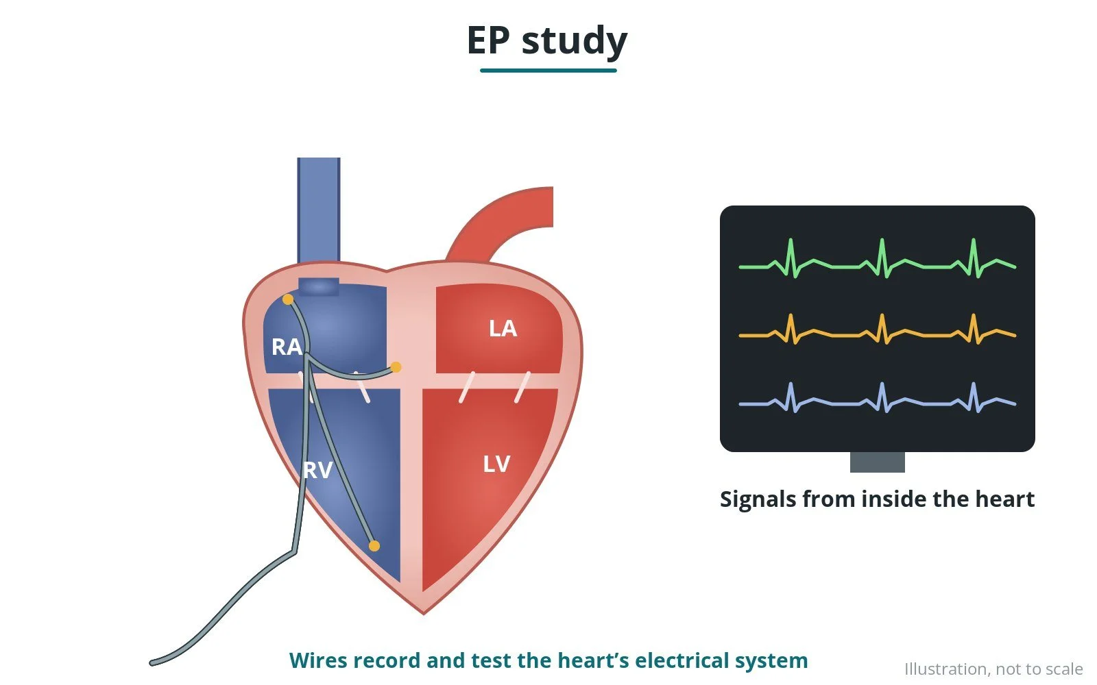
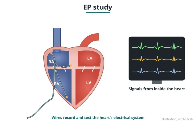
How thin catheters record and test the heart's electrical system from inside, to find the cause of a rhythm problem. Original diagrams; steps, kit, risks and recovery.
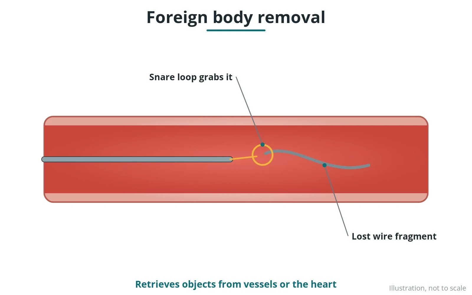
How snares and other tools are used by catheter to retrieve lost or broken equipment from the heart or blood vessels. Original diagrams; steps, kit, risks and recovery.
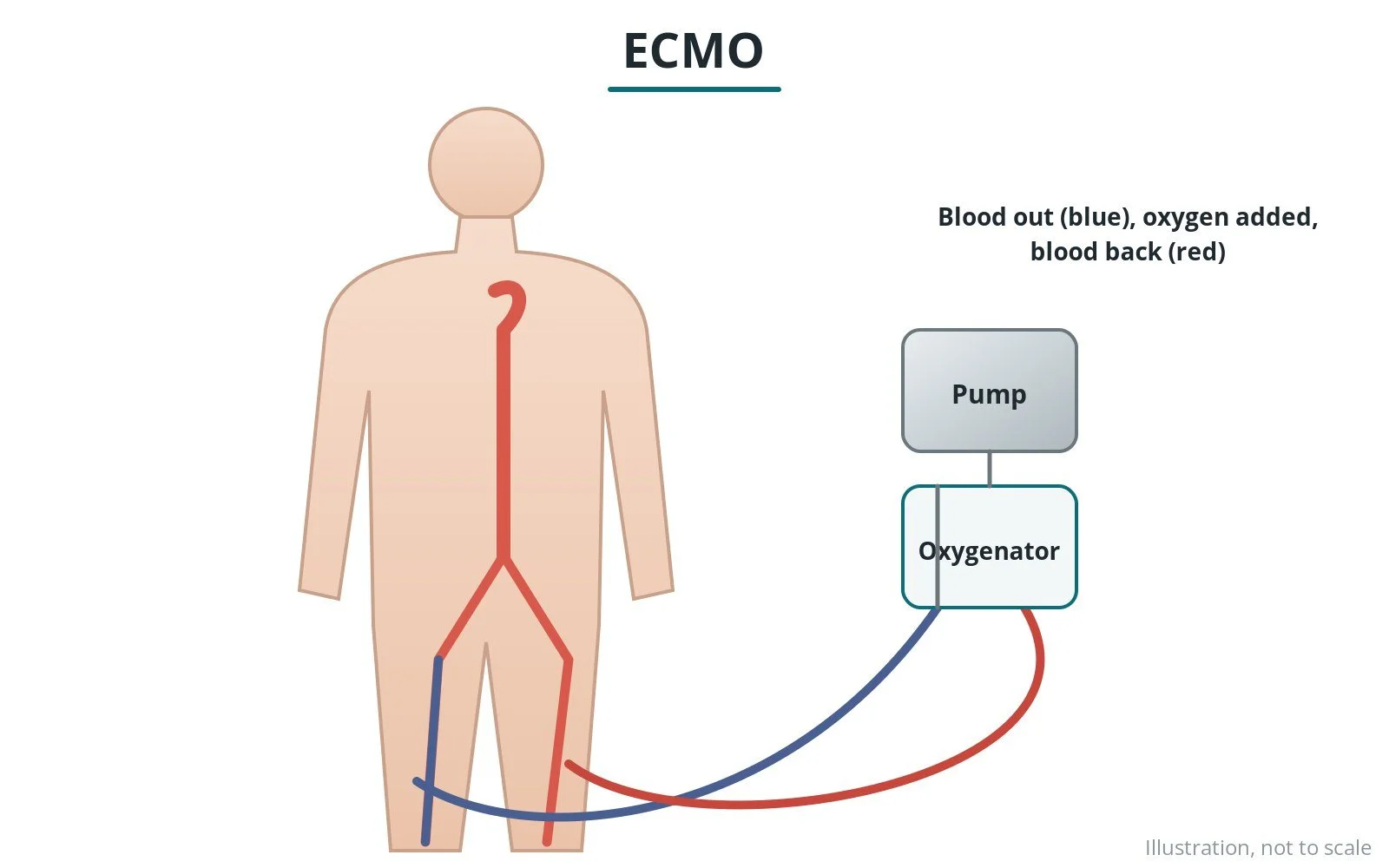
How a machine takes over the work of the heart and lungs for a time, connected through large tubes in the groin. Original diagrams; cannulation steps, kit, risks and care.
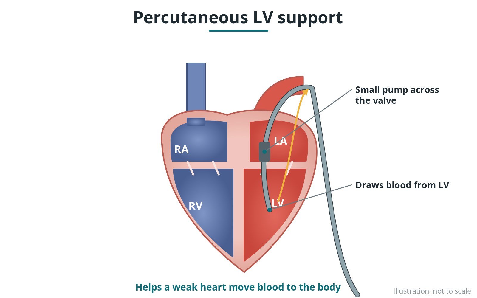
How a small pump placed across the aortic valve by catheter helps the left ventricle move blood in shock or high-risk procedures. Original diagrams; steps, kit, risks and care.
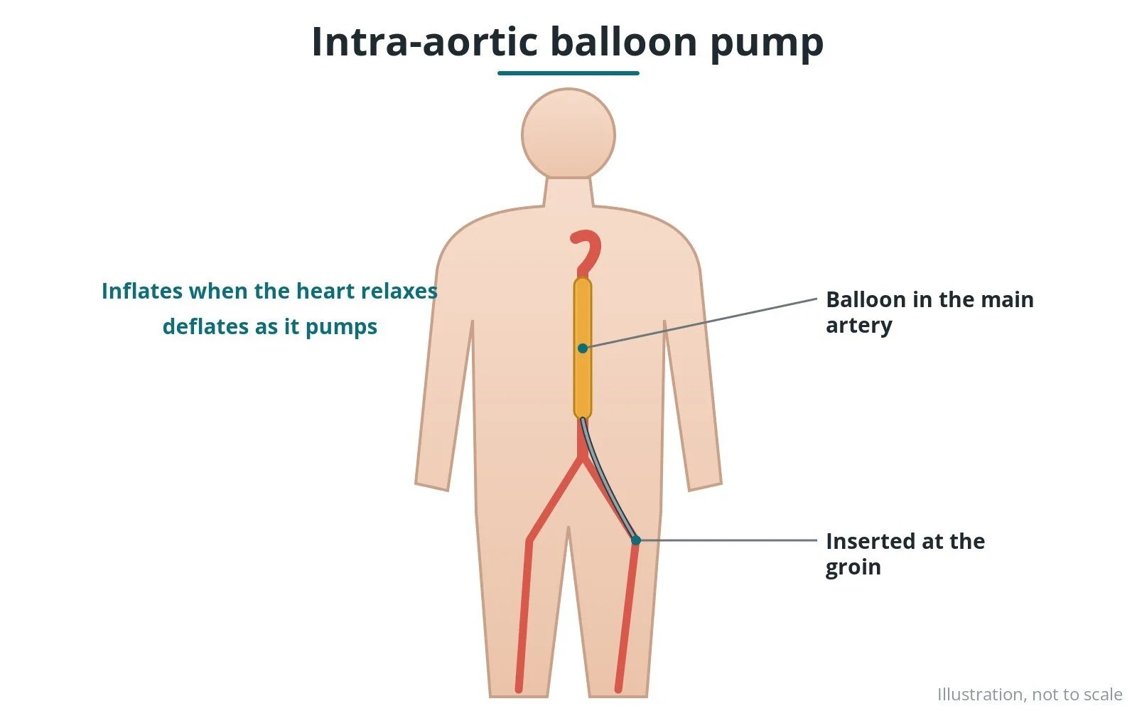
How a long balloon in the aorta inflates and deflates with each heartbeat to support the heart. Original diagrams; insertion steps, kit, risks and care.
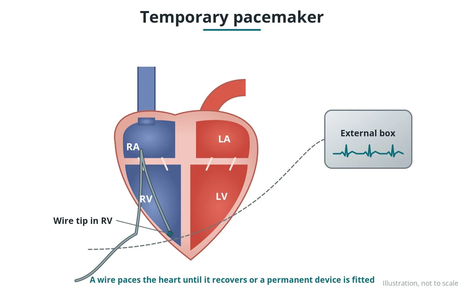
How a temporary pacing wire is passed through a vein into the heart and connected to an external box to keep the heart rate safe. Original diagrams; steps, kit and risks.
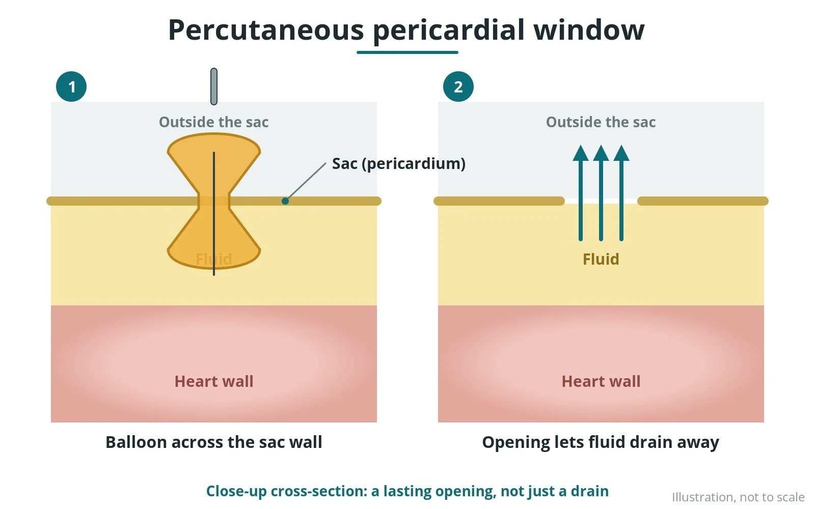
How a balloon makes an opening in the sac around the heart so recurring fluid can drain away. Original diagrams; steps, kit, risks and recovery.
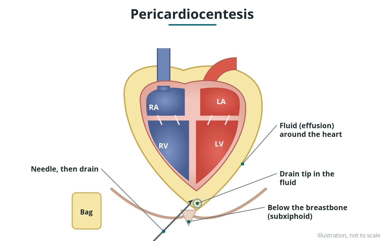
How fluid around the heart is drained with a needle and a small drain, guided by echo and X-ray. Original diagrams; steps, kit, risks and recovery.
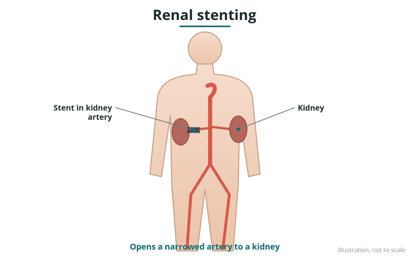
How a narrowed artery to the kidney is opened with a balloon-expandable stent, in selected patients. Original diagrams; steps, kit, risks and recovery.
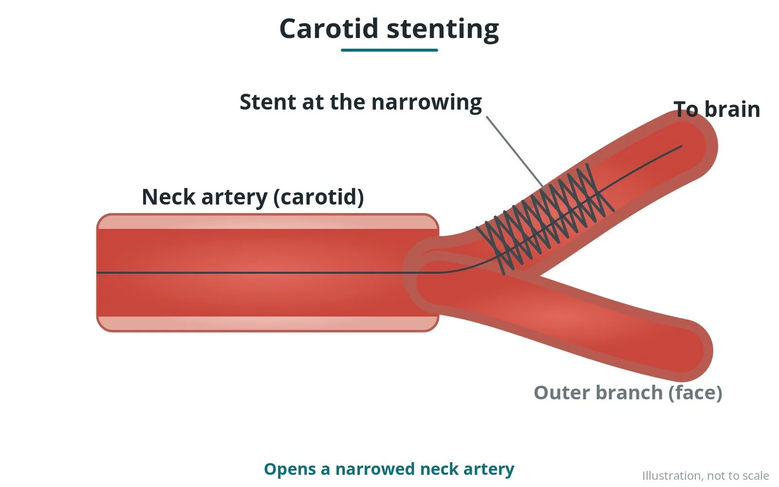
How a narrowed carotid artery in the neck is opened with a stent, usually with a filter to catch debris. Original diagrams; steps, kit, risks and recovery.
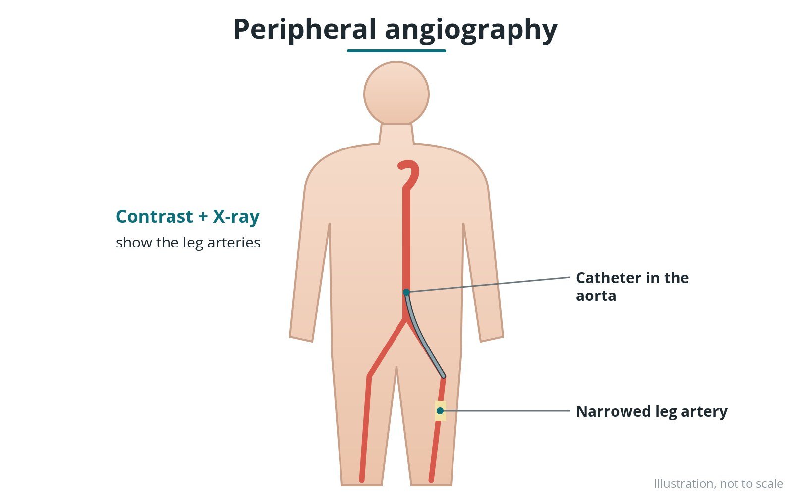
How contrast dye shows narrowings or blockages in arteries outside the heart, such as the legs. Original diagrams; steps, kit, risks and recovery.
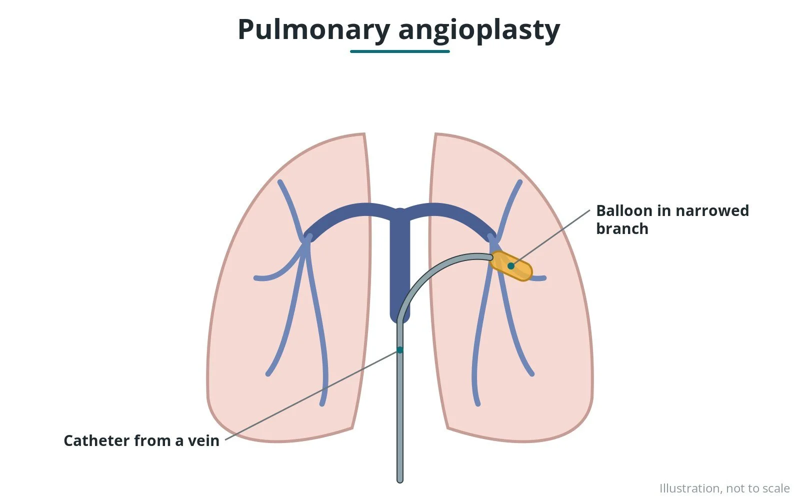
How narrowed arteries in the lungs are widened with a balloon, sometimes with a stent, in CTEPH or congenital heart disease. Original diagrams; steps, kit, risks and recovery.
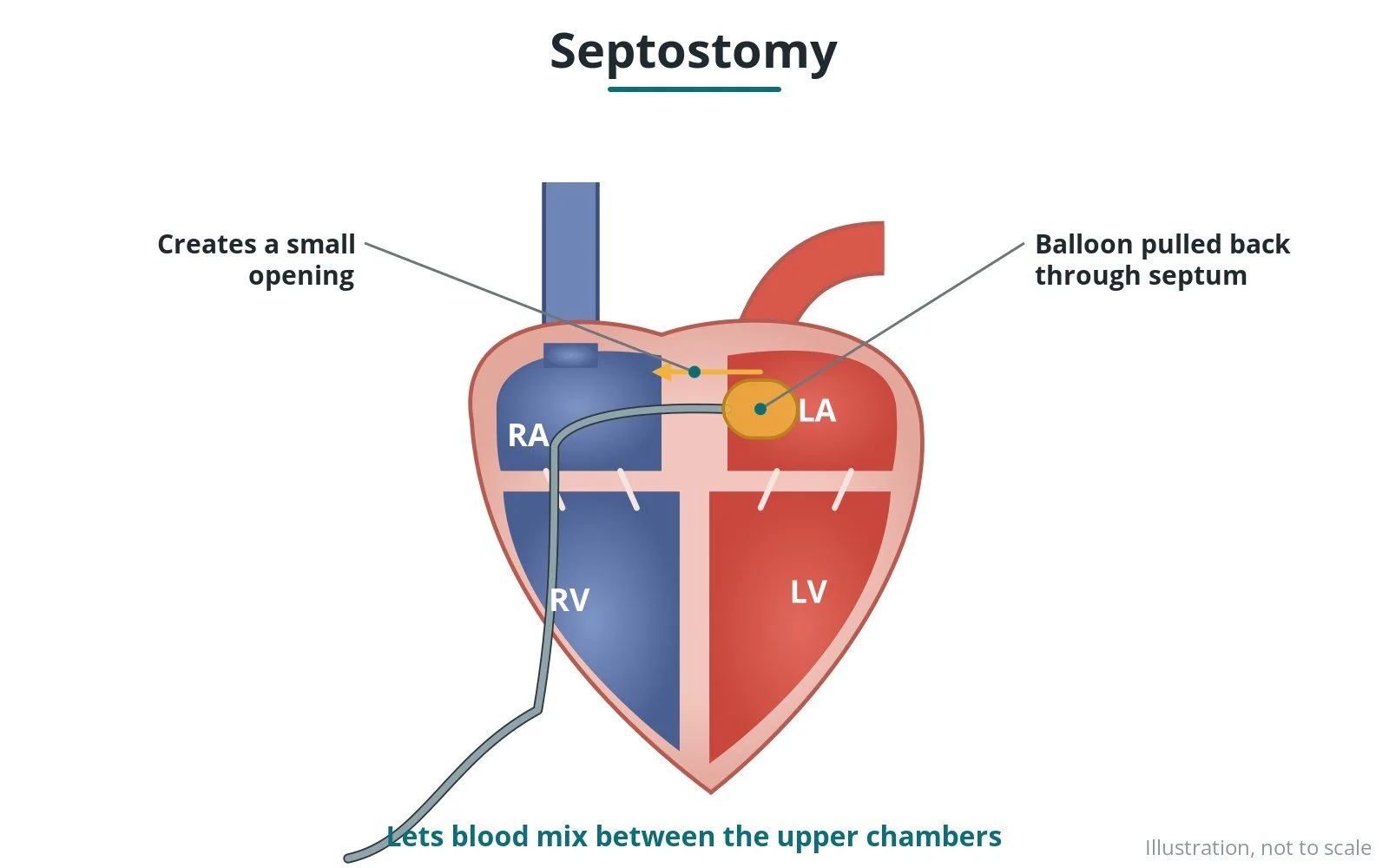
How a balloon catheter makes or enlarges an opening between the upper heart chambers, mainly in newborns with some congenital heart conditions. Original diagrams; steps and risks.
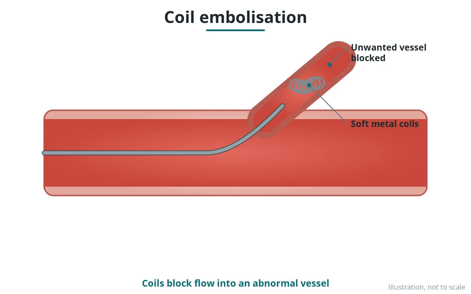
How small metal coils are placed through a catheter to block an unwanted vessel or connection. Original diagrams; steps, kit, risks and recovery.
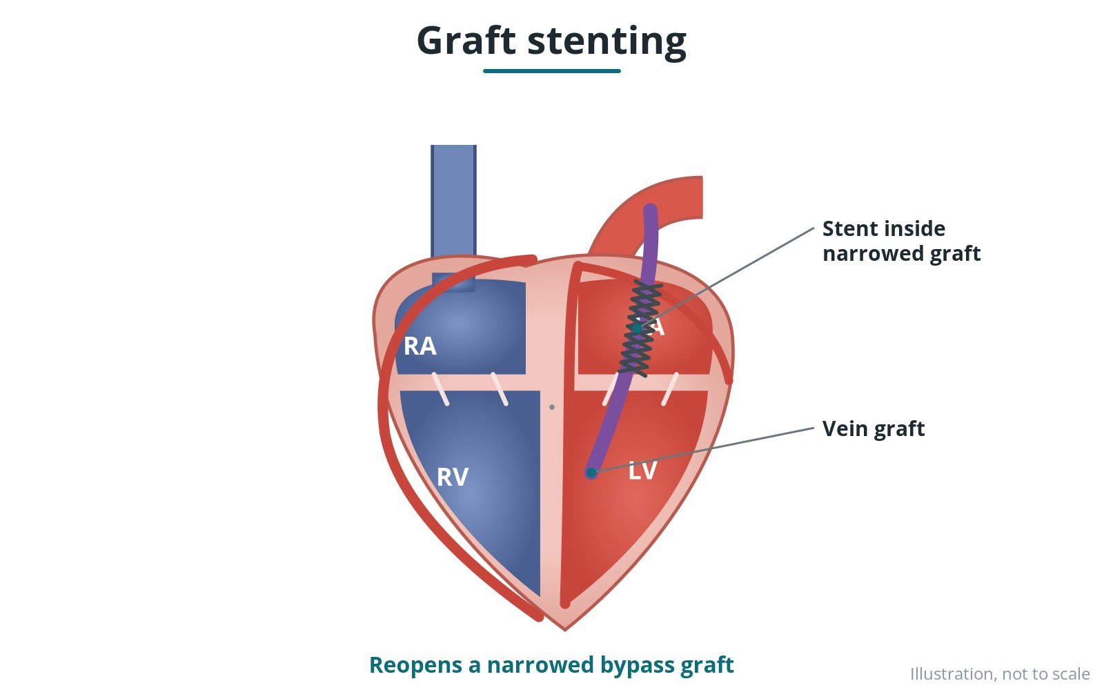
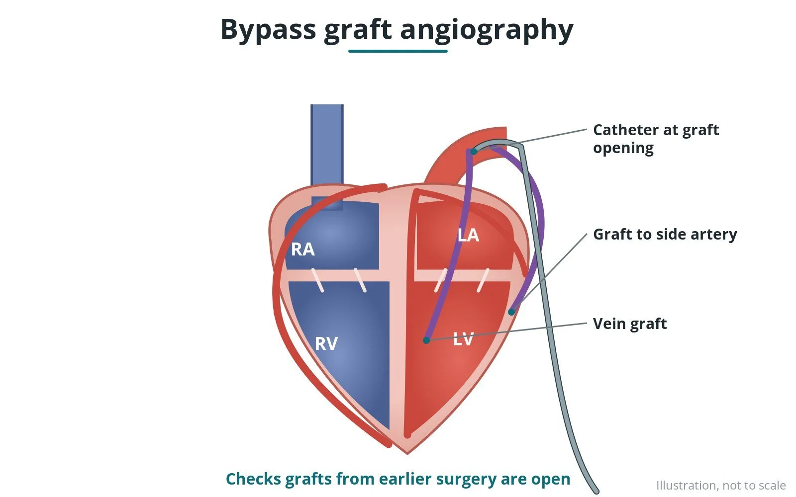
How a narrowed vein or artery graft from earlier bypass surgery is opened with a balloon and stent. Original diagrams; steps, kit, risks and recovery.
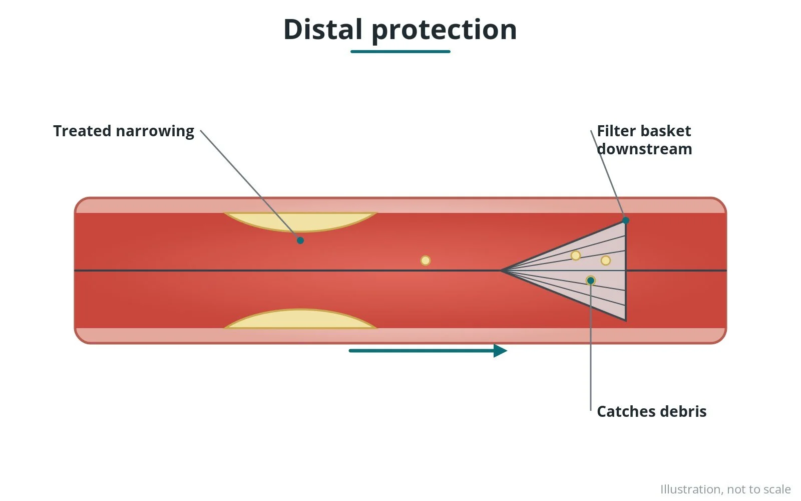
How a small filter or similar device placed beyond a narrowing catches debris released during treatment. Original diagrams; steps, kit, risks and recovery.
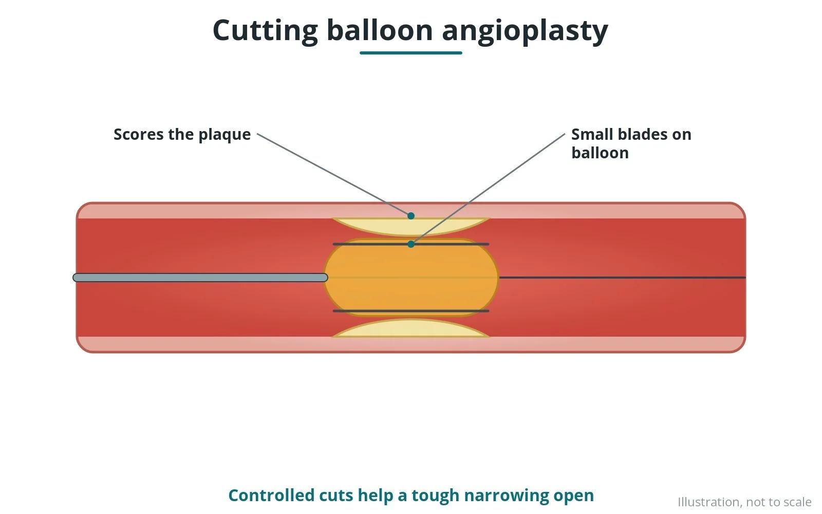
How a balloon with tiny blades or scoring wires makes controlled cuts in tough plaque so the artery opens evenly. Original diagrams; steps, kit, risks and recovery.
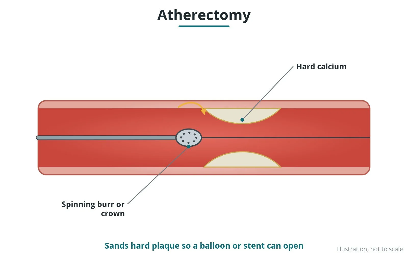
How a spinning burr or crown sands down hard, calcified plaque so a heart artery can be ballooned and stented. Original diagrams; steps, kit, risks and recovery.
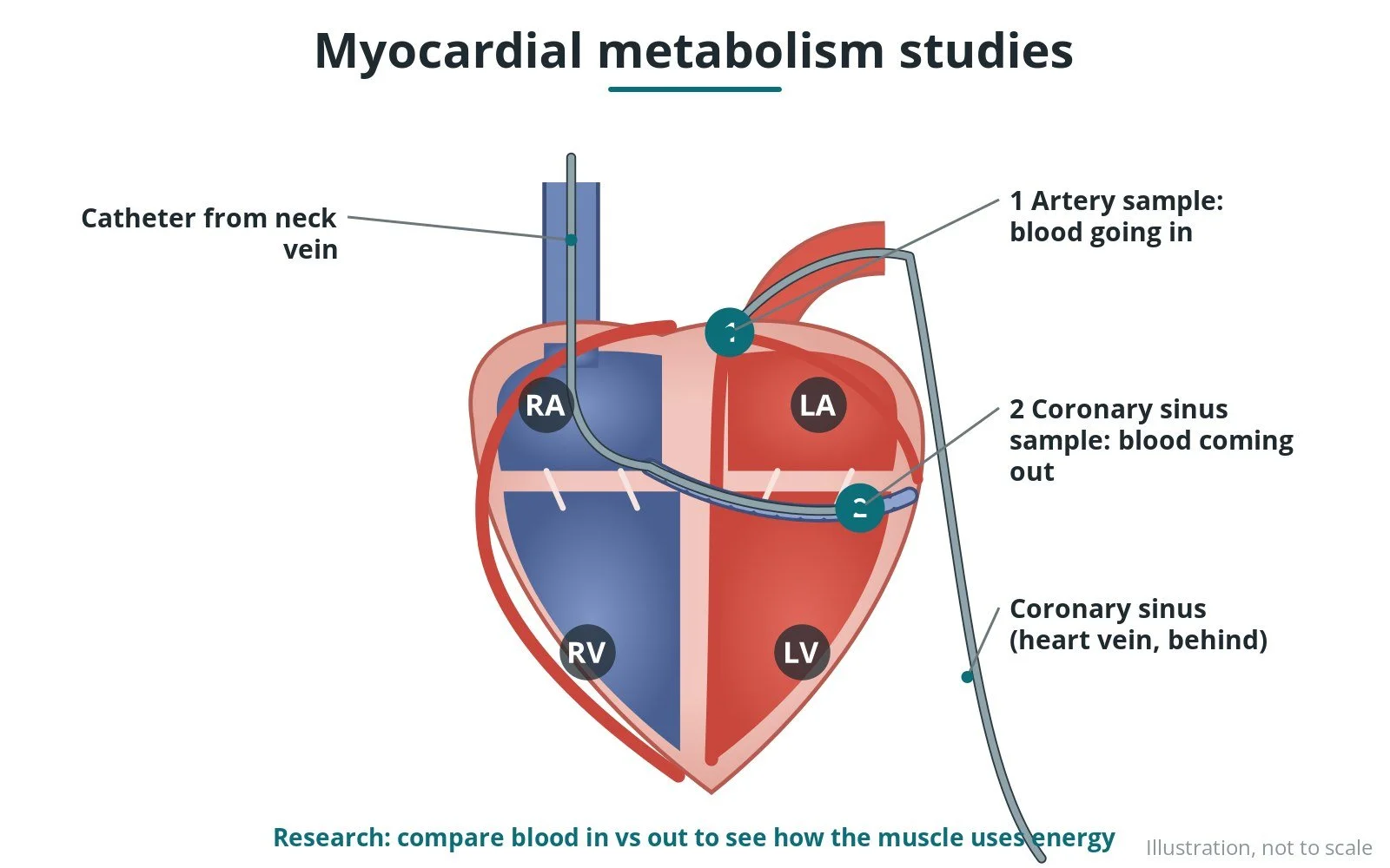
How blood samples from the artery and the heart's own vein (coronary sinus) are compared in research studies of how the heart uses energy. Original diagrams; steps and risks.
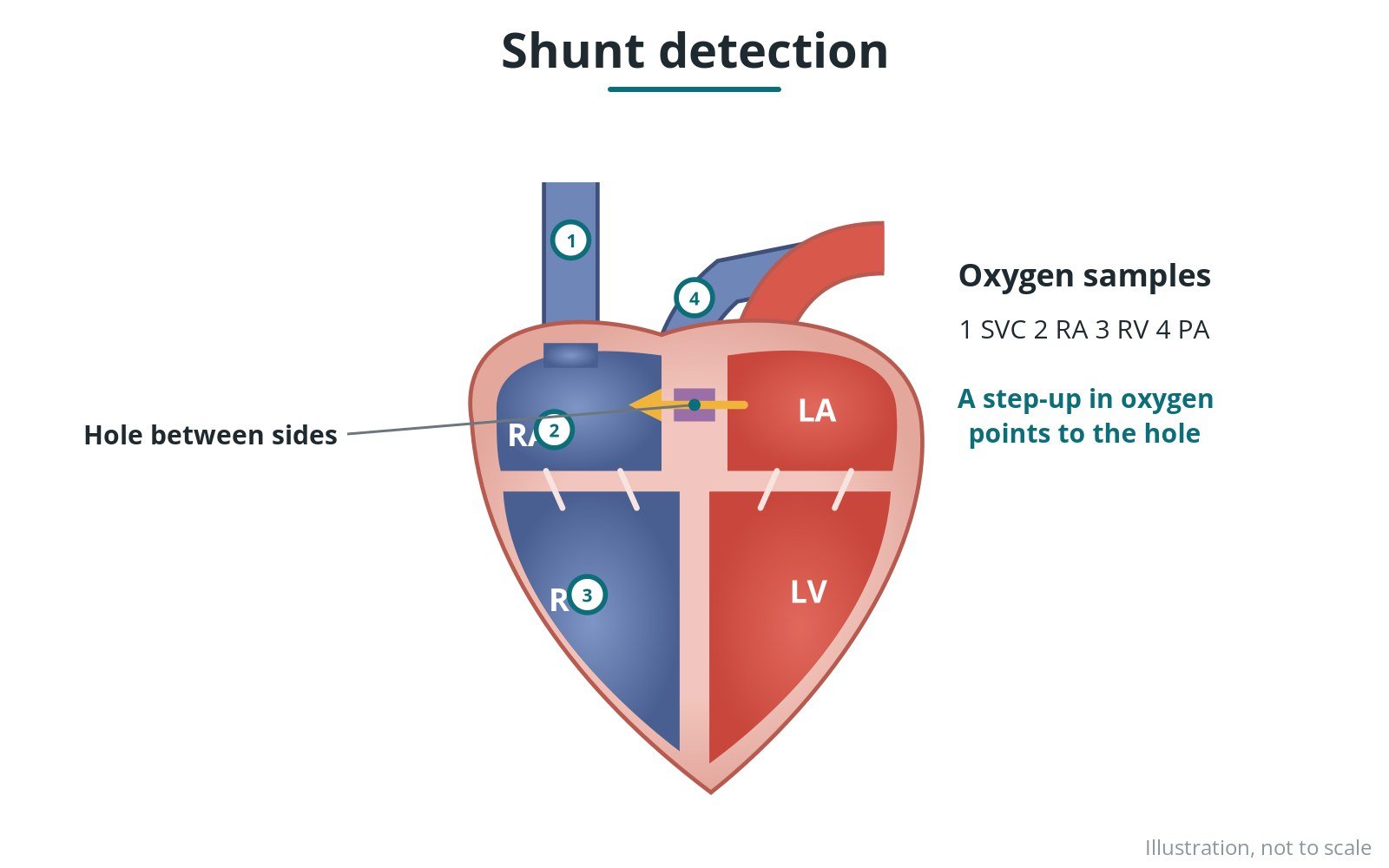
How oxygen samples from each heart chamber show blood flowing between the two sides. Original diagrams; steps, kit, risks and recovery.
How the grafts from earlier bypass surgery are found and filled with contrast dye to check they are open. Original diagrams; steps, kit, risks and recovery.
Plain-English guides to the procedures you'll see in the cardiac cath lab: what each one is, who it's for, the steps, the kit, the risks and the recovery. Written by a cath lab charge nurse for nurses, physiologists, radiographers and anyone new to the lab.
Start here
Cath Lab 101 · for iPhone
Interactive teaching illustrations you can tap, rotate and explore, alongside the guides and quizzes from this site.
Works offline. Every guide, illustration and quiz is built in, so it keeps working when the lab Wi-Fi drops.
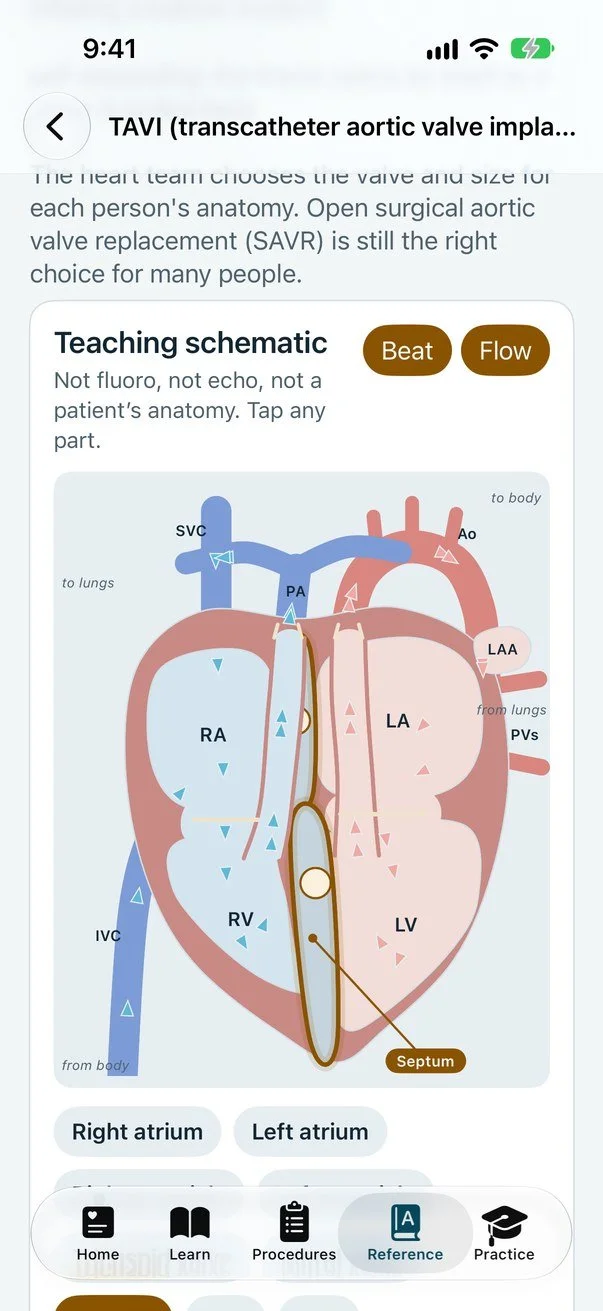
Tap any part to explore, with the heart beating and blood flowing.
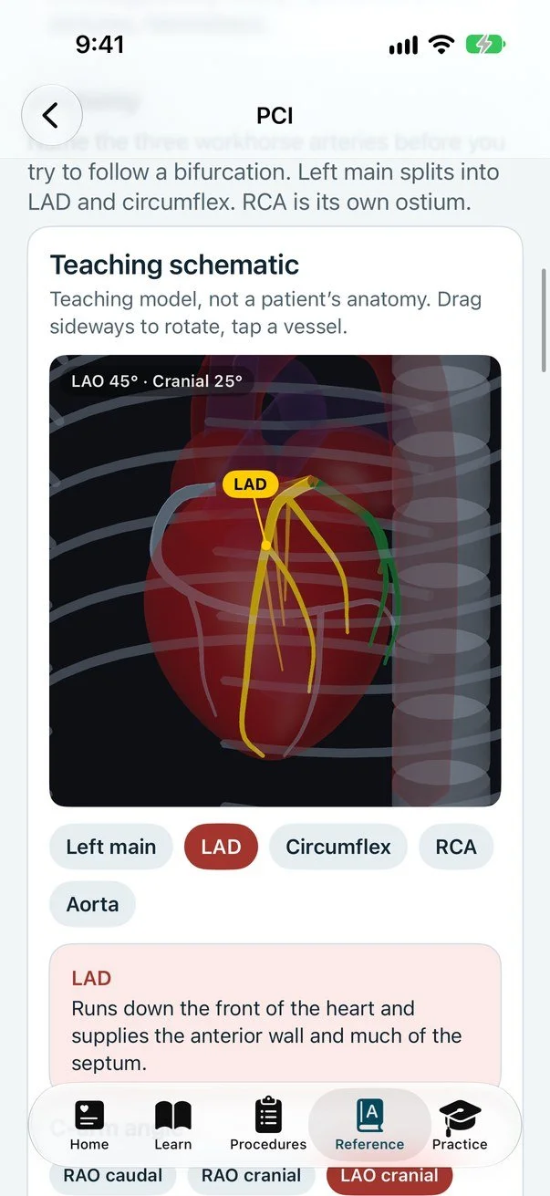
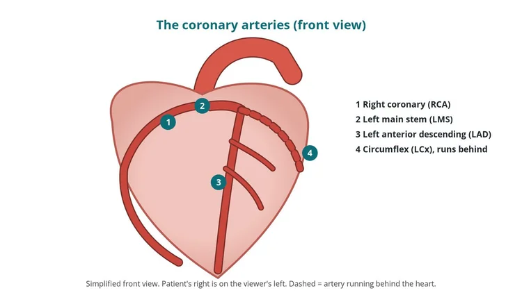
Drag to turn the heart, tap a vessel to name it.
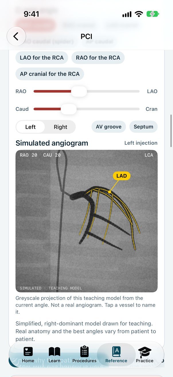
Change the angle and the teaching angiogram follows.
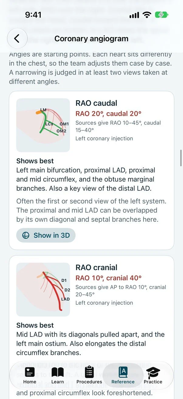
What each view shows best, with Show in 3D.
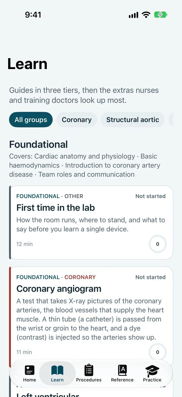
Foundational, Core and Advanced, filtered by group.
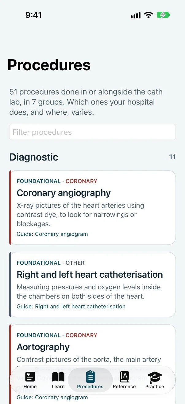
A plain-English line on each, linked to its guide.
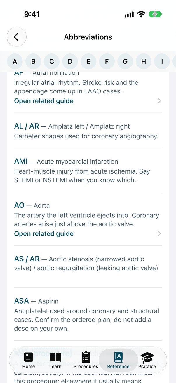
Searchable, with links to related guides.
Cath Lab 101Cath lab orientation & quizzes
For educational use only. Not for patient diagnosis or treatment. Published by Golden Shutter Ltd.
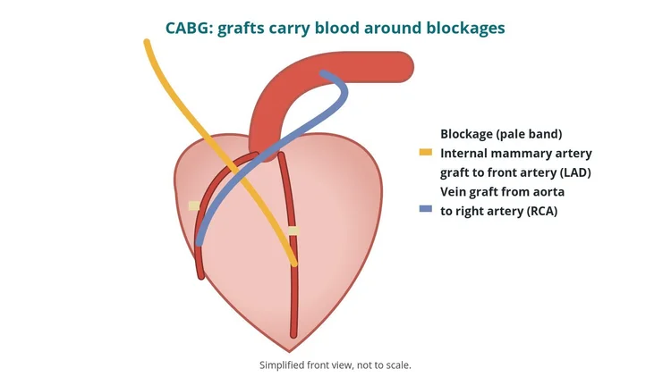
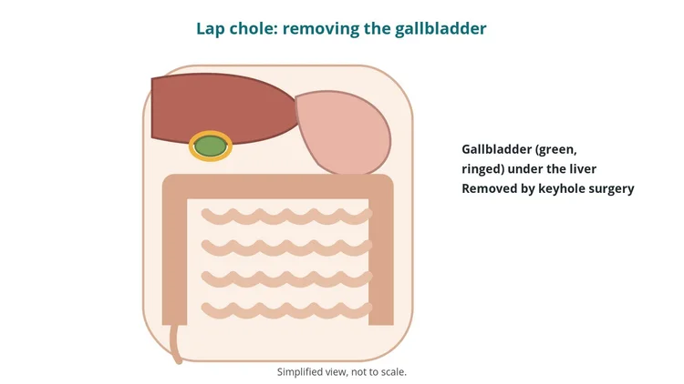
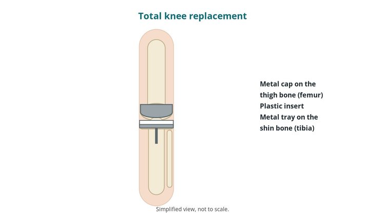
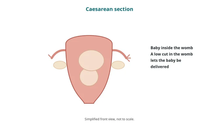
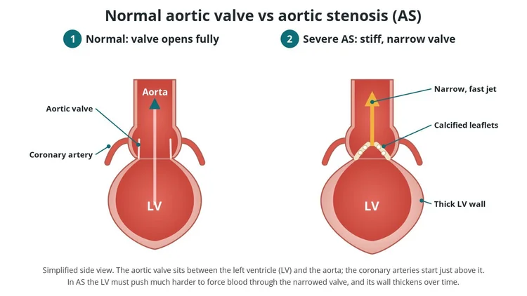
How a new valve is passed up an artery, usually from the groin, and opened inside a narrowed aortic valve. Who it helps, the steps, the main risks and what recovery looks like.
Read the guideAdvanced ECG guide for nurses and cath lab staff: abnormal ECGs explained, from SVT, AF, VT and heart block to bundle branch block, WPW, Brugada, STEMI equivalents, electrolytes, pacing problems and arrest rhythms, with causes, symptoms, treatment and what to call out in the lab.
ECG basics for student nurses and new cath lab staff: 12-lead electrode placement, the normal ECG waveform, reading a rhythm strip step by step, AF, flutter, heart block, VT, VF, STEMI patterns and what to watch for during procedures.
Education only. These pages are orientation for the team. They are not a protocol, and they don't replace your local policy, training or supervision. Always follow your centre's protocols.