Ductal (PDA) stenting in newborns: a Cath Lab 101 guide
Reviewed by Rodnie Oro, cath lab charge nurse · Last reviewed: November 2026 · Education only, not medical advice
Ductal stenting in newborns in one minute
What it is: The arterial duct (ductus arteriosus) joins the aorta to the pulmonary artery and normally closes in the first days of life. Some babies are born with heart conditions in which this duct is the only way blood reaches the lungs. A stent, a small metal mesh tube, is placed inside the duct by catheter so it stays open.
Who it is for: Newborns with duct-dependent lung blood flow, for example pulmonary atresia or very severe pulmonary stenosis, as decided by the congenital heart team. It is an alternative to a surgical shunt (BT shunt) in suitable duct shapes.
Main risks: The duct going into spasm with a sudden fall in oxygen levels, the stent blocking with clot, too much blood flow to the lungs, and damage to the artery used for access. Your team will explain your baby's own risk.
Usually: Under general anaesthetic, in the cath lab, about 1 to 3 hours. The baby returns to the neonatal or cardiac unit afterwards.
On this page: What is ductal stenting in newborns? · Who is it for? · When is it done? · Step by step · Recovery · Risks · Kit list · First time in the lab? · FAQ
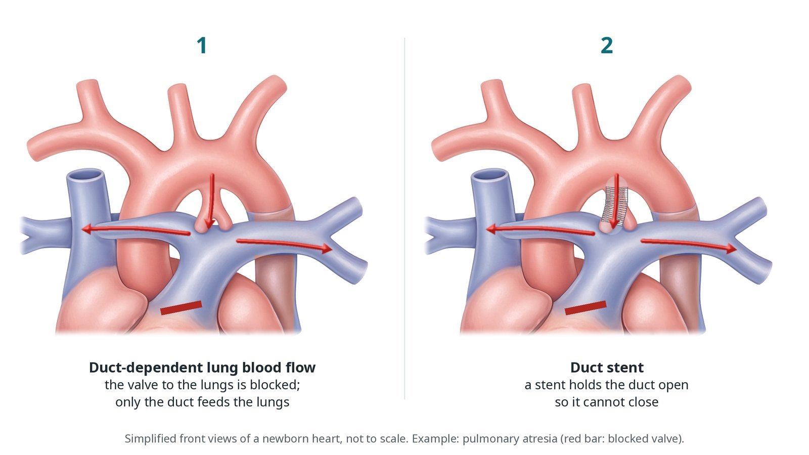
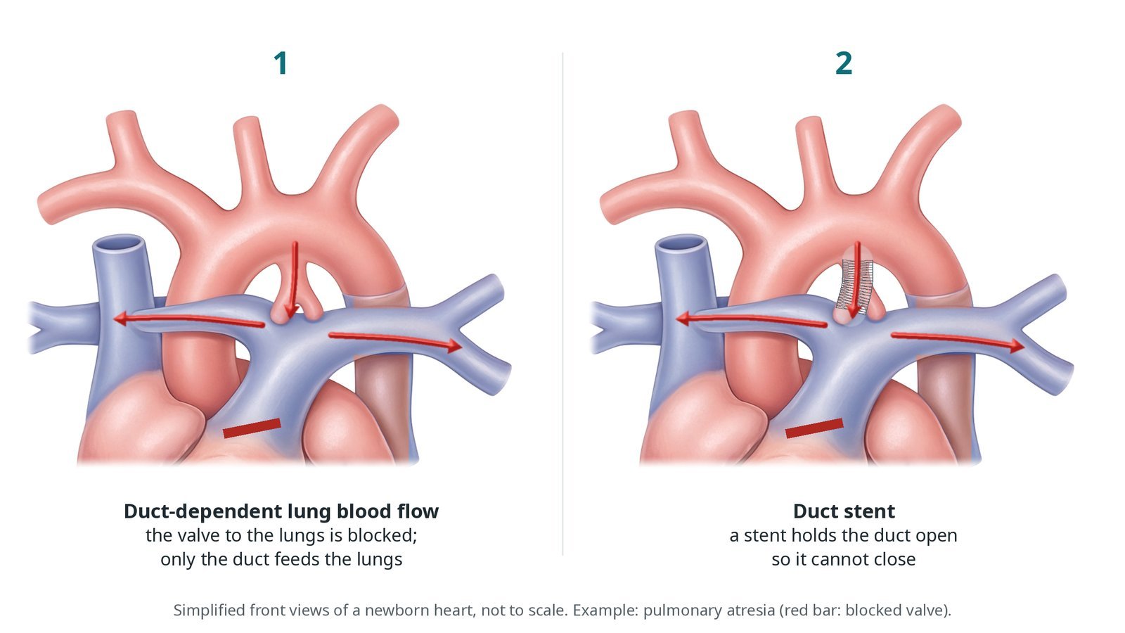
(1) Duct-dependent lung blood flow, for example pulmonary atresia: only the duct feeds the lungs. (2) A stent holds the duct open so it cannot close.
What is ductal stenting in newborns?
Before birth the arterial duct lets blood bypass the lungs, from the pulmonary artery to the aorta. After birth, as the baby breathes and oxygen levels rise, the duct normally narrows and closes within days.
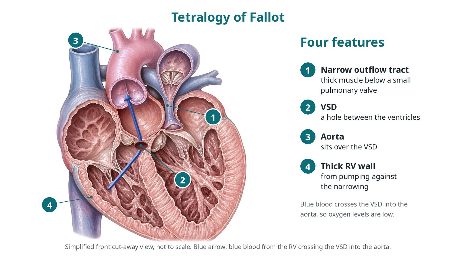
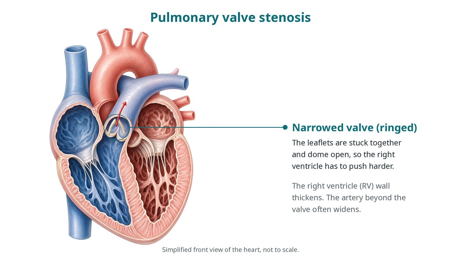
In some heart conditions the path from the right ventricle to the lungs is blocked or very narrow, for example pulmonary atresia, critical pulmonary stenosis, severe tetralogy of Fallot or tricuspid atresia. Blood then reaches the lungs backwards through the duct, from the aorta into the pulmonary arteries. This is duct-dependent pulmonary blood flow. If the duct closes, oxygen levels fall dangerously.
A prostaglandin infusion keeps the duct open after birth. It can cause pauses in breathing (apnoea), fever and low blood pressure, so it is a bridge, not a long-term answer.
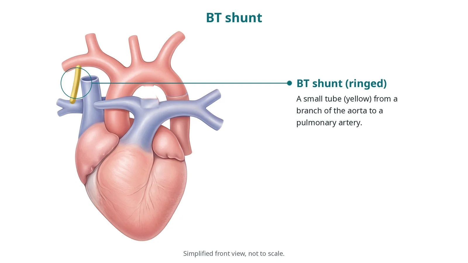
A longer-term source of lung blood flow is then needed until the next operation. Traditionally this is a surgical BT shunt, a tube from a branch of the aorta to a pulmonary artery. A duct stent does the same job without opening the chest. In these babies the duct is often long, twisting and steep (vertical), arising from under the aortic arch.
New to the acronyms? See the Cath Lab Abbreviations A–Z.
Who is it for?
Newborns with duct-dependent pulmonary blood flow, chosen by the congenital heart team after echo, and often CT, shows a duct shape that can be stented. The team weighs the stent against a surgical shunt. Some babies also have a stent as a bridge to growing before a full repair.
Who it may not suit: Some duct shapes are too long or twisted to stent safely, and duct tissue can narrow the branch pulmonary arteries. These babies may do better with a surgical shunt.
The decision is made by your heart team, who will talk through the reasons, the alternatives and the risks with you.
When is it done?
Usually in the first days or weeks of life, while the baby is on a prostaglandin infusion on the neonatal or cardiac unit. Whether prostaglandin is continued or paused before the case is decided by the team.
gives you a chance to ask questions and give consent
arranges blood tests, including kidney function and a blood count, and an ECG
Step by step
1. Getting the baby ready
The baby has a general anaesthetic and is ventilated. A warming mattress, a temperature probe and warm fluids keep the baby warm, because newborns lose heat quickly in a cool lab. Lines are checked and the prostaglandin infusion is managed as the team has planned. All drugs and flushes are weight-based, as per centre protocol. Blood for cross-matching is available.
2. Access
The route depends on the duct's shape and where it arises. A duct arising from the descending aorta is often reached from the femoral artery. A steep duct under the arch is often easier from the carotid or axillary artery, by ultrasound or a small surgical cut-down. A femoral vein is used in some anatomies. Sheaths are as small as possible, per operator. Heparin is given, dose as per centre protocol.
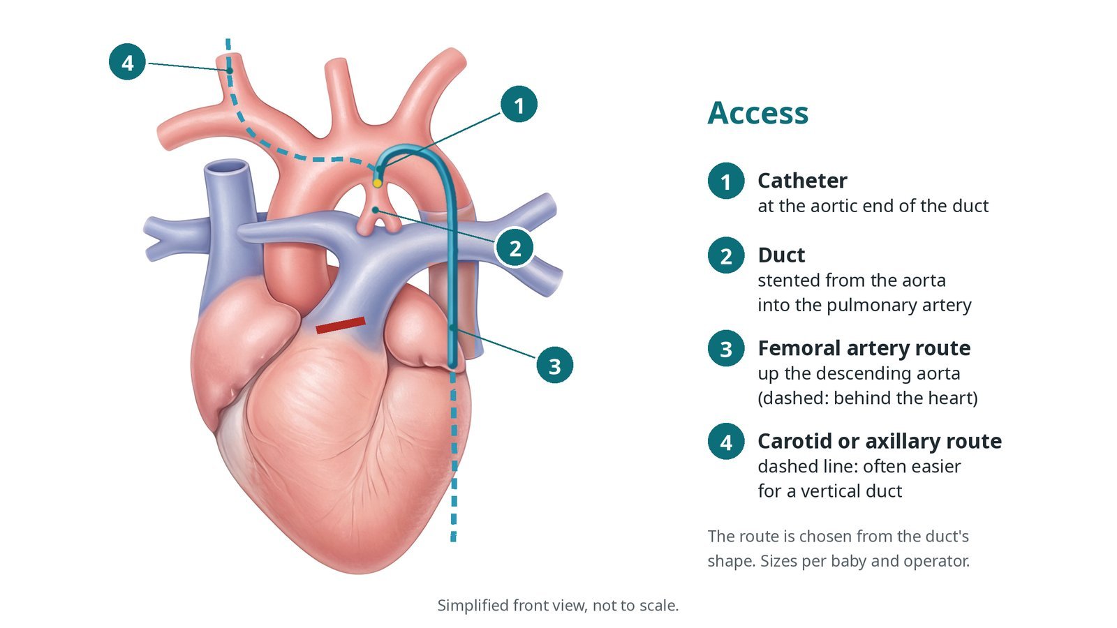
Access: (1) catheter at the aortic end of the duct; (2) the duct, stented from the aorta into the pulmonary artery; (3) femoral artery route; (4) carotid or axillary route (dashed), often easier for a vertical duct.
3. Stenting the duct
A. Angiogram. Contrast injected at the aortic end shows the duct's shape, length and narrowest point.
B. Wire across. A soft coronary-type wire is passed through the duct into a branch pulmonary artery.
C. Stent. A stent on a balloon is positioned to cover the whole duct, from the aortic end to the pulmonary artery end, and opened. Stent size and length are per operator; more than one stent may be needed.
D. Check. An angiogram checks that the whole duct is covered and both lungs fill. Oxygen levels rise towards the team's target.
Watch: A sudden fall in oxygen levels or heart rate as the duct is crossed suggests spasm: tell the operator at once. The team supports ventilation and moves quickly to open the stent.
4. Oxygen targets and medicines
In this circulation normal adult oxygen levels are not the aim. Blue (venous) and red blood still mix in the heart, so the team sets a target range for oxygen saturation that balances lung and body blood flow. Very high saturations can mean too much blood is going to the lungs. An antiplatelet medicine, and sometimes a heparin infusion, is started to stop the stent clotting, per team. Prostaglandin is stopped once the stent is in, per team.
Watch: Falling oxygen levels after the case may mean the stent is clotting: tell the team at once.
5. Finishing
Sheaths are removed and pressure applied; a cut-down is closed by the surgeon. The pulse, colour and warmth of the limb used for access are checked. The baby returns to the neonatal or cardiac unit with a handover covering oxygen targets, medicines and access site.
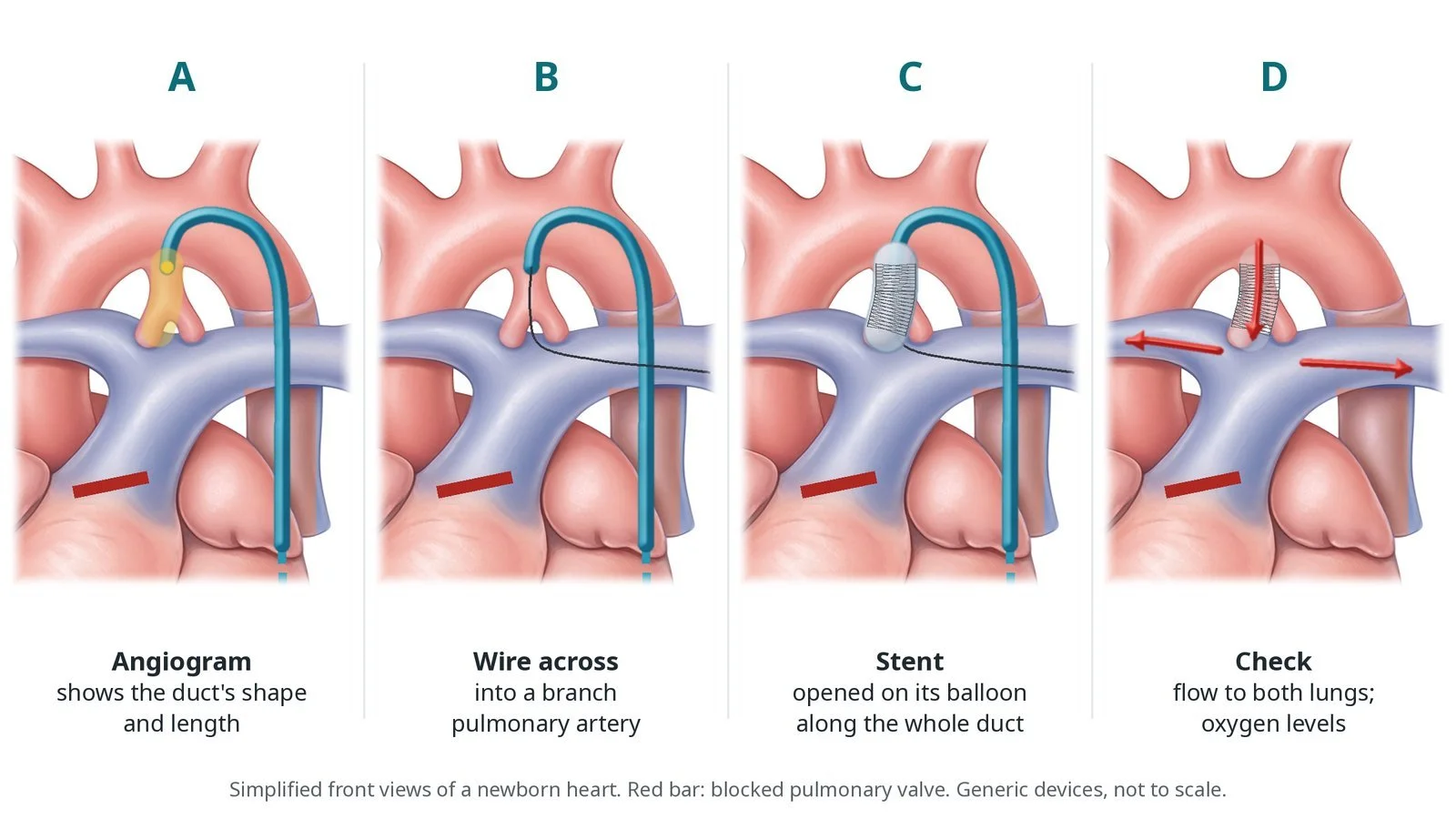
A: angiogram of the duct. B: wire across into a branch pulmonary artery. C: stent opened on its balloon along the whole duct. D: flow to both lungs and oxygen levels checked.
Recovery and aftercare
First hours: oxygen levels, heart rate, blood pressure, temperature and the access limb are checked closely on the neonatal or cardiac unit, as per centre protocol.
Medicines: an antiplatelet medicine is usually given every day; do not miss doses, because it helps keep the stent open.
Echo: checks flow through the stent before going home.
Going home: many babies go home after days to weeks, depending on feeding, weight gain and the heart condition.
At home: parents are shown what normal colour and breathing look like for their baby; some families have home oxygen-level monitoring.
Follow-up: close review with the congenital heart team until the next planned surgery.
Call 999 if your baby is more blue than usual, floppy, breathing fast or struggling, or very sleepy and not feeding.
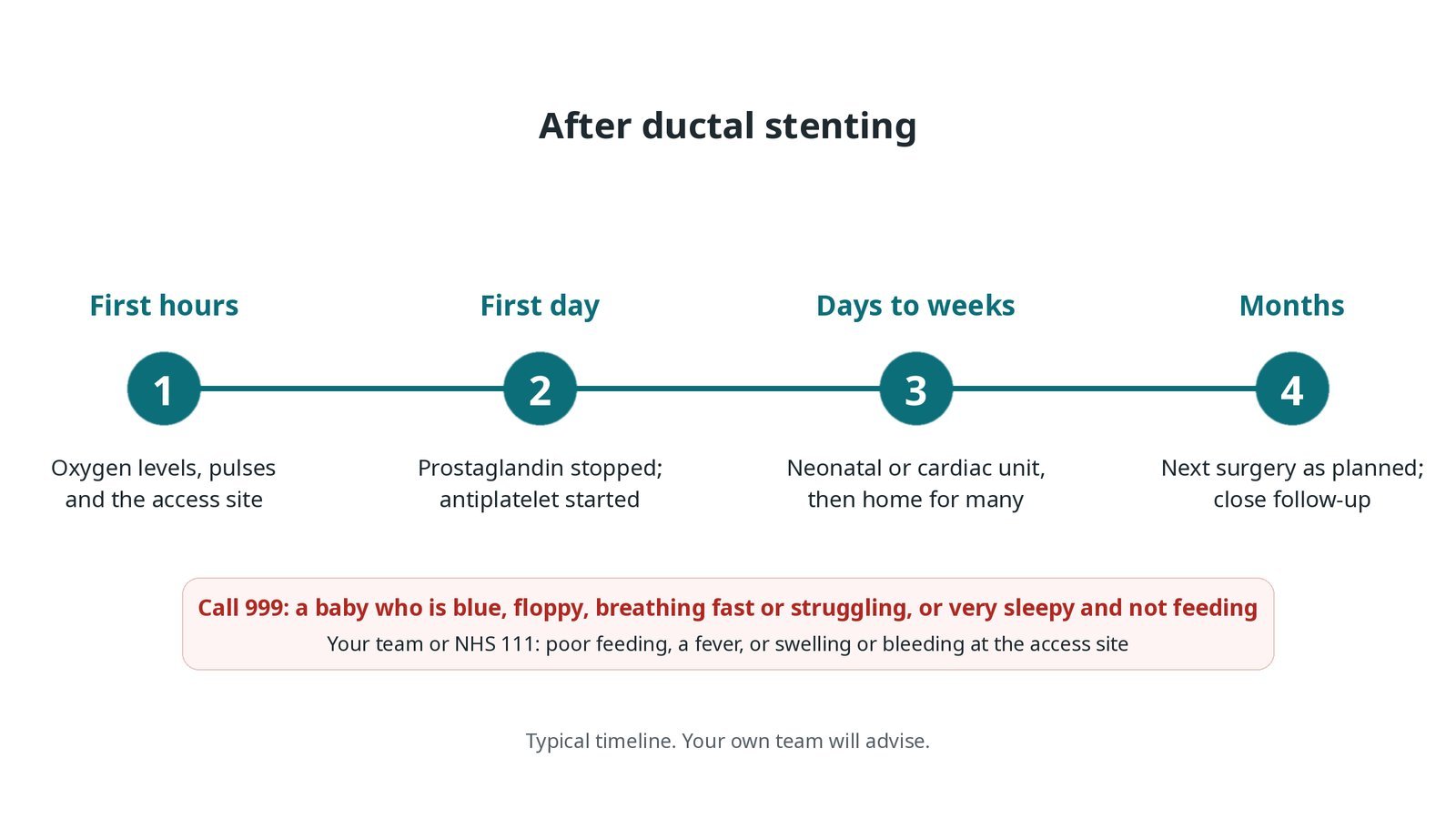
After ductal stenting: (1) oxygen levels, pulses and access site; (2) prostaglandin stopped, antiplatelet started; (3) neonatal or cardiac unit, then home for many; (4) next surgery as planned.
Risks and complications
Your team will explain the risks that apply to you. Possible problems include:
Duct spasm with a sudden fall in oxygen levels
Clot blocking the stent (uncommon, an emergency)
Too much blood flow to the lungs, causing breathing or heart strain
Damage to, or a blocked, access artery in the leg, neck or arm (uncommon)
The stent moving or not covering the whole duct, needing another stent (uncommon)
Narrowing of a branch pulmonary artery or of the stent over time
Cath lab kit list (printable)
Barts example kit list
An example from one UK centre, written generically. Every lab has its own protocol, and sizes and doses vary.
small arterial sheath (or venous), per baby and operator
ultrasound for access; cut-down set for the carotid or axillary route
shaped catheters and guide catheters, per operator
soft coronary-type guidewires
coronary-type stents on balloons in several lengths, per operator
inflation device
prostaglandin infusion, managed as per team plan
warming mattress, temperature probe and warmed fluids
heparin and antiplatelet medicines, weight-based, as per centre protocol
snare and spare stents for a stent that moves
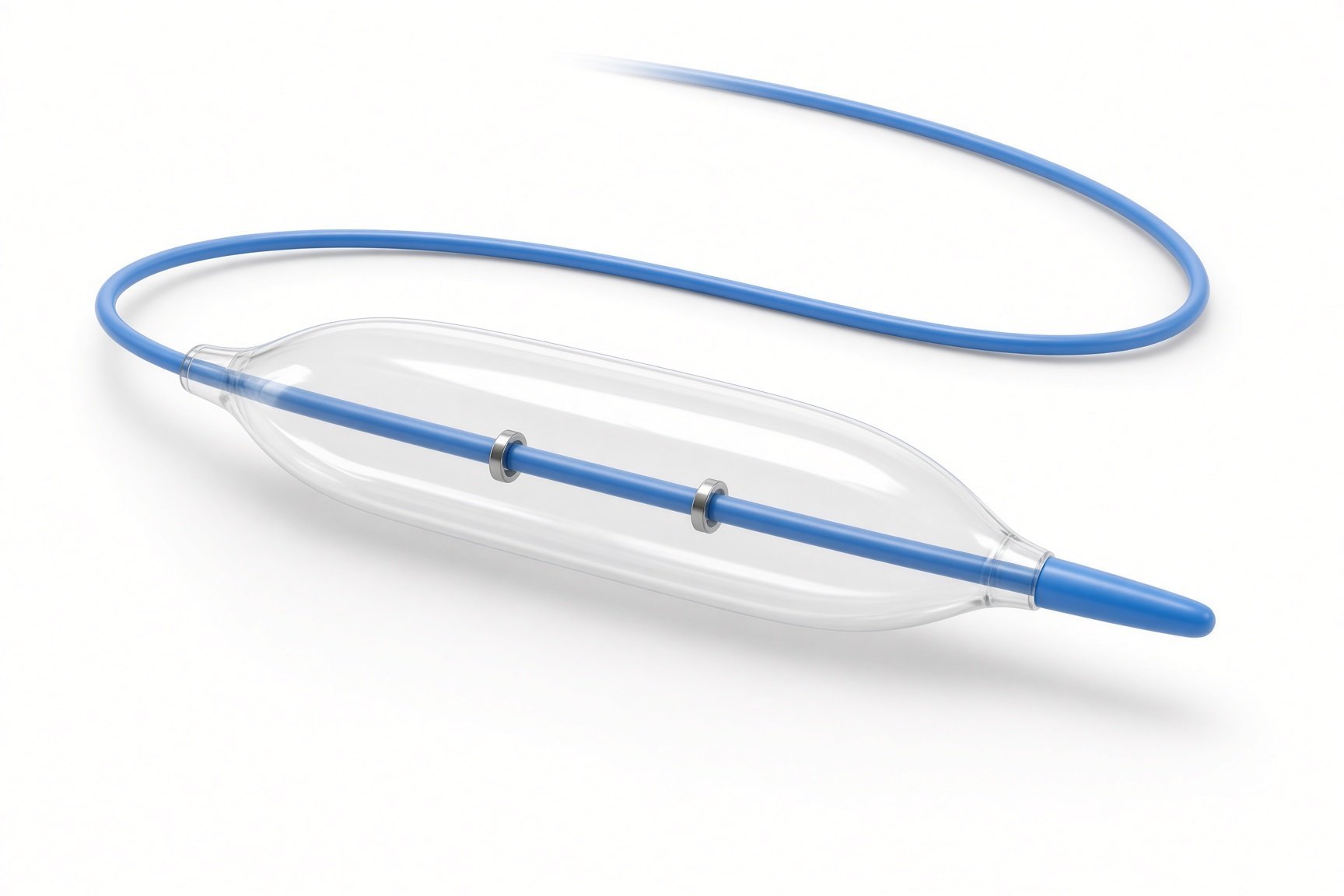
Coronary stents on balloons.
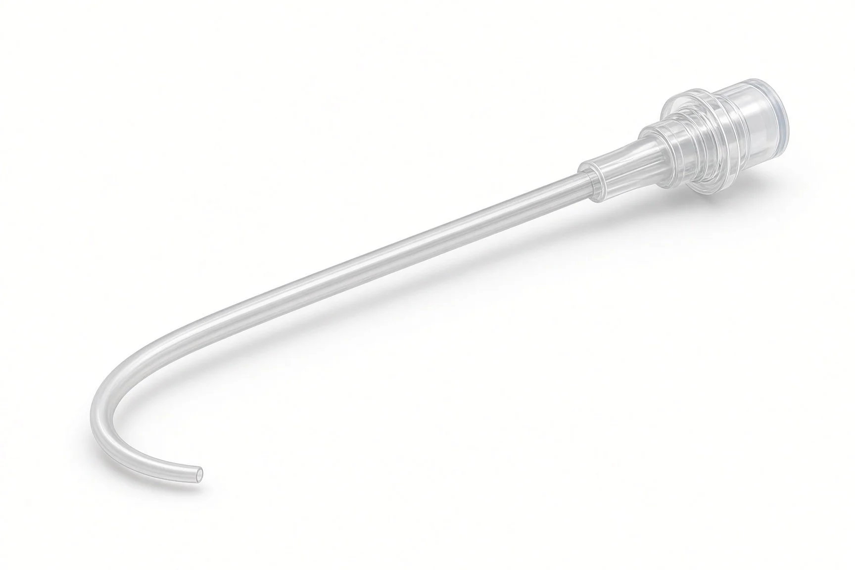
Shaped catheters.
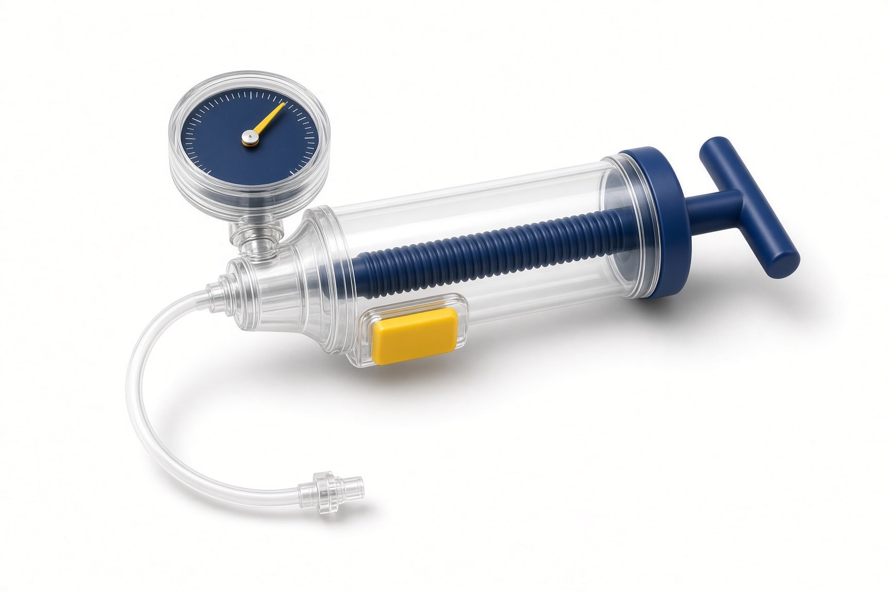
Inflation device.
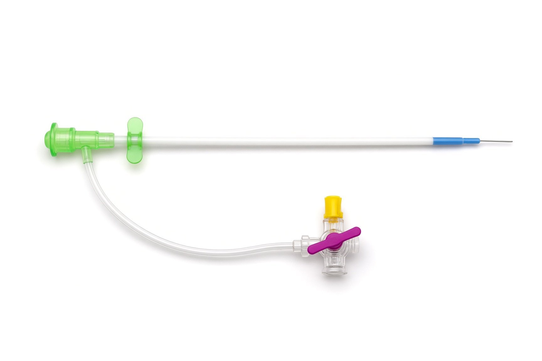
Small arterial sheath.
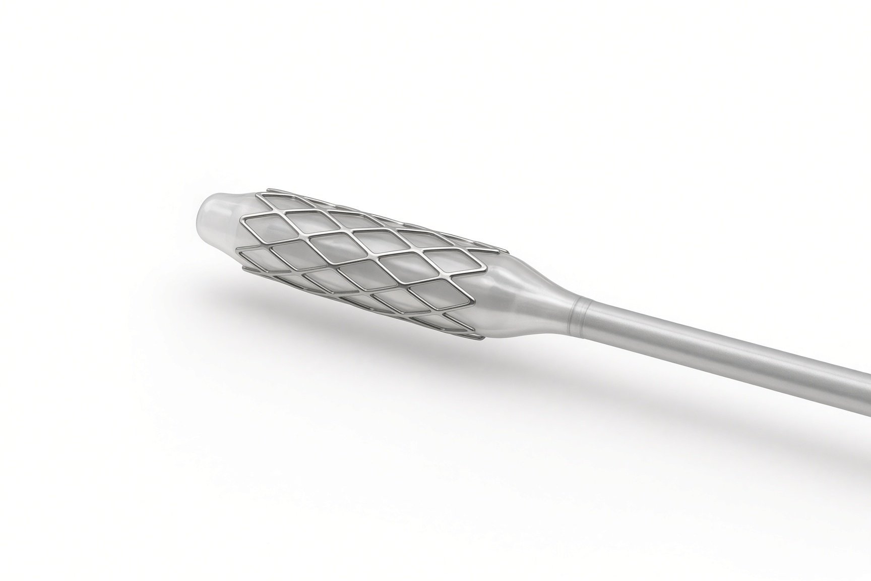
Coronary stents on balloons.
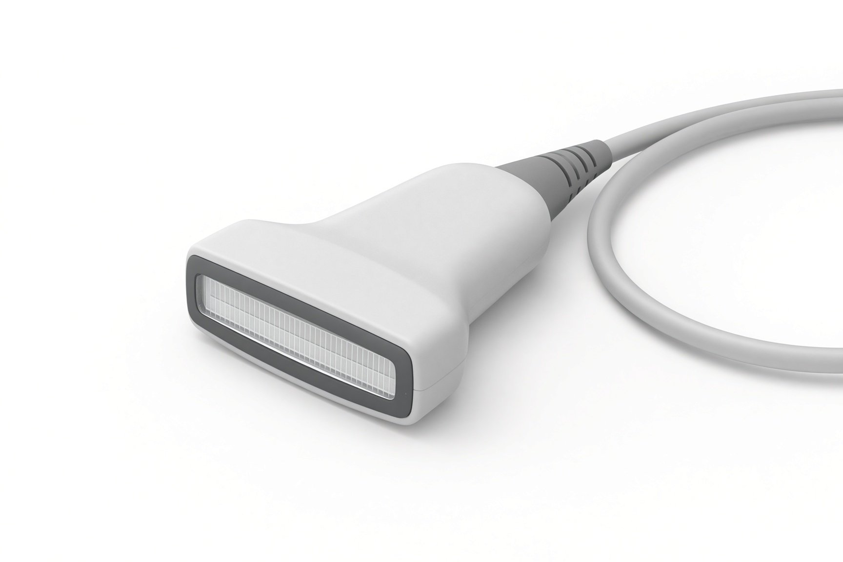
Ultrasound for access.

Prostaglandin infusion.

Warming mattress.
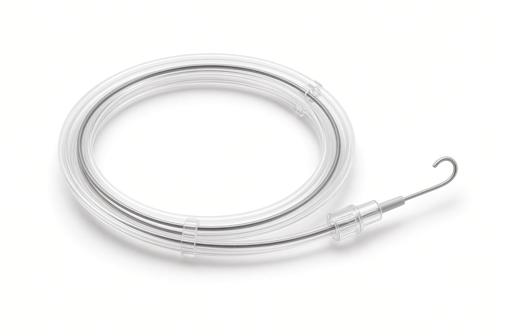
Coronary guidewires.
Procedural steps (Barts example)
Getting the baby ready
Access
Stenting the duct
Oxygen targets and medicines
Finishing
First time in the cath lab?
Keep the baby warm. Newborns cool quickly under the X-ray tube. Check the warming mattress and temperature probe before the drapes go on.
Protect the prostaglandin. Know which line it runs in and never pause it without a clear instruction.
Watch the saturations, not just the pictures. Spasm or a clotting stent shows first as falling oxygen levels.
Support the family. Parents are often waiting nearby after a very hard week. Make sure someone updates them.
Label and protect the prostaglandin line. Stopping it by accident can close the duct.
Call out oxygen levels as the wire crosses. They can fall fast if the duct goes into spasm.
Ask the team for the target saturation range and write it down for handover.
FAQ
Why a stent instead of a shunt operation?
A stent avoids opening the chest. Whether it suits your baby depends on the duct's shape, and the team will explain the choice.
Will my baby be pink afterwards?
Usually not fully. Blue and red blood still mix in the heart, so oxygen levels lower than normal are expected and are the team's target.
Does the stent stay in for ever?
It works for months until the next operation, when the surgeon usually removes or ties off the duct.
Why is my baby on aspirin-type medicine?
An antiplatelet medicine lowers the chance of clot forming in the stent.
Related Cath Lab 101 guides
RVOT stenting in tetralogy of Fallot (coming soon)
Balloon pulmonary valvuloplasty (coming soon)
Sources
All checked on 12 November 2026. Each page's own date is shown where it gives one.
Feltes TF, et al. Indications for cardiac catheterization and intervention in pediatric cardiac disease: a scientific statement from the American Heart Association. Circulation 2011;123(22):2607–2652
StatPearls (NCBI Bookshelf), search: pulmonary atresia. ncbi.nlm.nih.gov/books
StatPearls (NCBI Bookshelf), search: patent ductus arteriosus. ncbi.nlm.nih.gov/books
Disclaimer
Education only. This page is for general education for healthcare students, cath lab staff and curious readers. It isn't medical advice and doesn't replace advice from your own doctor, heart team or local unit protocols. Equipment and medicines are described generically. No product or manufacturer is recommended. If you think someone is having a heart emergency, call 999.
Disclosure: I work for a company that makes a device used in this procedure. This page is independent education, isn't sponsored or reviewed by any company, and doesn't recommend any product.
All diagrams on this page are original illustrations created for rodnieoro.com.