Hybrid stage 1 for hypoplastic left heart: a Cath Lab 101 guide
Reviewed by Rodnie Oro, cath lab charge nurse · Last reviewed: November 2026 · Education only, not medical advice
Hybrid stage 1 for hypoplastic left heart in one minute
What it is: In hypoplastic left heart syndrome (HLHS) the left side of the heart is too small to pump blood to the body. The hybrid stage 1 combines surgery and catheter work without a heart-lung machine: the surgeon places bands on both pulmonary arteries through the breastbone, and the cath team places a stent in the arterial duct. The opening between the atria may also be enlarged.
Who it is for: Selected newborns with HLHS or similar single-ventricle hearts, often those who are small, premature or unwell, or as a bridge to a later decision, as chosen by the congenital heart team. Practice varies between centres.
Main risks: HLHS is a serious, life-threatening condition. Risks include low blood pressure or an imbalance between lung and body blood flow, bleeding, narrowing of blood flow around the small aortic arch, problems with the duct stent or atrial opening, infection, and death. The team will discuss your baby's own risks openly.
Usually: Under general anaesthetic in a hybrid theatre, a few hours, then care on the cardiac intensive care unit. A larger operation (comprehensive stage 2) follows at a few months of age.
On this page: What is hybrid stage 1 for hypoplastic left heart? · Who is it for? · When is it done? · Step by step · Recovery · Risks · Kit list · First time in the lab? · FAQ
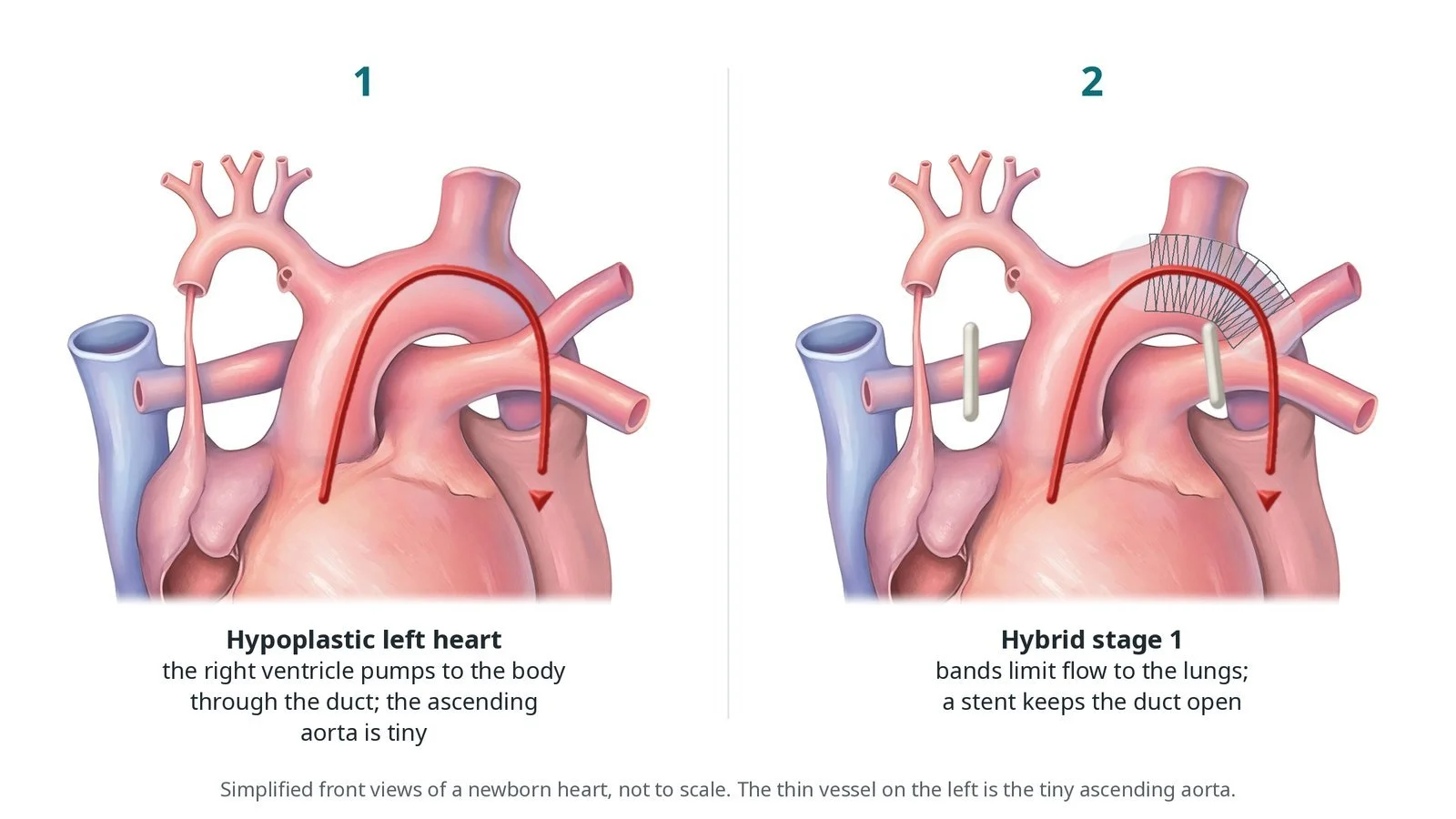
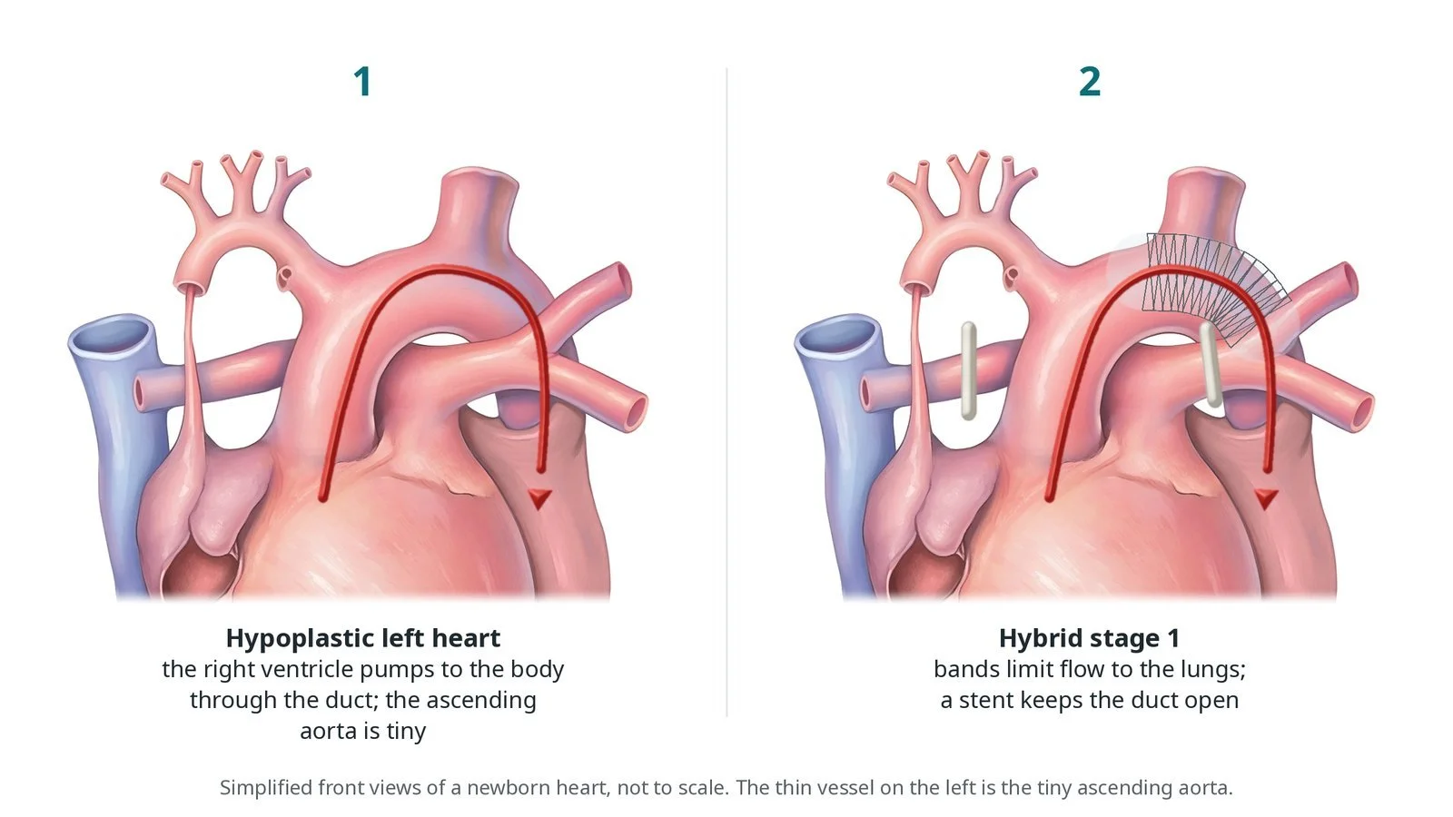
(1) Hypoplastic left heart: the right ventricle pumps to the body through the duct, and the ascending aorta is tiny. (2) Hybrid stage 1: bands limit flow to the lungs and a stent keeps the duct open.
What is hybrid stage 1 for hypoplastic left heart?
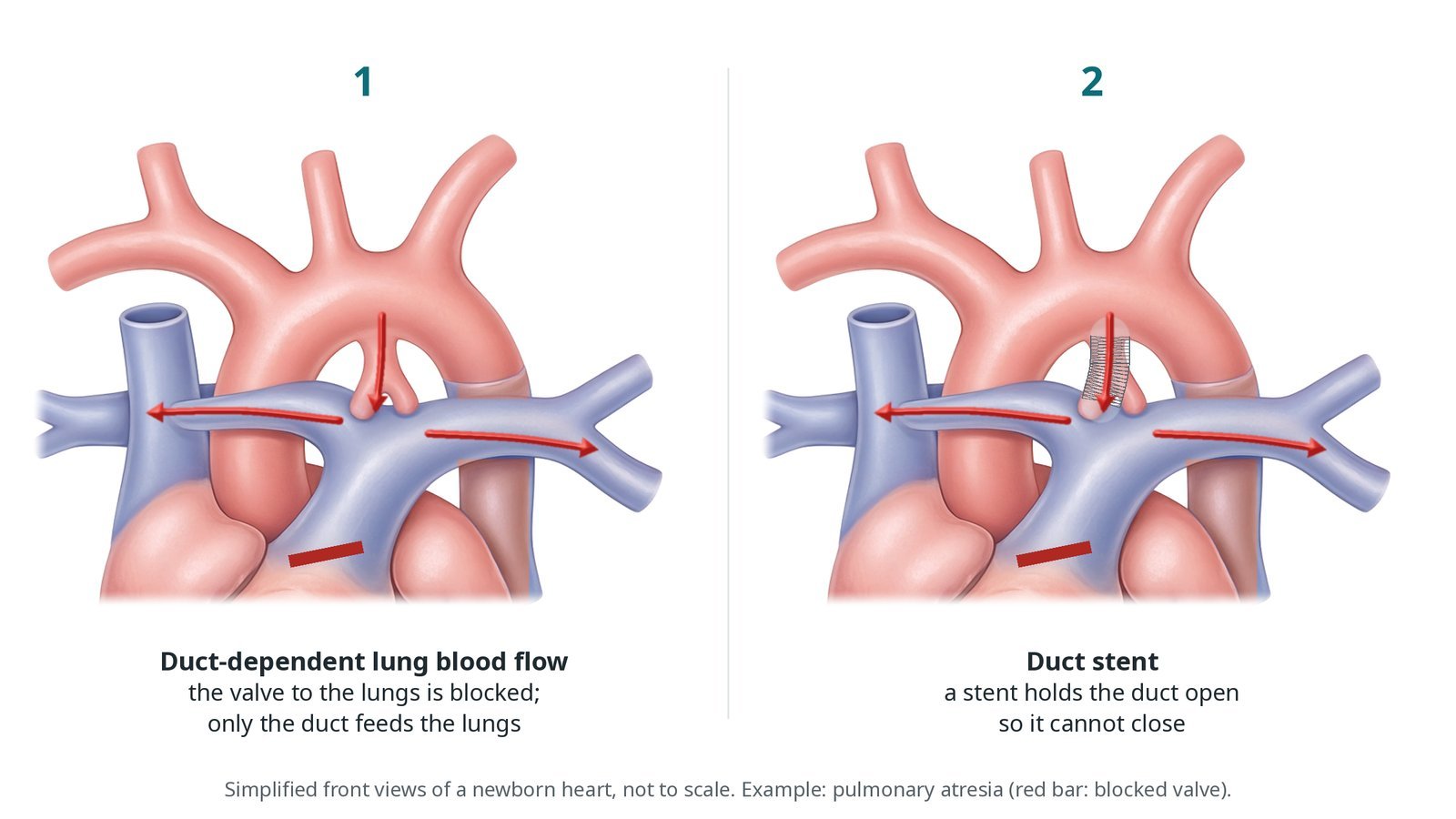
In HLHS the left ventricle, mitral valve, aortic valve and ascending aorta are small or blocked. The right ventricle has to pump blood to both the lungs and the body. Blood reaches the body through the arterial duct, from the pulmonary artery into the aorta, so the circulation is duct-dependent and a prostaglandin infusion keeps the duct open after birth.
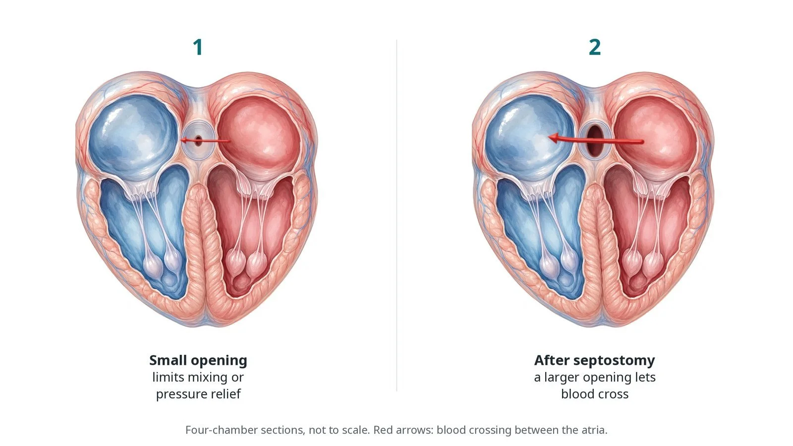
Blood returning from the lungs to the left atrium has to cross the atrial septum to reach the right side. If this opening is small (restrictive), the left atrium becomes congested and oxygen levels fall.
Blood flow to the heart muscle and brain travels backwards from the duct, round the small arch, into the tiny ascending aorta and coronary arteries. Anything that narrows this path, such as a stent that sticks out into the aorta, can be dangerous.
Treatment is staged. Stage 1 is either the Norwood operation, on bypass, or the hybrid approach: bands on both pulmonary arteries limit lung blood flow and pressure, a duct stent replaces the prostaglandin, and the atrial septum is opened if needed. Stage 2 (arch reconstruction with a Glenn) and stage 3 (Fontan) follow.
New to the acronyms? See the Cath Lab Abbreviations A–Z.
Who is it for?
The congenital heart team chooses between the Norwood and hybrid approaches. Hybrid stage 1 may be chosen for:
babies with low birth weight or prematurity
babies with other illnesses that make bypass riskier
babies in whom a decision about later two-ventricle repair or transplant is still open
some centres use it more widely; practice varies
Who it may not suit: Some anatomies, such as a very small ascending aorta with poor backward flow, may favour a Norwood operation. The team will explain the options for each baby.
The decision is made by your heart team, who will talk through the reasons, the alternatives and the risks with you.
When is it done?
In the first days or weeks of life, while the baby is on prostaglandin on the neonatal or cardiac unit. HLHS is often diagnosed on the antenatal scan, so families may meet the team before birth.
gives you a chance to ask questions and give consent
arranges blood tests, including kidney function and a blood count, and an ECG
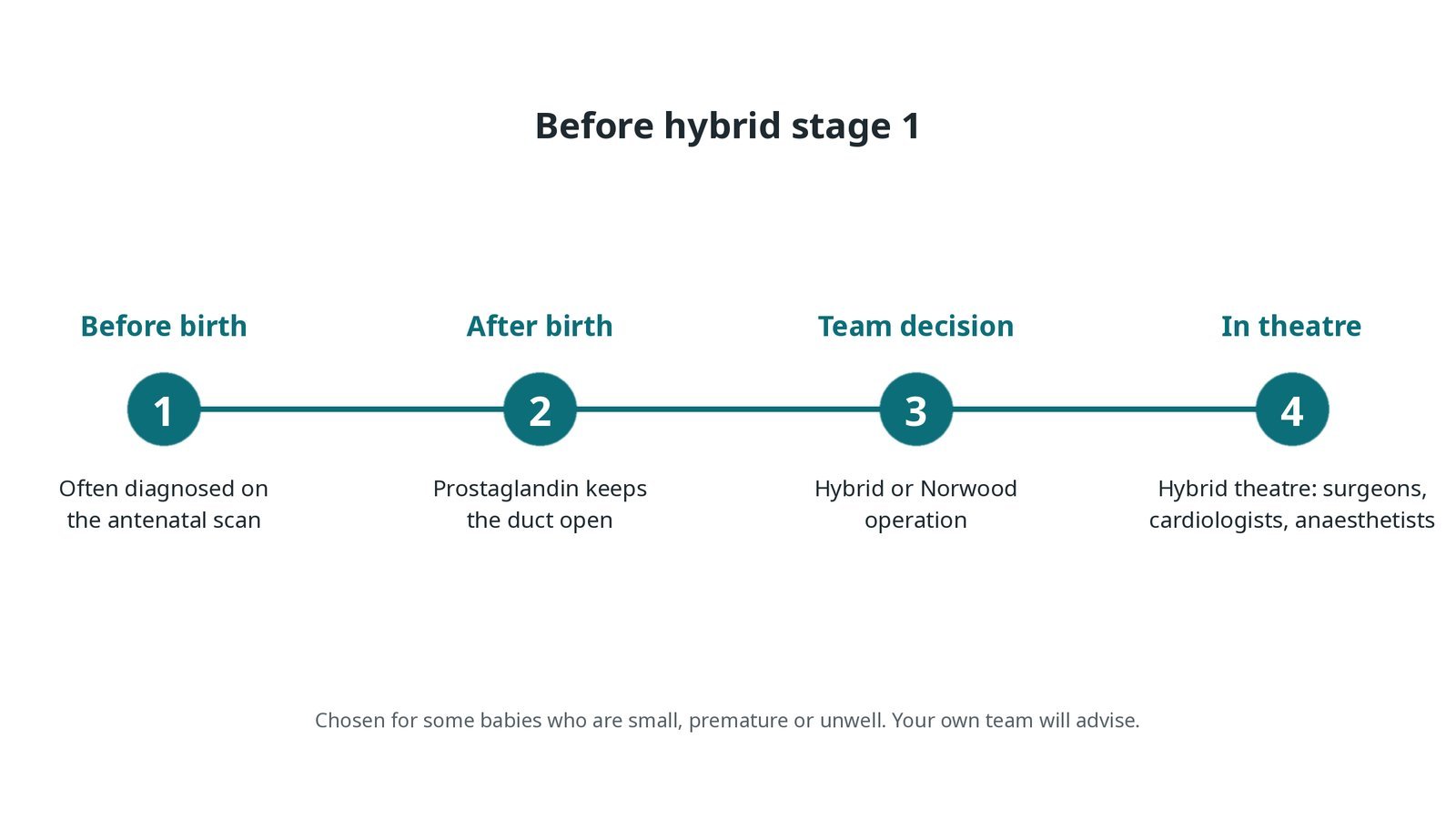
Before hybrid stage 1: often an antenatal diagnosis; prostaglandin keeps the duct open after birth; team decision between hybrid and Norwood; surgeons, cardiologists and anaesthetists in a hybrid theatre.
Step by step
1. The hybrid theatre and team
The procedure takes place in a hybrid theatre that has both surgical equipment and X-ray imaging. The team includes the congenital surgeon, interventional cardiologist, anaesthetist, perfusionist on standby, scrub and cath lab nurses, radiographer and physiologist. Roles, the sterile field, imaging positions and radiation protection are agreed at the team brief and WHO checklist. The baby is kept warm, and all drugs are weight-based, as per centre protocol.
2. Bands on the pulmonary arteries
The surgeon opens the chest through the breastbone (sternotomy) and places a band around each branch pulmonary artery. As the bands are tightened, oxygen levels fall a little and blood pressure rises, showing that more blood is going to the body. Band tightness is judged by the surgeon and team.
Watch: A big fall in oxygen levels or blood pressure as the bands go on: tell the surgeon and anaesthetist at once.
3. Stenting the duct and atrial septum
A. Bands. Both branch pulmonary arteries are banded by the surgeon.
B. Sheath. The surgeon places a short sheath in the main pulmonary artery through a purse-string stitch. An angiogram outlines the duct and its length is measured.
C. Duct stent. A stent is placed to cover the whole duct, without sticking out into the aorta, so the backward flow to the arch is not narrowed. Stent type and size are per operator. Prostaglandin is then stopped, per team.
D. Atrial septum. If the opening between the atria is small, it is enlarged with a balloon (septostomy) or a stent, in the same session or later in the cath lab, guided by echo.
Watch: Sudden low blood pressure or ECG changes after the duct stent may mean the backward flow round the arch is narrowed: tell the team at once.
4. Finishing
The sheath is removed and the purse-string tied. The chest is closed or, if the baby is unstable, left open with a dressing and closed later on intensive care. The baby goes to the cardiac intensive care unit with a full handover from both teams.
5. Interstage and next steps
The weeks between stage 1 and stage 2 (the interstage) are a high-risk time. Many babies go home with close monitoring of oxygen levels, feeding and weight. Echo checks the duct stent, the bands and the atrial opening. The comprehensive stage 2 operation, at a few months of age, rebuilds the arch, removes the bands and stent and creates a Glenn connection.
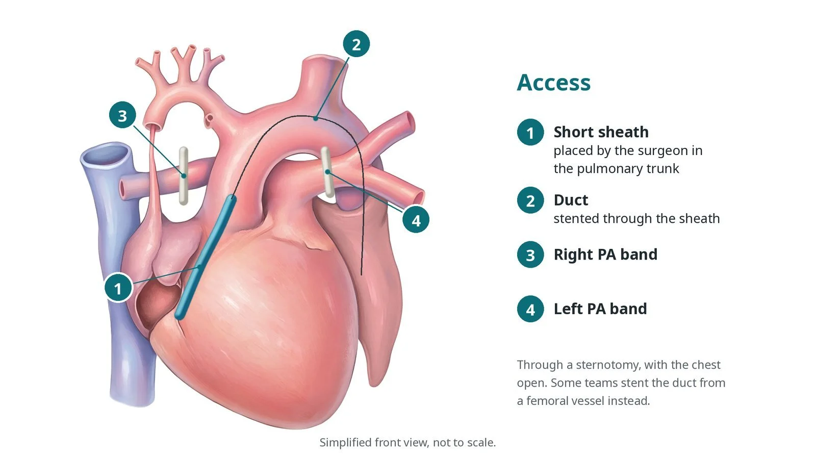
Access: (1) short sheath placed by the surgeon in the pulmonary trunk; (2) the duct, stented through the sheath; (3) right and (4) left pulmonary artery bands.
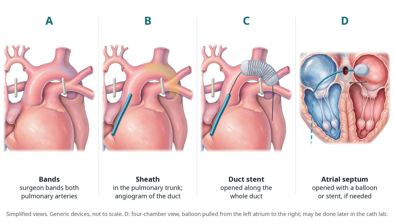
A: the surgeon bands both pulmonary arteries. B: sheath in the pulmonary trunk and an angiogram of the duct. C: stent opened along the whole duct. D: the atrial septum opened with a balloon or stent if needed.
Recovery and aftercare
First days: care on the cardiac intensive care unit, with breathing support, monitoring and the chest closed when the baby is ready.
Medicines: prostaglandin is stopped after the duct stent; antiplatelet and other medicines are started as the team advises.
Feeding and growth: feeding is built up carefully; weight gain matters for the next operation.
Going home: many babies go home after weeks, with close interstage monitoring of oxygen levels, feeding and weight.
Echo: regular checks of the duct stent, bands and atrial opening.
Follow-up: frequent contact with the congenital heart team until the comprehensive stage 2 operation at a few months of age.
Call 999 if your baby is more blue than usual, floppy, breathing fast or struggling, or very sleepy and not feeding.
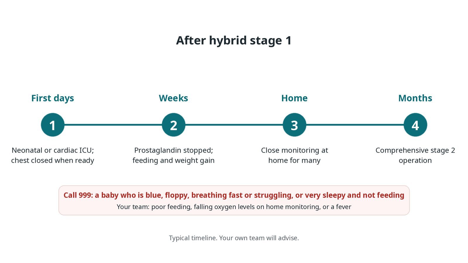
After hybrid stage 1: (1) neonatal or cardiac ICU, chest closed when ready; (2) prostaglandin stopped, feeding and weight gain; (3) close home monitoring for many; (4) comprehensive stage 2 operation at a few months.
Risks and complications
Your team will explain the risks that apply to you. Possible problems include:
Low blood pressure or an imbalance between lung and body blood flow
Bleeding needing transfusion or a return to theatre
Narrowing of the backward blood flow round the small aortic arch (uncommon)
Duct stent narrowing or moving, needing more treatment
A small atrial opening needing a further procedure
Infection, including of the wound
Death; HLHS is a high-risk condition and the team will discuss this openly
Cath lab kit list (printable)
Barts example kit list
An example from one UK centre, written generically. Every lab has its own protocol, and sizes and doses vary.
short sheath for the pulmonary trunk, per operator
guidewires and angiography catheter
duct stents, self-expanding or balloon-expandable, in several lengths, per operator
septostomy balloon or stent for the atrial septum
surgical banding material and sternotomy set (surgical team)
echo machine, transthoracic or epicardial probe
prostaglandin infusion, managed as per team plan
warming mattress, temperature probe and warmed fluids
weight-based emergency drugs and blood for transfusion, as per centre protocol
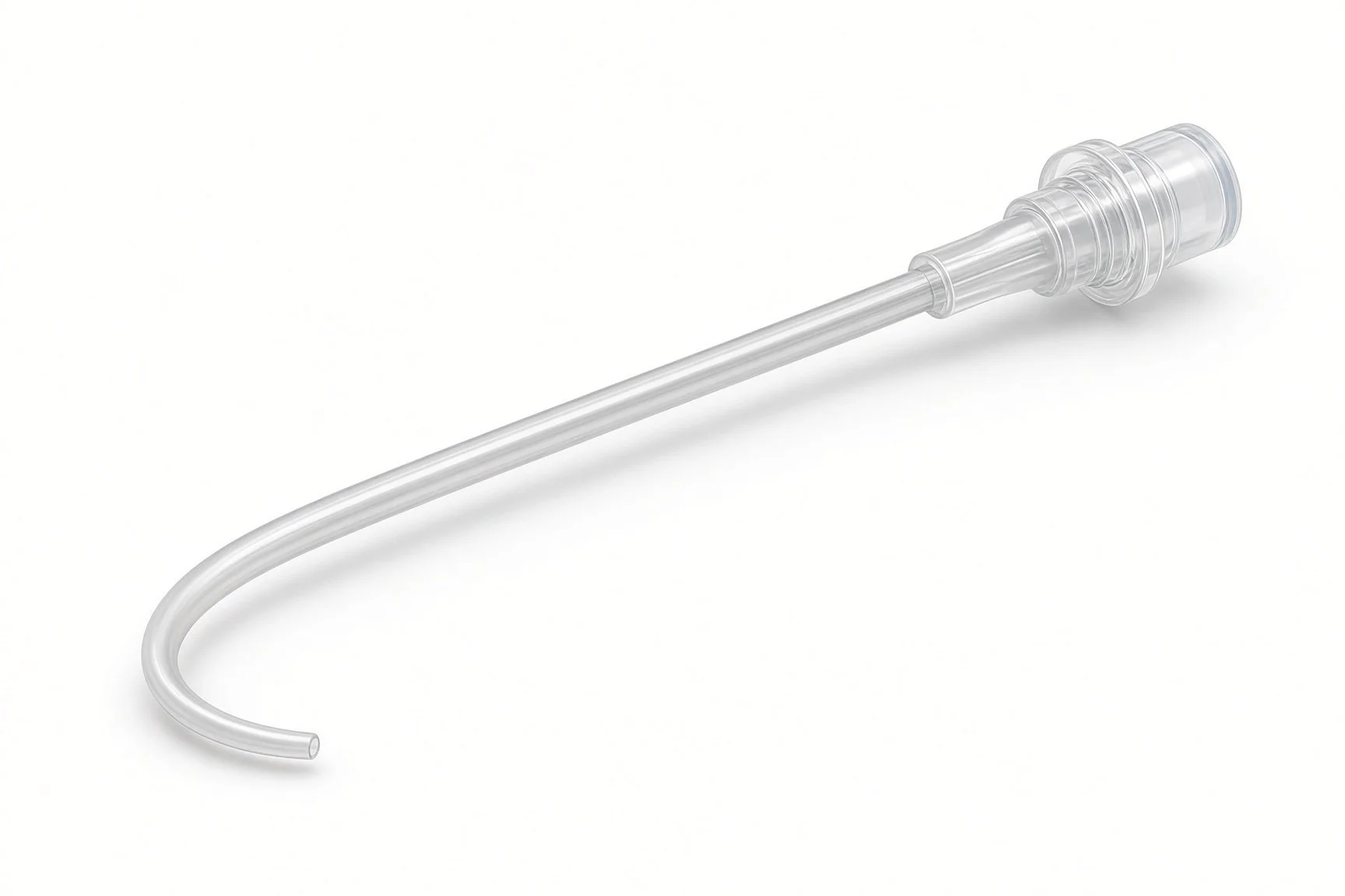
Angiography catheter.
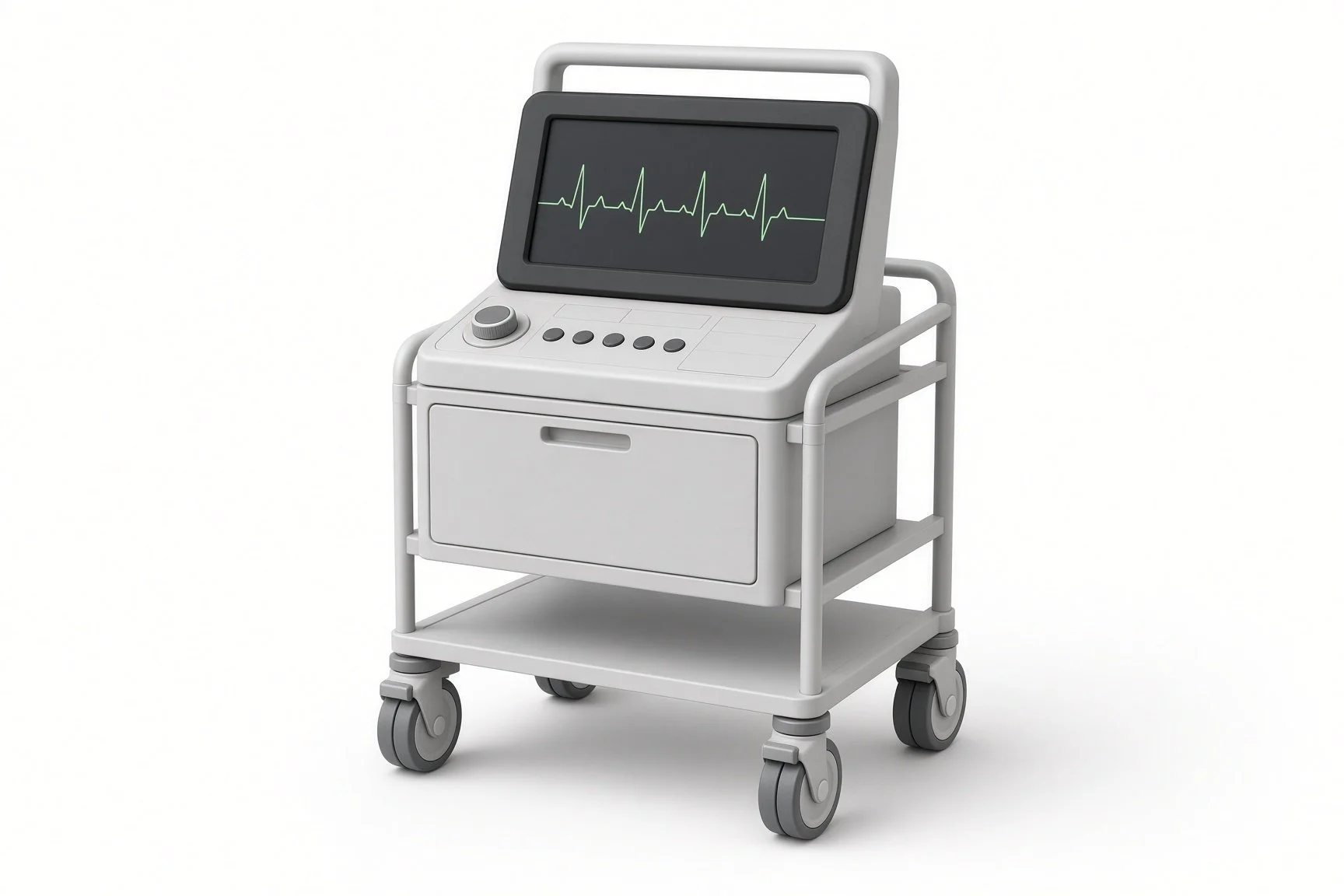
Echo machine.
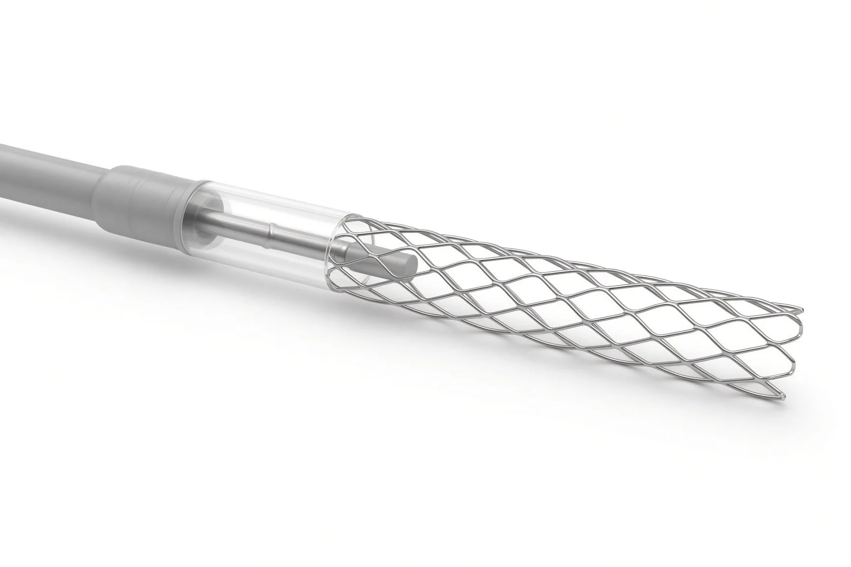
Self-expanding or balloon stents.
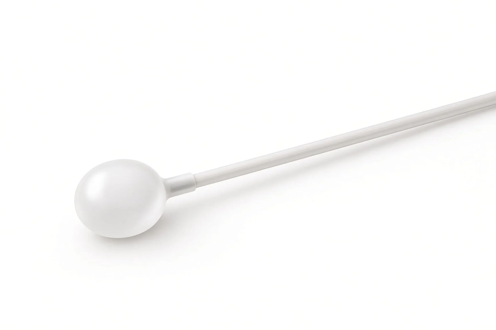
Septostomy balloon.
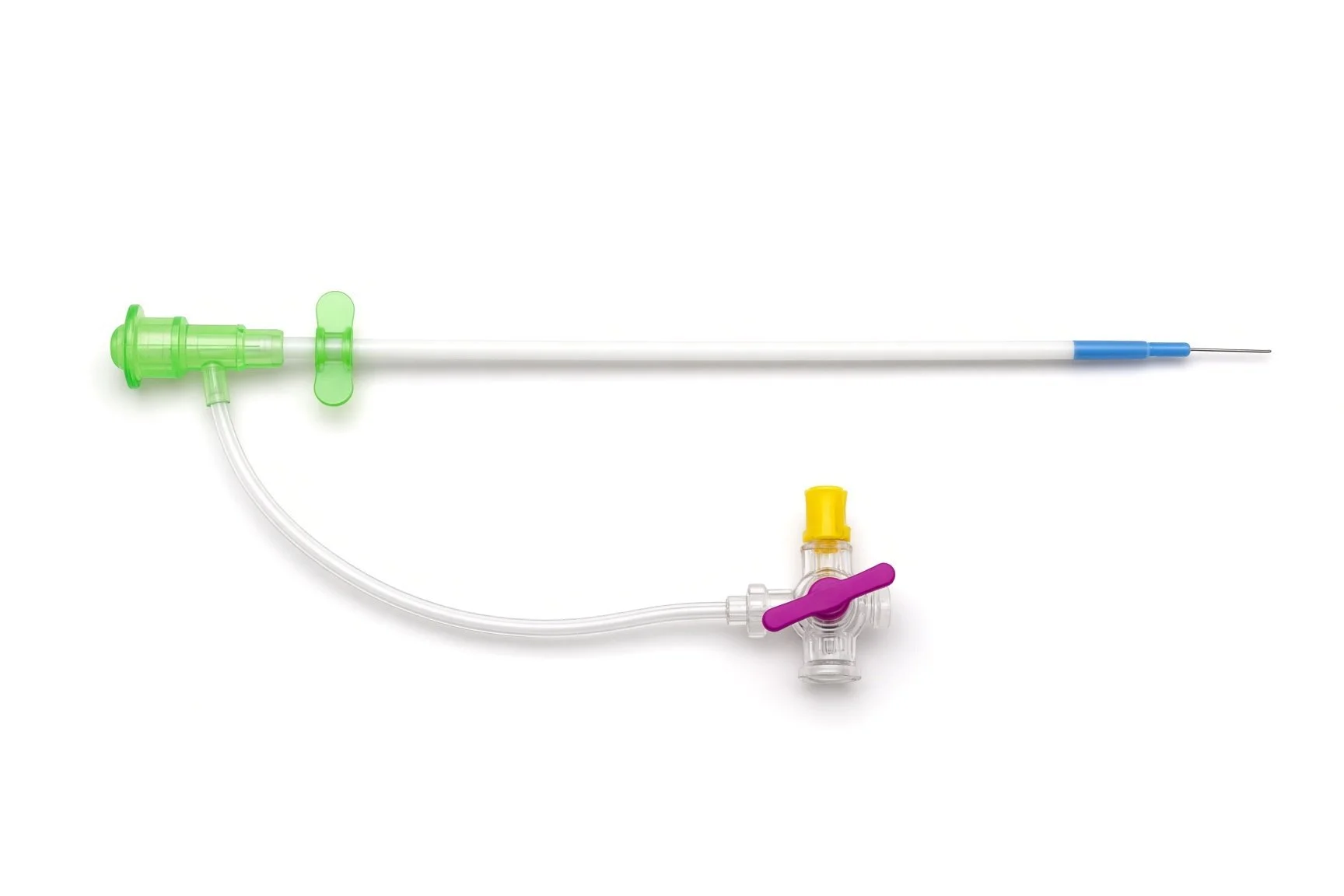
Short sheath for the pulmonary trunk.
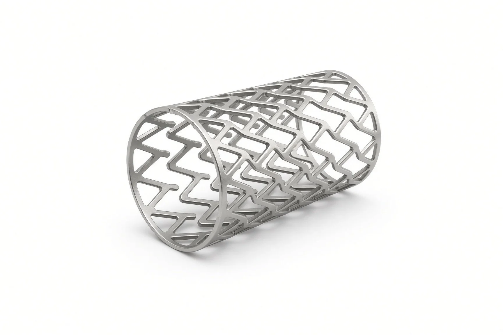
Self-expanding or balloon stents.

Prostaglandin infusion.

Warming mattress.
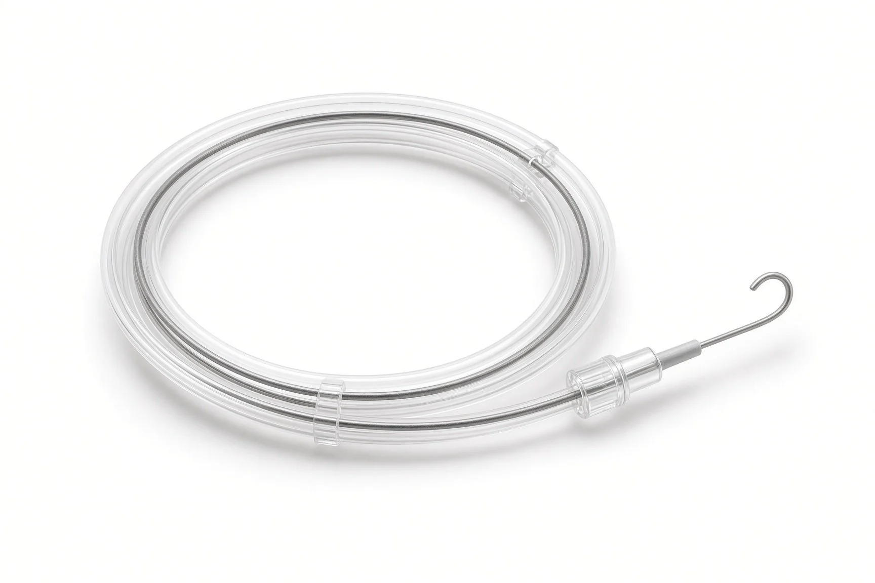
Guidewires.
Procedural steps (Barts example)
The hybrid theatre and team
Bands on the pulmonary arteries
Stenting the duct and atrial septum
Finishing
Interstage and next steps
First time in the cath lab?
Join the brief. Hybrid cases mix two teams. Know who leads each phase and how the catheter kit reaches the sterile field.
Mind the radiation. The surgeons are close to the beam. Remind the room before screening and keep shielding in place.
Watch oxygen levels and blood pressure together. In HLHS a balance between lung and body flow matters more than high oxygen levels.
Remember the family. Parents have often known about this since pregnancy. Make sure they are updated as each stage finishes.
Know which trolley is surgical and which is catheter. Agree in the brief who opens and counts catheter kit on the sterile field.
Have stents of several lengths open-ready. The duct length is measured on the angiogram just before.
FAQ
Why a hybrid rather than a Norwood operation?
The hybrid avoids the heart-lung machine in a newborn. It may suit babies who are small or unwell. The team will explain why they suggest one or the other.
Who will be in the room?
Heart surgeons, cardiologists, anaesthetists and specialist nurses, radiographers and physiologists, working together.
Is this the last operation?
No. HLHS needs staged treatment: stage 2 at a few months and usually a Fontan operation in early childhood.
What should we watch for at home?
The team will teach you, often including home oxygen-level checks, feeding and weight. Call your team early with any worry.
Related Cath Lab 101 guides
Ductal (PDA) stenting in newborns (coming soon)
Sources
All checked on 12 November 2026. Each page's own date is shown where it gives one.
Feltes TF, et al. Indications for cardiac catheterization and intervention in pediatric cardiac disease: a scientific statement from the American Heart Association. Circulation 2011;123(22):2607–2652
StatPearls (NCBI Bookshelf), search: hypoplastic left heart syndrome. ncbi.nlm.nih.gov/books
Disclaimer
Education only. This page is for general education for healthcare students, cath lab staff and curious readers. It isn't medical advice and doesn't replace advice from your own doctor, heart team or local unit protocols. Equipment and medicines are described generically. No product or manufacturer is recommended. If you think someone is having a heart emergency, call 999.
Disclosure: I work for a company that makes a device used in this procedure. This page is independent education, isn't sponsored or reviewed by any company, and doesn't recommend any product.
All diagrams on this page are original illustrations created for rodnieoro.com.