Ruptured sinus of Valsalva aneurysm closure: a Cath Lab 101 guide
Reviewed by Rodnie Oro, cath lab charge nurse · Last reviewed: November 2026 · Education only, not medical advice
Ruptured sinus of Valsalva aneurysm closure in one minute
What it is: The sinuses of Valsalva are three small bulges in the aortic root, just above the aortic valve. A sinus can have a thin, weak wall that balloons out (an aneurysm) and may burst (rupture) into a right heart chamber. Blood then pours from the aorta into the heart. In catheter closure, a folding mesh occluder device is placed across the hole.
Who it is for: Selected adults and older children with a ruptured sinus of Valsalva aneurysm whose anatomy suits a device, as decided by a heart team with surgeons. Surgery remains the standard treatment, especially when there is a VSD or a significant aortic valve leak.
Main risks: A remaining leak, the device moving, a new or worse leak of the aortic valve, pressure on a coronary artery, heart rhythm problems, and problems at the access sites. Your team will explain your own risk.
Usually: Under general anaesthetic with transoesophageal echo (TOE), about 2 to 3 hours, usually home after 1 to 3 days.
On this page: What is ruptured sinus of Valsalva aneurysm closure? · Who is it for? · When is it done? · Step by step · Recovery · Risks · Kit list · First time in the lab? · FAQ
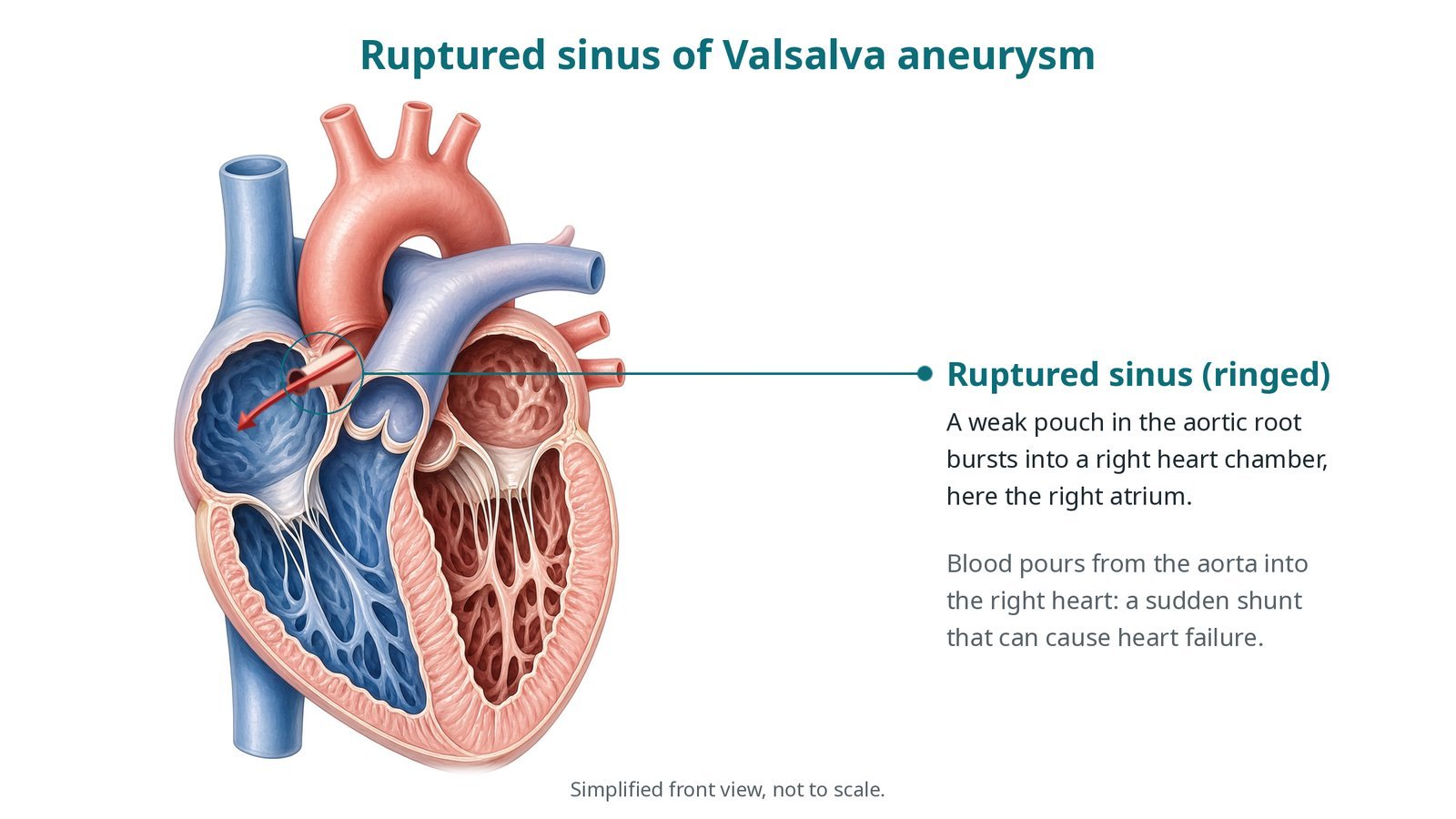
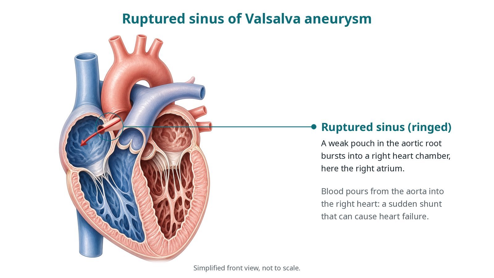
Ruptured sinus of Valsalva aneurysm (ringed): a weak pouch in the aortic root bursts into a right heart chamber, here the right atrium, causing a sudden shunt from the aorta into the right heart.
What is ruptured sinus of Valsalva aneurysm closure?
The aortic root has three sinuses behind the three cusps of the aortic valve: the right coronary sinus and left coronary sinus, where the coronary arteries begin, and the non-coronary sinus.
A sinus of Valsalva aneurysm is a thin-walled pouch, usually present from birth and most often in the right coronary sinus. It may be found by chance. If it bursts, it usually ruptures into a low-pressure right heart chamber: from the right coronary sinus into the right ventricle, or from the non-coronary sinus into the right atrium.
Rupture causes a sudden large shunt from the aorta into the right heart. People may feel sudden chest pain or breathlessness, and a loud continuous murmur appears. Heart failure can follow within days to weeks. A VSD or aortic valve leak may also be present.
Closure seals the hole. With a catheter, a wire is passed from the aorta through the rupture, snared from the vein side to make a loop, and the device is delivered from the vein side. TOE and aortograms check that the device does not touch the aortic valve or block a coronary artery.
New to the acronyms? See the Cath Lab Abbreviations A–Z.
Who is it for?
A heart team with congenital or structural cardiologists and surgeons decides, using echo, TOE and CT. A device may suit when:
the rupture is a single, suitably sized hole
it sits far enough from the aortic valve cusps and the coronary arteries
there is no significant aortic valve leak and no VSD needing surgery
Who it may not suit: People with a VSD, a significant aortic valve leak, several holes, or a hole close to the valve or a coronary artery usually need surgery.
The decision is made by your heart team, who will talk through the reasons, the alternatives and the risks with you.
When is it done?
Often soon after diagnosis, because the heart can fail quickly. Heart failure is treated with medicines first where possible.
gives you a chance to ask questions and give consent
arranges blood tests, including kidney function and a blood count, and an ECG
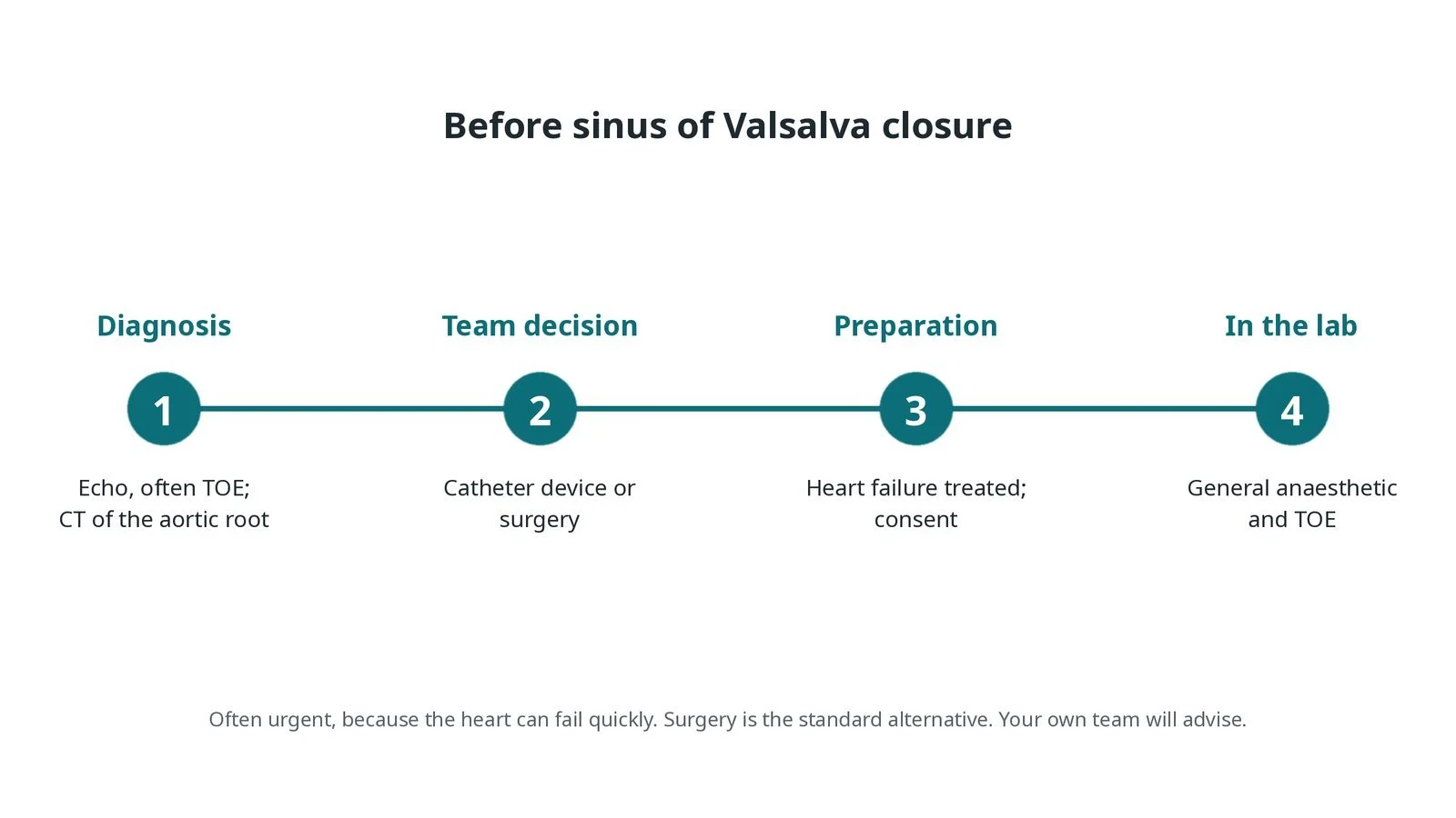
Before closure: echo, often TOE, and CT of the aortic root; team decision between a catheter device and surgery; heart failure treated; general anaesthetic and TOE in the lab. Often urgent.
Step by step
1. Getting ready
A general anaesthetic is given and a TOE probe placed. ECG, invasive blood pressure and oxygen saturation are monitored. Antibiotics are given as per centre protocol.
2. Access and pictures
Sheaths are placed in the femoral artery and femoral vein under ultrasound. Heparin is given, dose as per centre protocol. A pigtail aortogram and TOE show the rupture, its size and its distance from the valve and coronary arteries. Coronary angiograms may be taken.
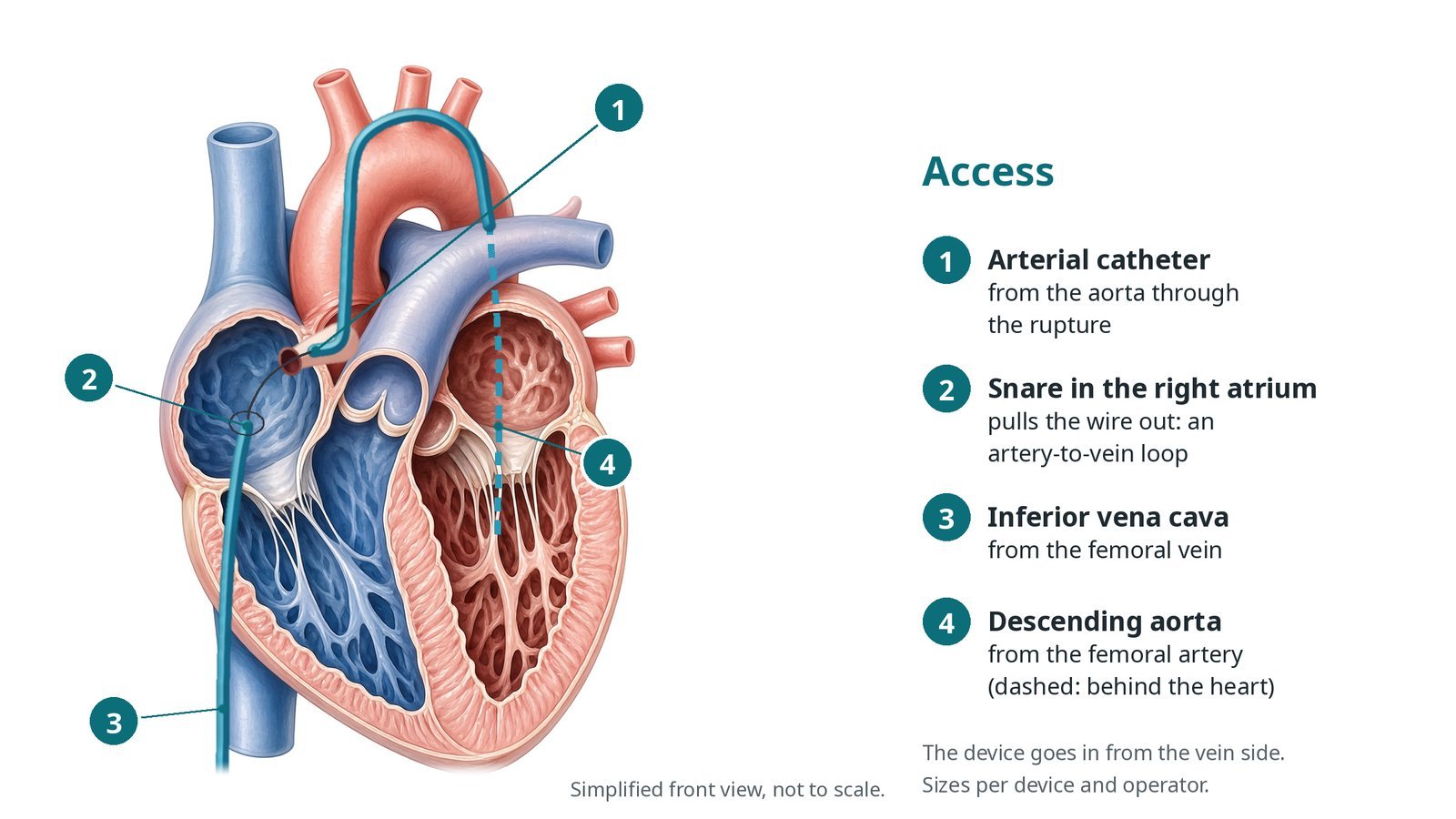
Access: (1) arterial catheter from the aorta through the rupture; (2) snare in the right atrium forms an artery-to-vein loop; (3) inferior vena cava from the femoral vein; (4) descending aorta from the femoral artery.
3. Closing the rupture
A. Cross and snare. A catheter and wire from the aortic side are passed through the rupture into the right heart, and the wire is snared from the vein side and pulled out, making an artery-to-vein loop.
B. Sheath. A long delivery sheath is advanced over the loop from the vein side, through the rupture, into the aortic root.
C. Aortic disc. The occluder's aortic end is opened and pulled back against the hole. TOE and an aortogram check that it does not touch the aortic valve or narrow a coronary artery. Device type and size are per operator.
D. Release. The right heart end is opened, the position is checked again, and the device is released from its cable.
Watch: Tension on the wire loop can pull on the heart: watch for a slow heart rate, low blood pressure or a new valve leak, and tell the operator. The loop is usually protected with a catheter over it.
4. Final checks
TOE checks for any remaining leak, a new aortic valve leak and the device position. An aortogram or coronary angiogram confirms the coronary arteries fill normally. The ECG is checked for new heart block.
Watch: New ST changes or chest pain after release may mean coronary compression: tell the operator straight away.
5. Finishing
Sheaths are removed and the access sites closed with pressure or a closure device, per operator. The patient goes to a monitored bed with ECG monitoring.
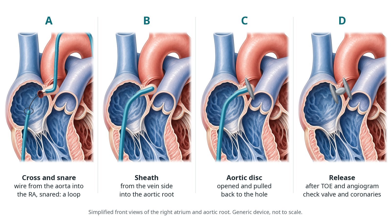
A: wire from the aorta into the right atrium, snared. B: delivery sheath from the vein side into the aortic root. C: aortic disc opened and pulled back. D: release after TOE and an angiogram check the valve and coronaries.
Recovery and aftercare
First hours: bed rest with checks of the groin, ECG and blood pressure, as per centre protocol.
Echo: before going home, to check the device, any leak and the aortic valve.
Going home: usually after 1 to 3 days, once heart failure symptoms are settling.
Medicines: an antiplatelet medicine is usually prescribed for some months, plus any heart failure medicines; take them as advised.
Driving: ask your team; DVLA rules depend on the procedure and your heart condition. Do not drive until the groin has healed.
Follow-up: echo and clinic review over the following months and years. Tell your dentist about the device and look after your teeth.
Call 999 if you have chest pain, sudden breathlessness, fainting, signs of a stroke, or heavy bleeding from the groin.
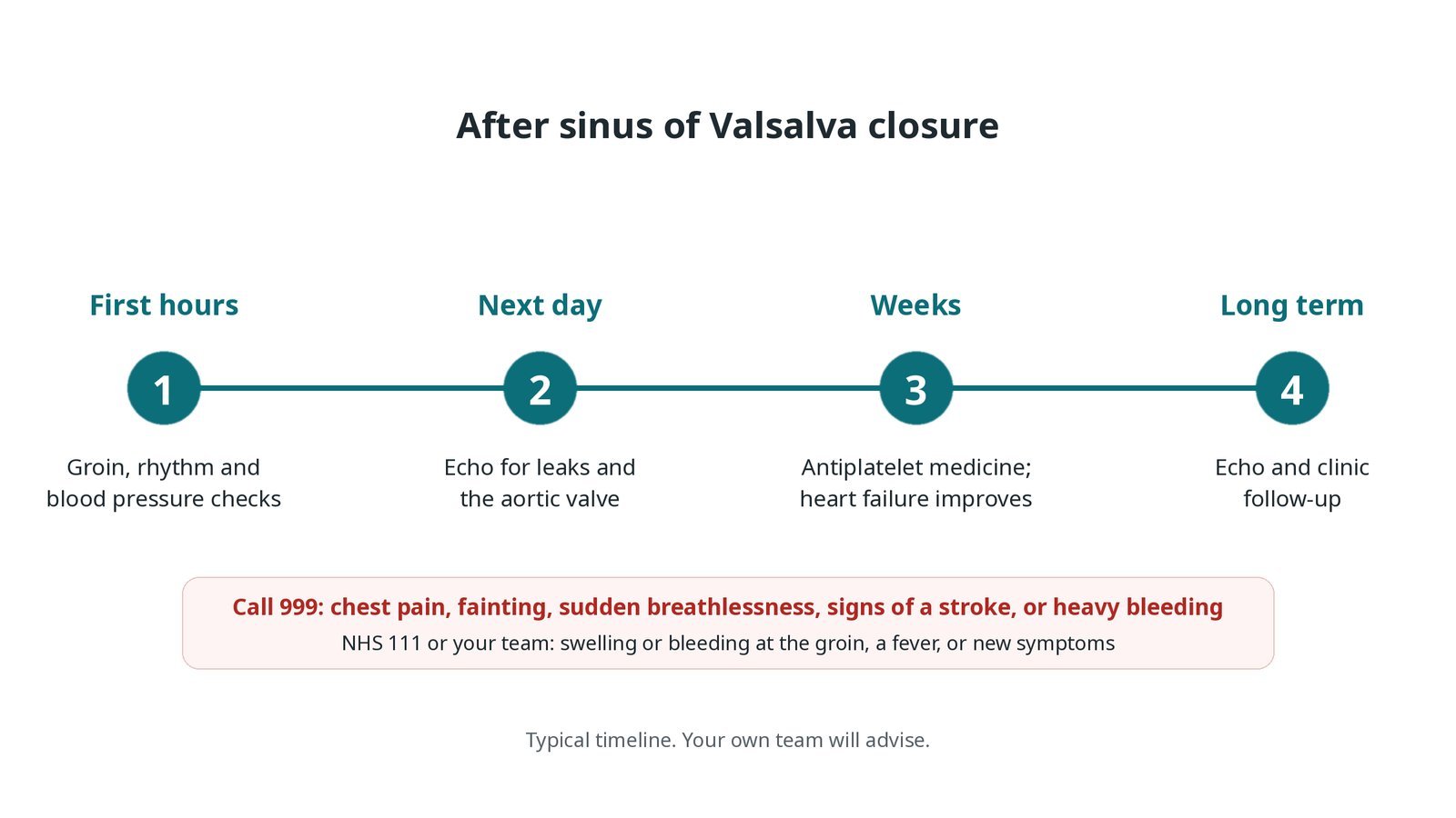
After sinus of Valsalva closure: (1) groin, rhythm and blood pressure checks; (2) echo for leaks and the aortic valve; (3) antiplatelet medicine as heart failure improves; (4) long-term echo and clinic follow-up.
Risks and complications
Your team will explain the risks that apply to you. Possible problems include:
A remaining leak around the device
The device moving, needing retrieval or surgery (uncommon)
A new or worse leak of the aortic valve (uncommon)
Pressure on a coronary artery (rare)
Heart block or other rhythm problems (uncommon)
Bleeding, bruising or vessel damage at the groin
Damage to red blood cells from a fast remaining leak (rare)
Cath lab kit list (printable)
Barts example kit list
An example from one UK centre, written generically. Every lab has its own protocol, and sizes and doses vary.
femoral arterial and venous sheaths, per operator
pigtail catheter for aortograms; coronary catheters
shaped catheters and exchange-length guidewires
snare for the artery-to-vein loop
long delivery sheath, per device
occluder devices in several sizes, per operator
transoesophageal echo (TOE) machine and probe
sizing balloon, if used
heparin and antibiotics as per centre protocol
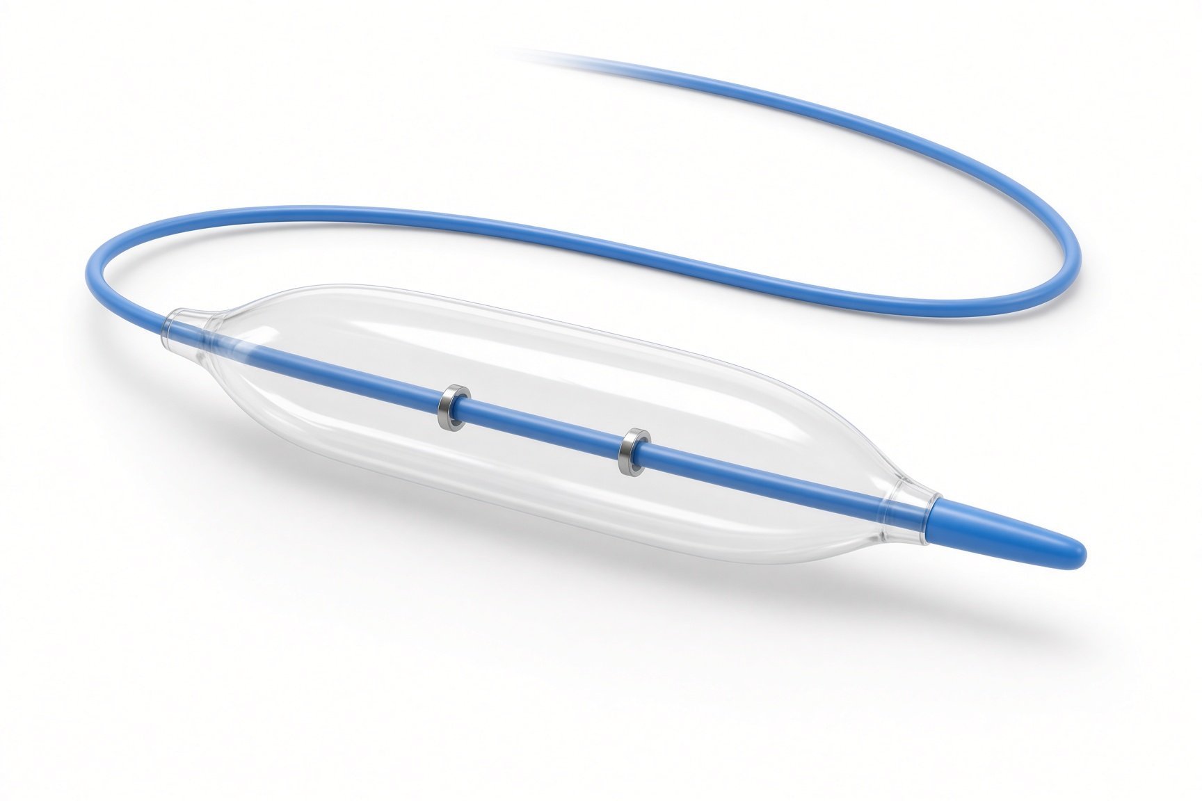
Sizing balloon if used.
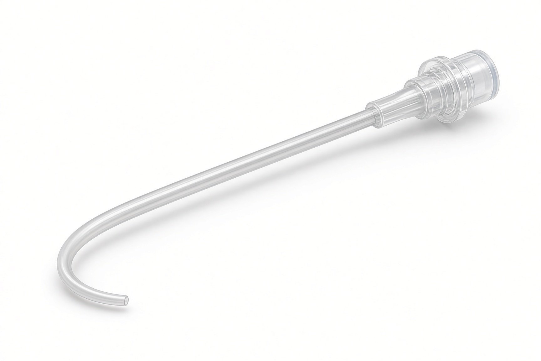
Pigtail catheter for aortograms.
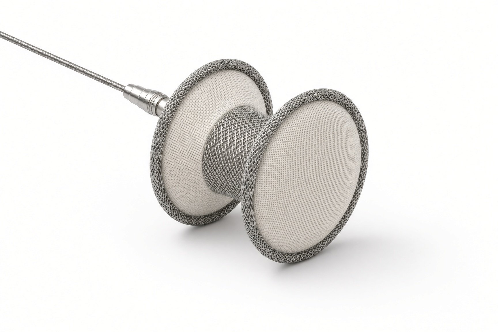
Occluder device.
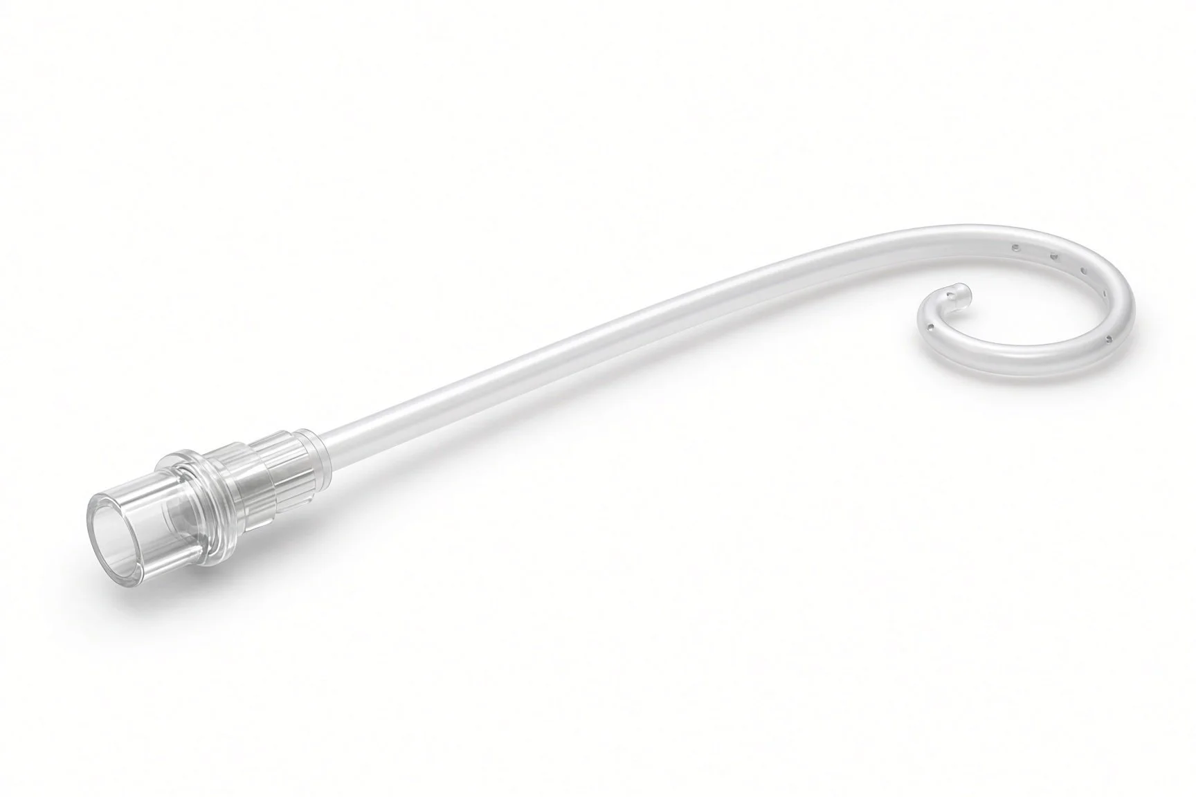
Pigtail for aortograms.
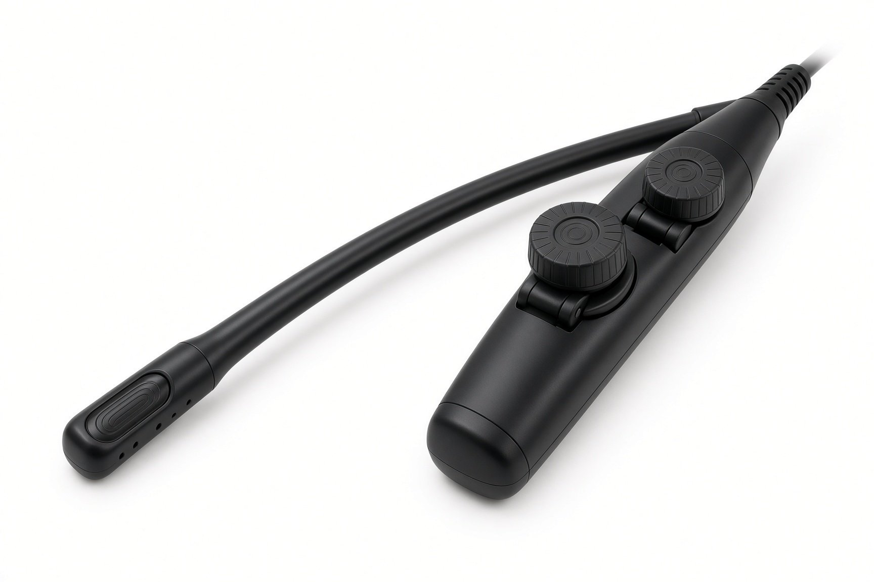
TOE probe.
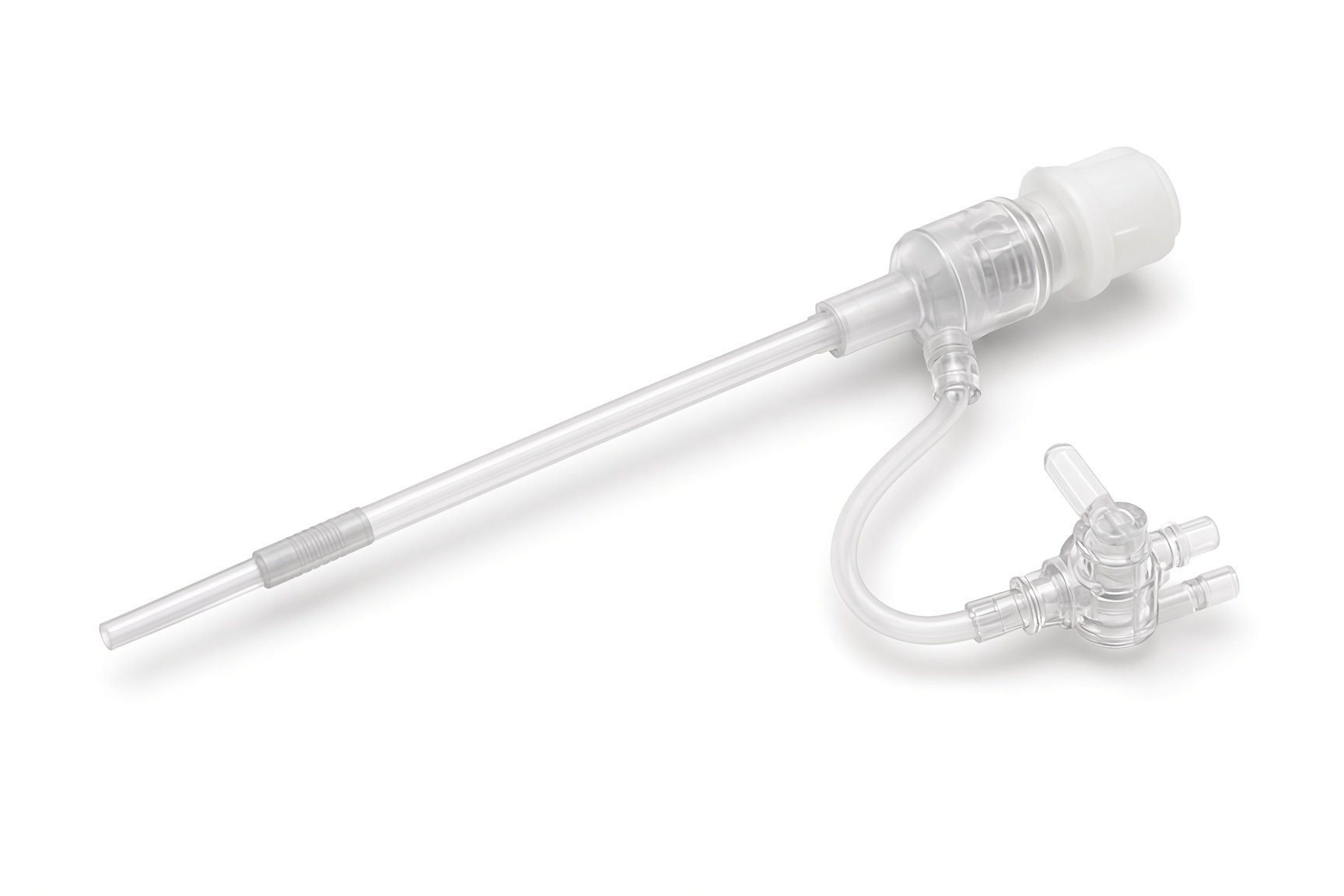
Long delivery sheath.
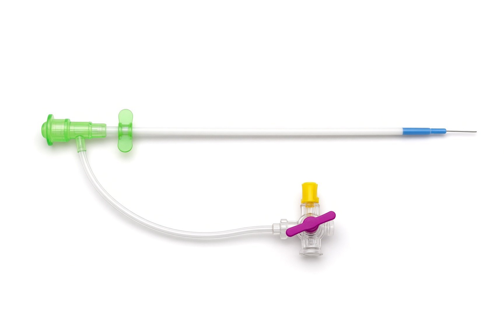
Arterial and venous sheaths.
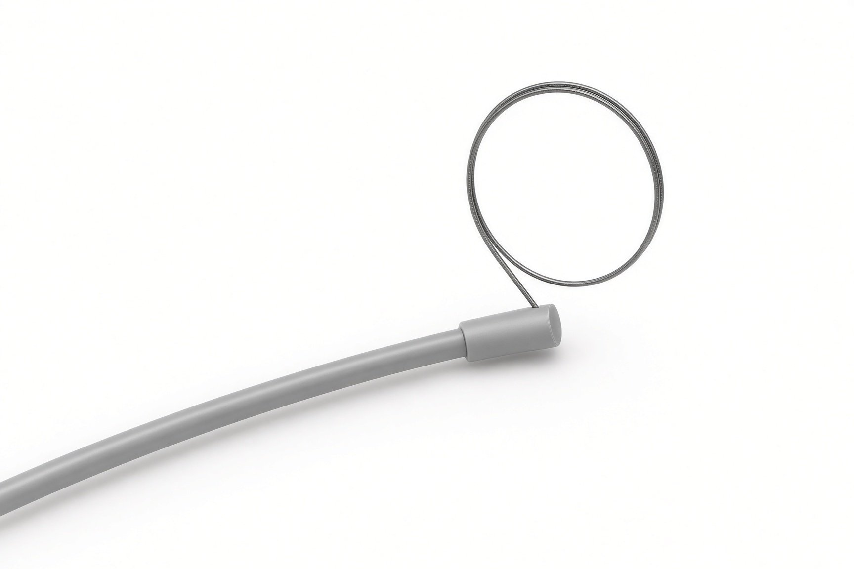
Snare.
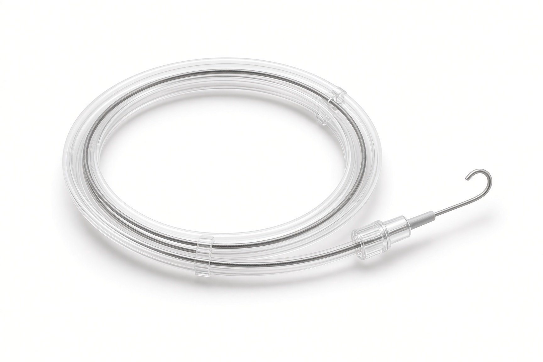
Catheters and guidewires.
Procedural steps (Barts example)
Getting ready
Access and pictures
Closing the rupture
Final checks
Finishing
First time in the cath lab?
Understand the loop. The wire runs from the groin artery, through the heart, and out of the groin vein. Keep both ends visible and still.
Watch the ECG and pressure during loop work. Tension can slow the heart or drop the blood pressure.
TOE drives the decisions. Make sure the echo screen is visible to the operator and the room is quiet at the check steps.
Record the device. Note the device type, size and lot number for the implant record.
Set up the snare and the long delivery sheath early. The loop and delivery steps follow each other quickly.
FAQ
Why did the sinus burst?
Most aneurysms are present from birth with a weak wall. They can burst without warning, sometimes during exertion.
Is a catheter closure as good as surgery?
In selected people with suitable anatomy it works well. Surgery remains the standard, and the team will explain which suits you.
Will my breathlessness get better?
Once the leak is closed, heart failure symptoms usually improve over weeks.
Do I need antibiotics before dental work?
Ask your team. Guidance on antibiotics before dental treatment depends on your device and heart condition; good dental care matters for everyone with a heart device.
Related Cath Lab 101 guides
Sources
All checked on 12 November 2026. Each page's own date is shown where it gives one.
Baumgartner H, et al. 2020 ESC Guidelines for the management of adult congenital heart disease. Eur Heart J 2021;42(6):563–645
Stout KK, et al. 2018 AHA/ACC Guideline for the Management of Adults With Congenital Heart Disease. J Am Coll Cardiol 2019;73(12):e81–e192
StatPearls (NCBI Bookshelf), search: sinus of Valsalva aneurysm. ncbi.nlm.nih.gov/books
Disclaimer
Education only. This page is for general education for healthcare students, cath lab staff and curious readers. It isn't medical advice and doesn't replace advice from your own doctor, heart team or local unit protocols. Equipment and medicines are described generically. No product or manufacturer is recommended. If you think someone is having a heart emergency, call 999.
Disclosure: I work for a company that makes a device used in this procedure. This page is independent education, isn't sponsored or reviewed by any company, and doesn't recommend any product.
All diagrams on this page are original illustrations created for rodnieoro.com.