Collateral embolisation (MAPCAs and venovenous collaterals): a Cath Lab 101 guide
Reviewed by Rodnie Oro, cath lab charge nurse · Last reviewed: November 2026 · Education only, not medical advice
Collateral embolisation in one minute
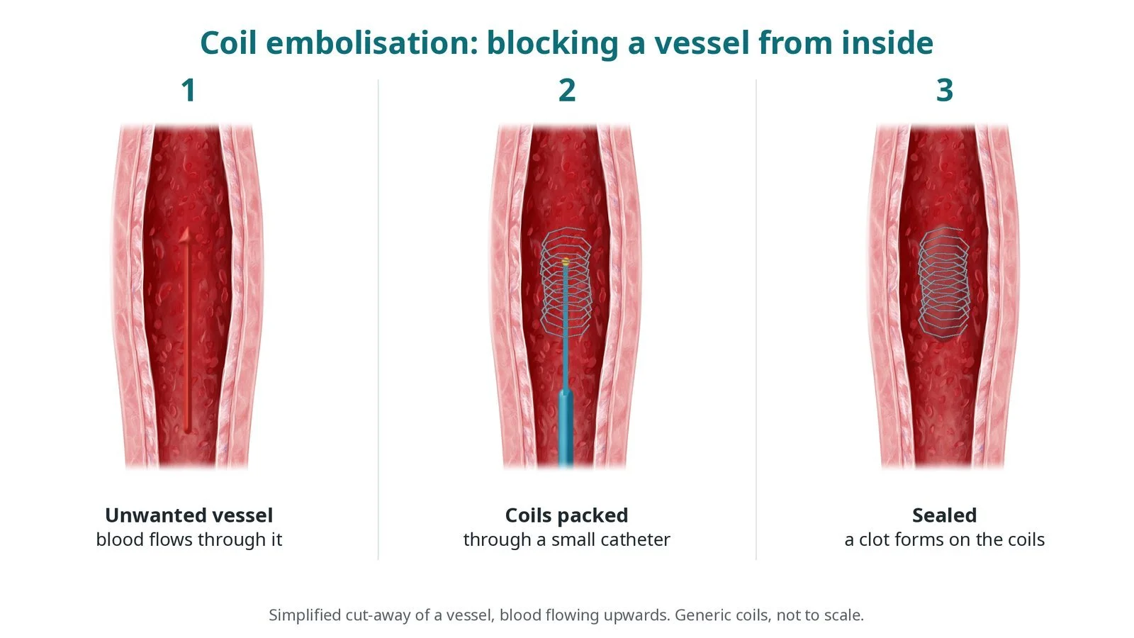
What it is: Collaterals are extra blood vessels. Aortopulmonary collaterals (including MAPCAs) carry blood from the aorta to the lungs; venovenous collaterals carry blue blood from the veins back to the heart, bypassing the lungs. Embolisation blocks selected collaterals with coils or vascular plugs passed through a catheter.
Who it is for: Children and adults with congenital heart disease, for example tetralogy with pulmonary atresia, or after a Glenn or Fontan operation, when collaterals are overloading the heart or lowering oxygen levels, as decided by the congenital heart team.
Main risks: A coil or plug lodging in the wrong place (non-target embolisation), a fall in oxygen levels if a needed vessel is closed, damage to the vessel, and problems at the access site. Your team will explain your own risk.
Usually: Under general anaesthetic for children, sedation or general anaesthetic for adults, about 1 to 3 hours, often home the same or next day. It is often part of a planned catheter study.
On this page: What is collateral embolisation? · Who is it for? · When is it done? · Step by step · Recovery · Risks · Kit list · First time in the lab? · FAQ
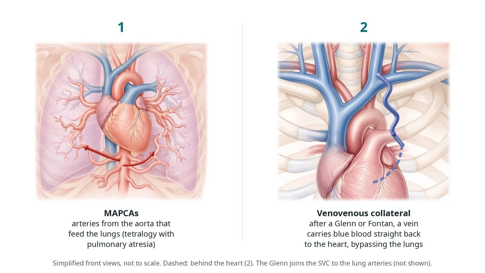
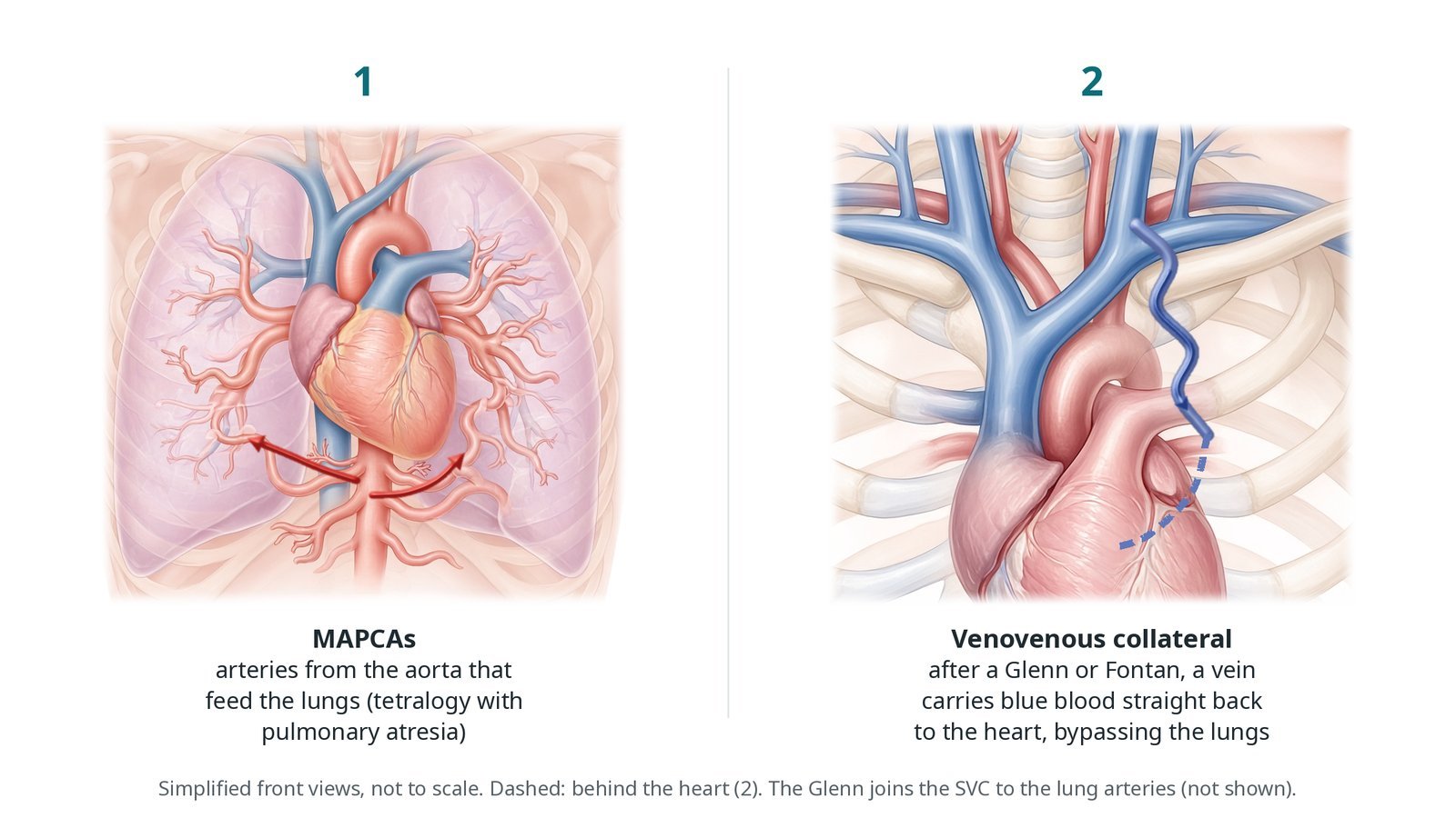
(1) MAPCAs: arteries from the aorta that feed the lungs in tetralogy with pulmonary atresia. (2) Venovenous collateral: after a Glenn or Fontan, a vein carries blue blood back to the heart, bypassing the lungs.
What is collateral embolisation?
MAPCAs (major aortopulmonary collateral arteries) are found in tetralogy of Fallot with pulmonary atresia. The normal pulmonary arteries are small or absent, and these arteries from the descending aorta supply the lungs instead. Some lung segments are fed only by a MAPCA, so it must be kept; the surgeon later joins these vessels to a new pulmonary artery (unifocalisation). Others duplicate a supply that the true pulmonary arteries already provide, and these can be closed.
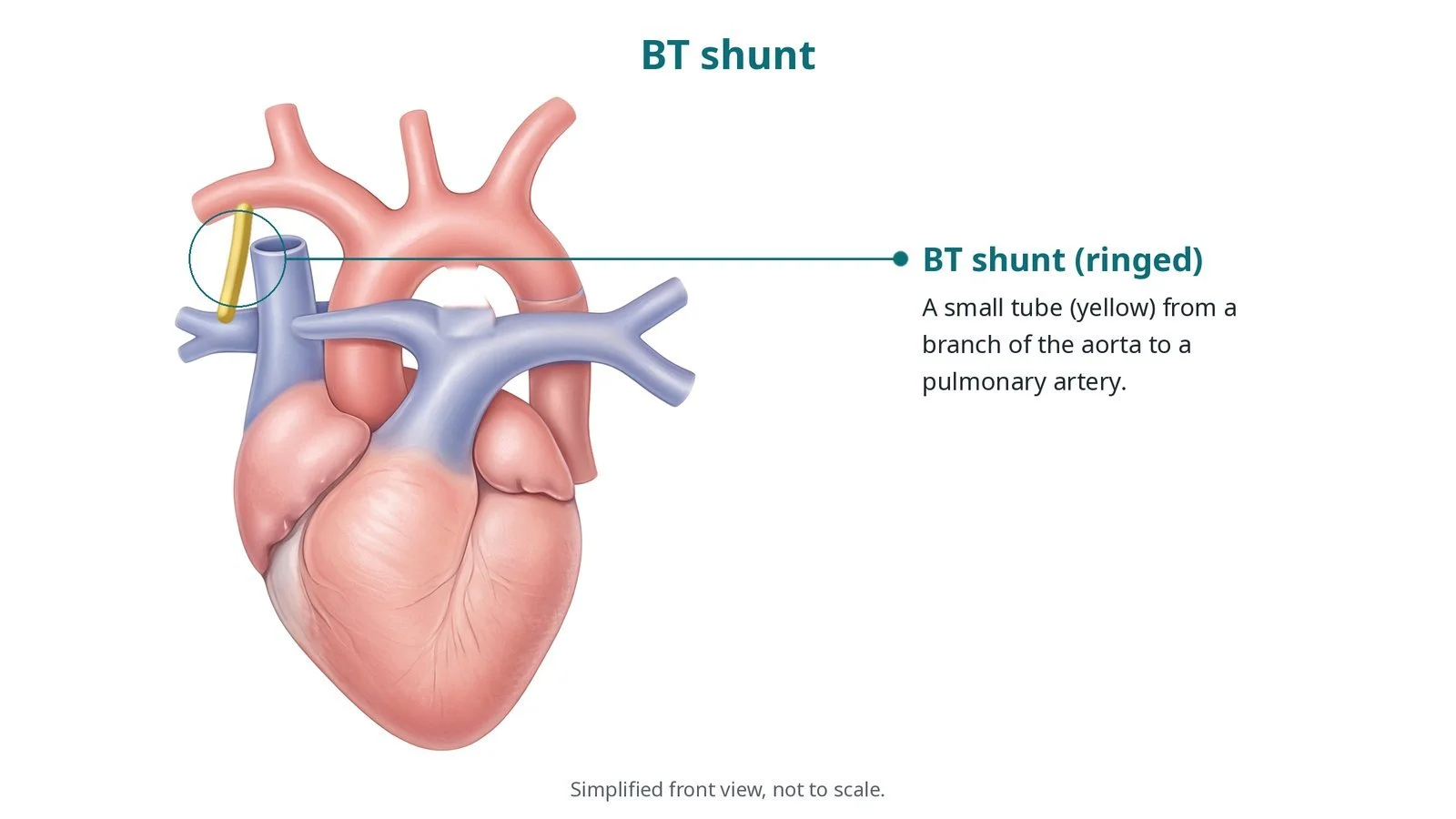
Smaller aortopulmonary collaterals also grow in other blue (cyanotic) conditions and after shunts. They send extra blood to the lungs, add work for the heart and compete with the planned blood supply after surgery.
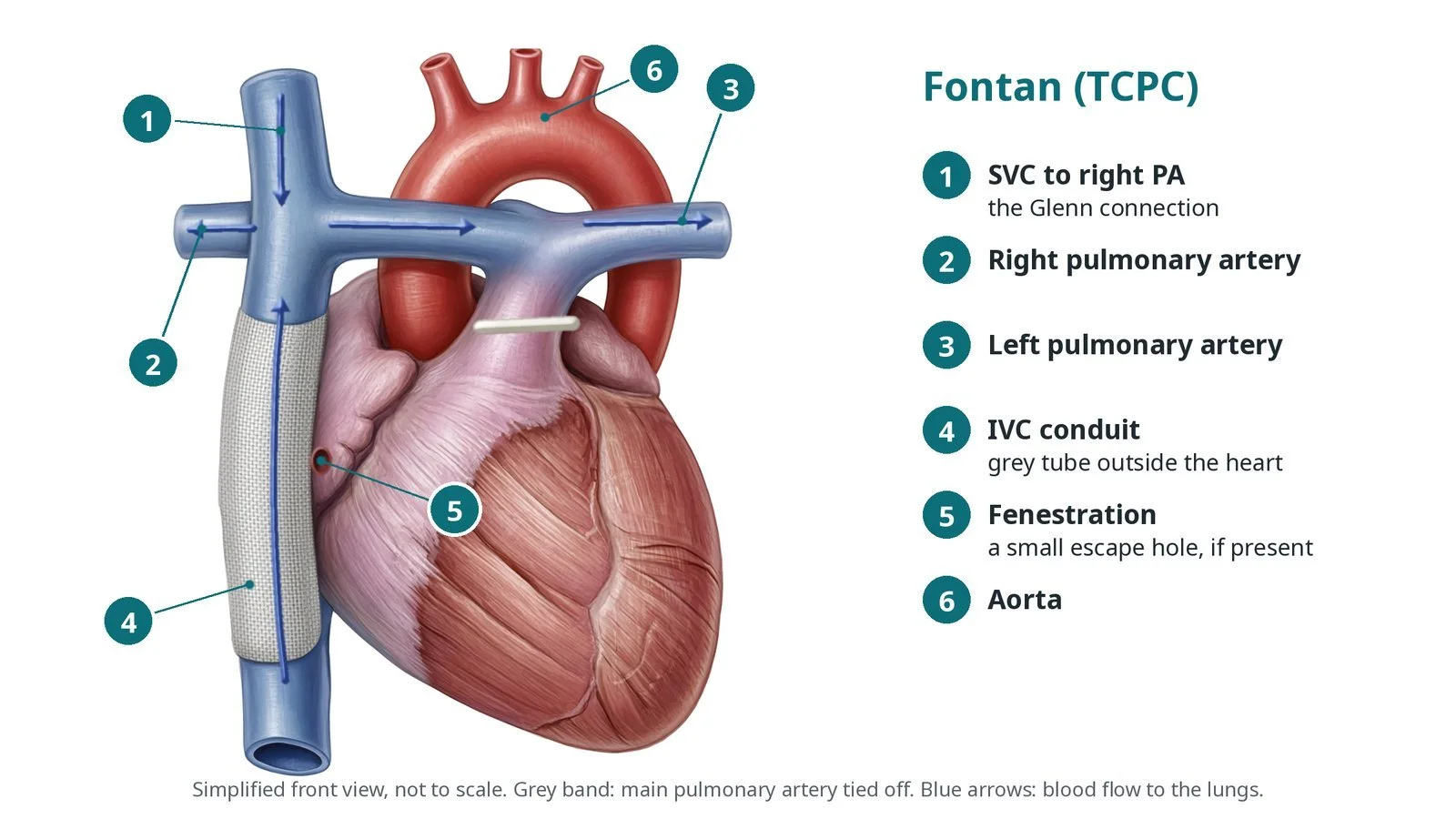
After a Glenn or Fontan operation, blood from the body flows to the lungs without a pumping chamber, so pressure in the veins is higher than normal. Venovenous collaterals can open from these veins to the low-pressure pulmonary veins or atrium. Blue blood then bypasses the lungs, and oxygen levels fall.
Coils and plugs block a collateral from inside. The key safety point is to place them exactly in the target vessel, so nothing drifts into the aorta, the lungs or the brain's circulation.
New to the acronyms? See the Cath Lab Abbreviations A–Z.
Who is it for?
The congenital heart team decides, using oxygen levels, echo, CT or MRI and angiograms. Typical reasons:
aortopulmonary collaterals that duplicate lung supply and overload the heart, often before or after surgery
venovenous collaterals causing low oxygen levels after a Glenn or Fontan
less often, collaterals causing bleeding, such as coughing up blood
Who it may not suit: A MAPCA that is the only blood supply to part of a lung is not closed. In some Fontan patients a venovenous collateral acts as a pressure release; closing it may raise venous pressure, so the team weighs this carefully.
The decision is made by your heart team, who will talk through the reasons, the alternatives and the risks with you.
When is it done?
Often during a planned catheter study, for example before Fontan completion, or when oxygen levels have fallen. Test occlusion with pressure and oxygen measurements may guide the decision during the case.
gives you a chance to ask questions and give consent
arranges blood tests, including kidney function and a blood count, and an ECG
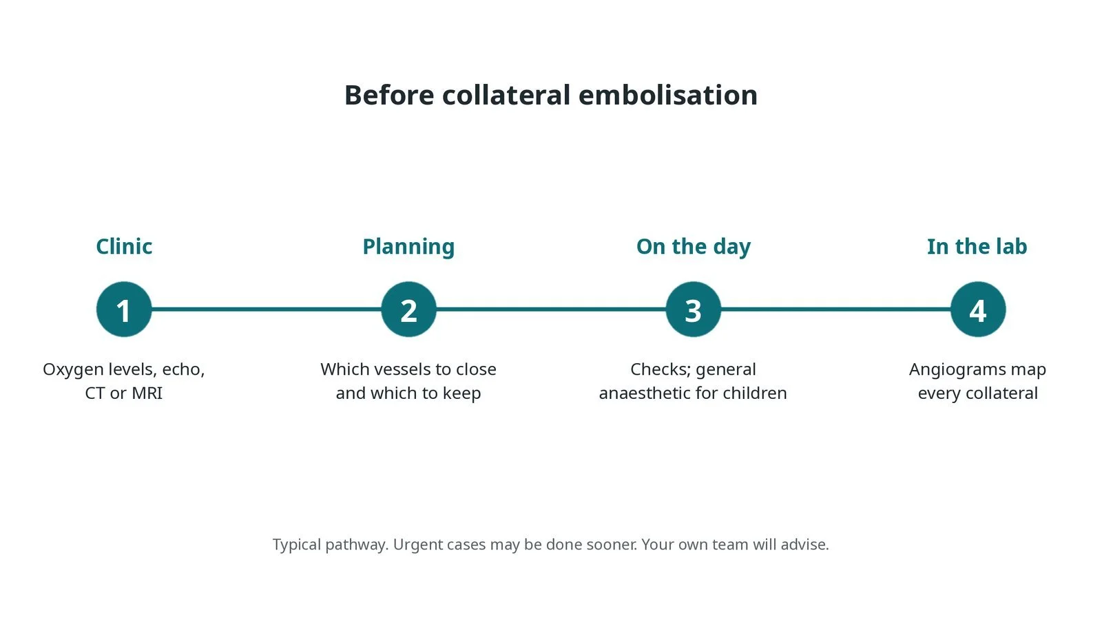
Before collateral embolisation: oxygen levels, echo and CT or MRI; planning which vessels to close and which to keep; checks on the day; angiograms map every collateral.
Step by step
1. Getting ready
Children have a general anaesthetic; adults have sedation or a general anaesthetic. Oxygen saturation is monitored throughout and recorded at the start. In children all drugs and contrast are weight-based, as per centre protocol, and babies are kept warm.
2. Access
A femoral artery sheath is used for MAPCAs and other arterial collaterals; a femoral vein sheath for venous ones. After a Glenn, the neck or arm veins may give a better route to the upper body veins. Heparin is given, dose as per centre protocol.
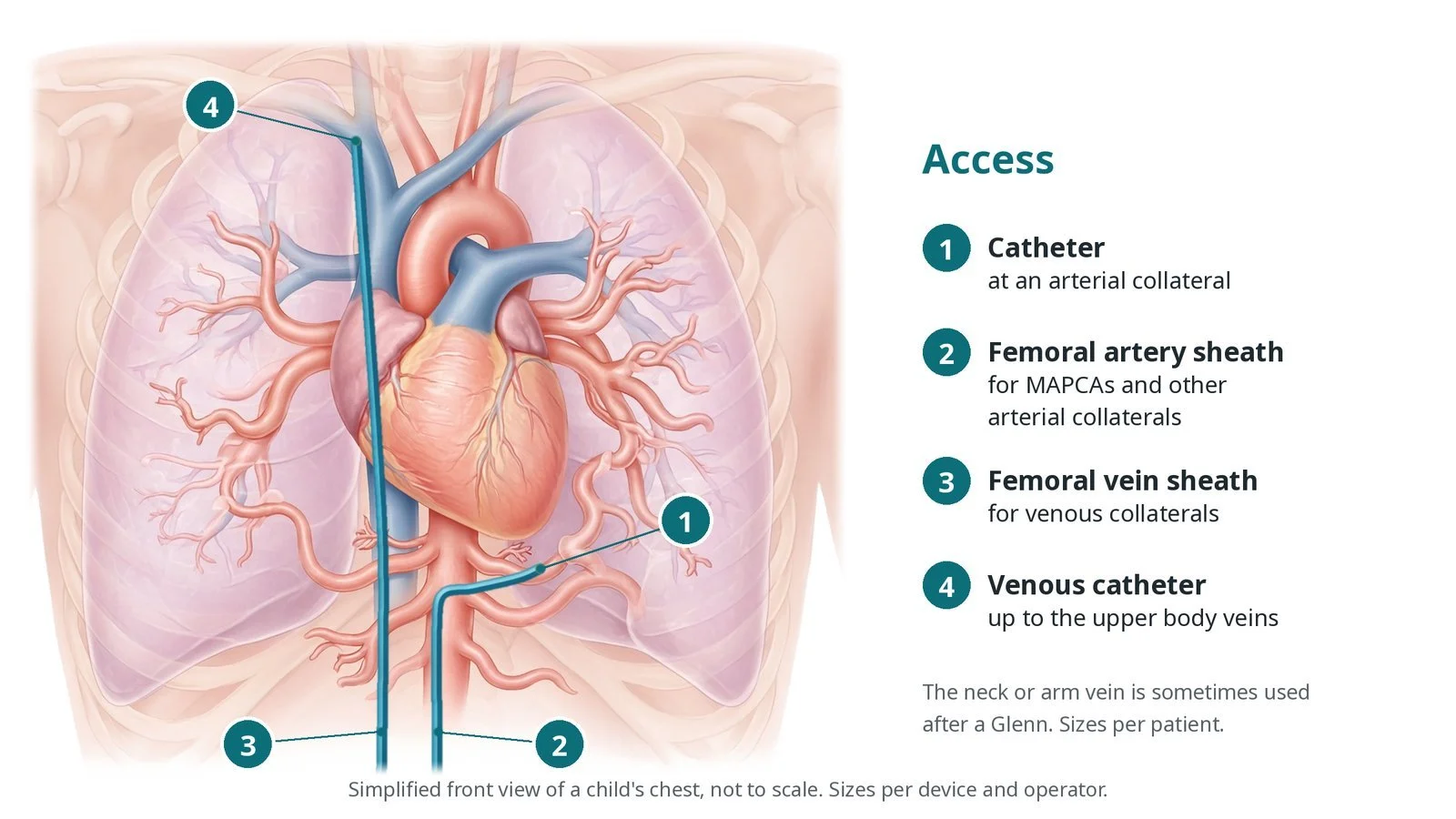
Access: (1) catheter at an arterial collateral; (2) femoral artery sheath; (3) femoral vein sheath for venous collaterals; (4) venous catheter up to the upper body veins.
3. Blocking the collateral
A. Map. An aortogram or venogram, then selective angiograms, show each collateral and what it supplies. For MAPCAs the team checks whether each lung segment has another supply.
B. Microcatheter. A catheter and microcatheter are placed deep in the target vessel, away from its origin, so the device sits securely.
C. Coils or plug. Coils are packed, or a vascular plug opened, until flow stops. Device type and size are per operator.
D. Check. A repeat angiogram confirms the vessel is closed and nothing has moved. Oxygen levels and, after a Fontan, venous pressures are rechecked.
Watch: A coil or plug moving out of the target vessel risks blocking a vessel elsewhere. Tell the operator at once and check leg pulses and, for venous collaterals, any neurological change.
4. Oxygen levels and pressures
Closing a venovenous collateral usually raises oxygen levels. Closing an aortopulmonary collateral may lower them a little, as expected, but a large fall can mean a needed lung supply was closed. After a Fontan, venous pressure is watched because it can rise.
Watch: A large, unexpected fall in oxygen levels after closure: tell the operator straight away.
5. Finishing
The sheaths are removed and pressure applied. In children the leg's colour, warmth and pulse are checked on both sides. The number and sizes of devices are recorded.
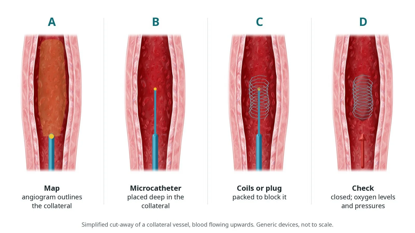
A: angiogram outlines the collateral. B: microcatheter placed deep in it. C: coils or a plug block it. D: closed; oxygen levels and pressures checked.
Recovery and aftercare
First hours: bed rest with checks of the access site, leg pulses, oxygen levels and heart rhythm, as per centre protocol.
Going home: often the same or next day.
Medicines: continue your usual heart medicines unless told otherwise; any changes will be explained.
Activity: avoid heavy lifting and rough play for a few days while the groin heals, or as advised.
Driving (adults): ask your team; do not drive until the groin has healed and any sedation has worn off.
Follow-up: oxygen levels and clinic review with the congenital heart team; some patients need more sessions or surgery.
Call 999 if there is sudden breathlessness, chest pain, fainting, signs of a stroke, heavy bleeding from the groin, or a child who is more blue than usual or floppy.
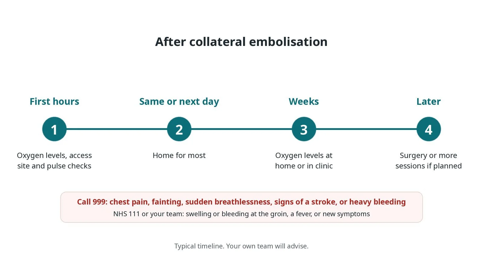
After collateral embolisation: (1) oxygen levels, access site and pulse checks; (2) home the same or next day for most; (3) oxygen levels over the following weeks; (4) surgery or more sessions if planned.
Risks and complications
Your team will explain the risks that apply to you. Possible problems include:
A coil or plug moving to the wrong place (uncommon)
A fall in oxygen levels if a needed vessel is closed (uncommon)
Raised venous pressure after closing a collateral in a Fontan patient
Damage to or rupture of the vessel (rare)
Bleeding or a blocked vessel at the access site; in babies, the leg pulse (uncommon)
Collaterals growing again, needing a further procedure
Cath lab kit list (printable)
Barts example kit list
An example from one UK centre, written generically. Every lab has its own protocol, and sizes and doses vary.
arterial and venous sheaths, per patient and operator
ultrasound for vascular access
shaped catheters and long sheaths, per operator
microcatheters and microwires
embolisation coils, pushable and detachable, in a range of sizes
vascular plugs, per operator
balloon catheter for test occlusion
snare for retrieval
contrast; heparin and other drugs, weight-based, as per centre protocol
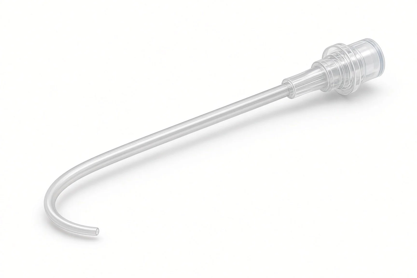
Shaped catheters.
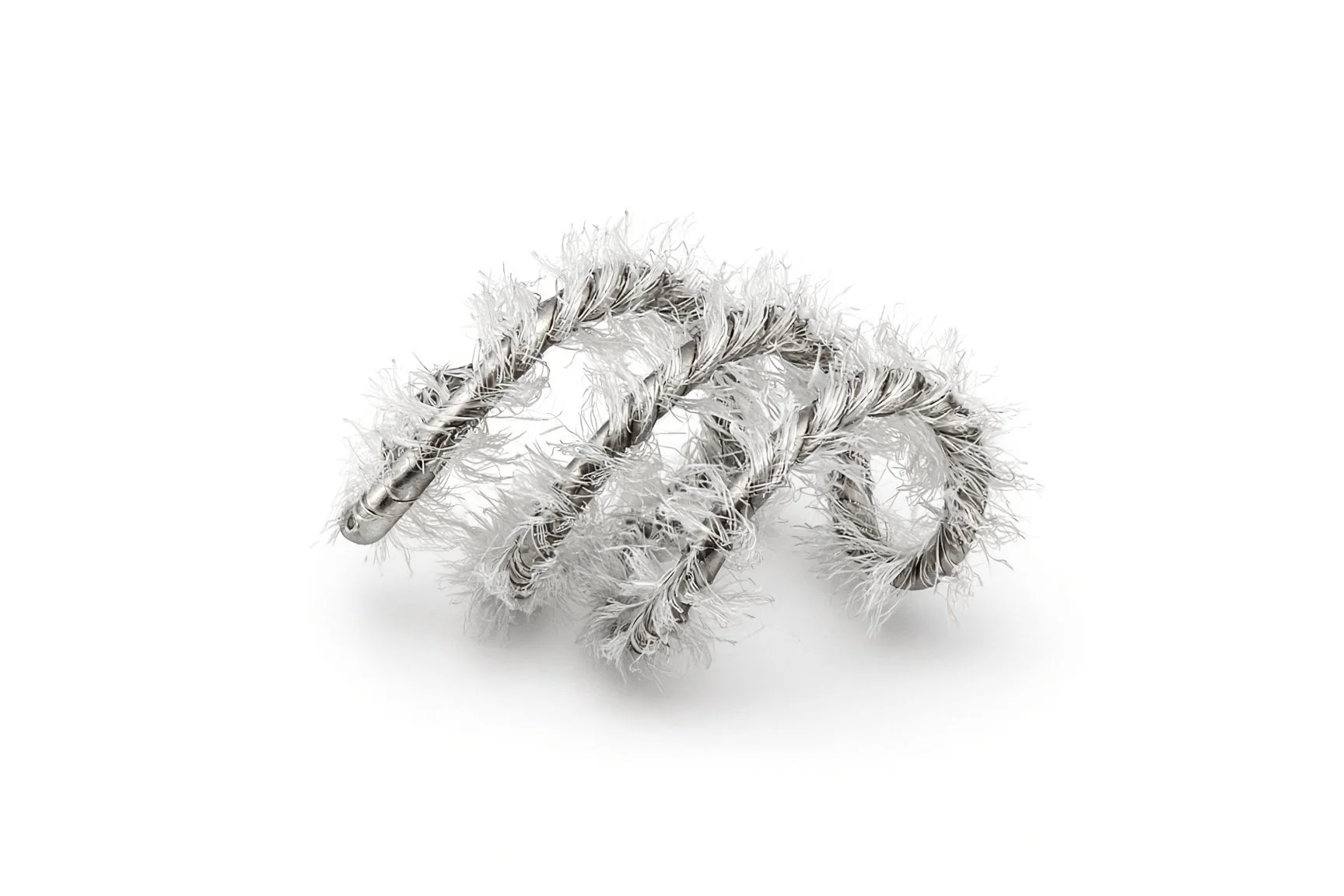
Embolisation coils.
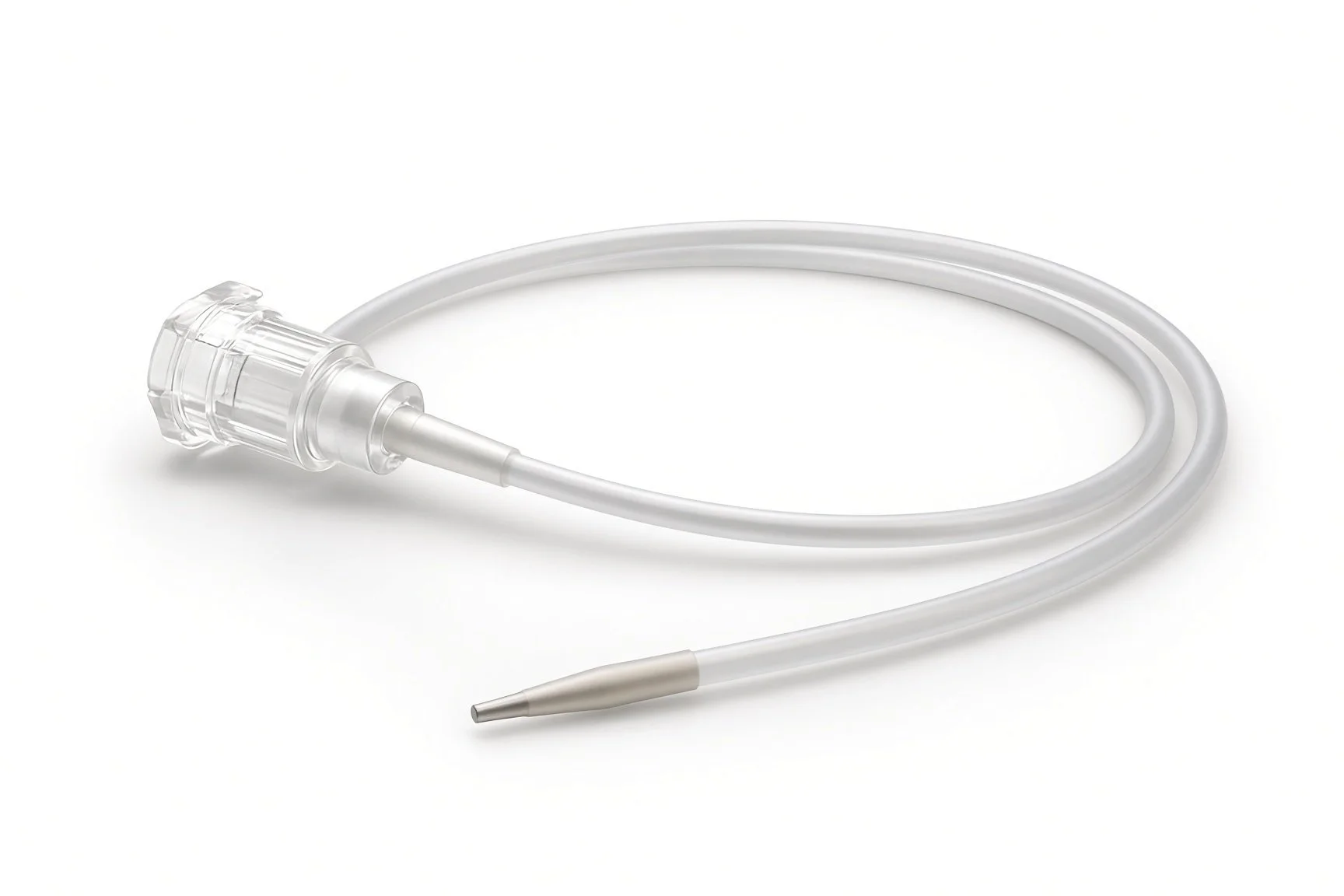
Microcatheter.
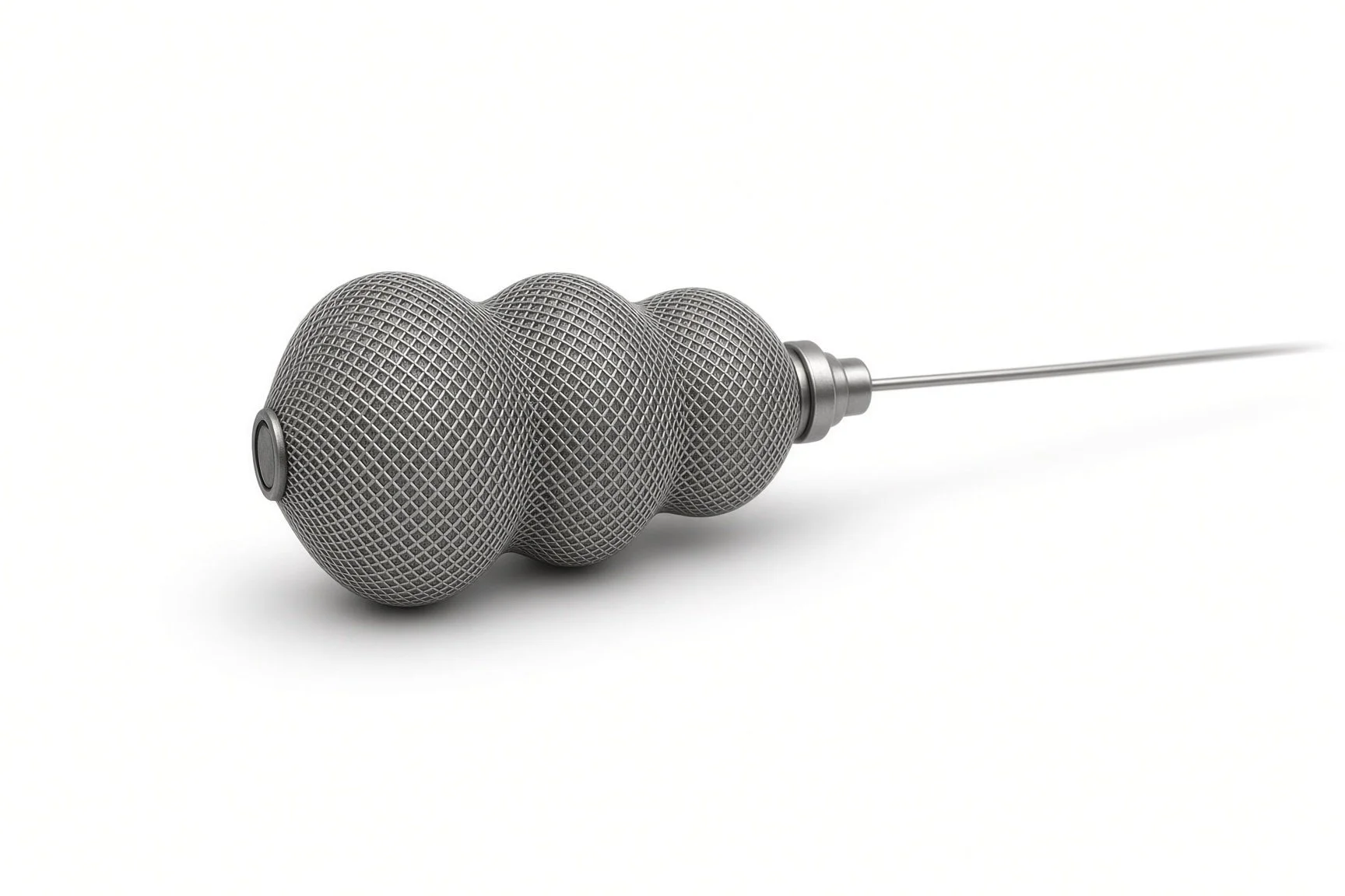
Vascular plugs.
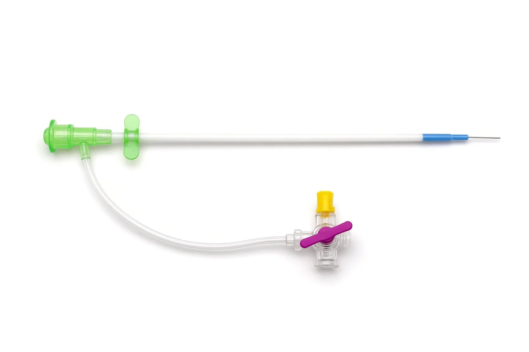
Arterial and venous sheaths.
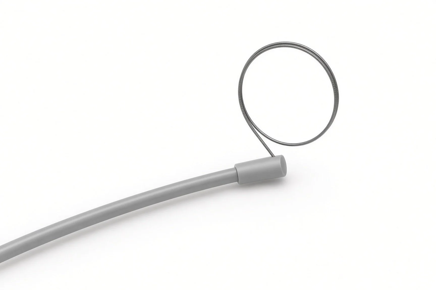
Snare for retrieval.
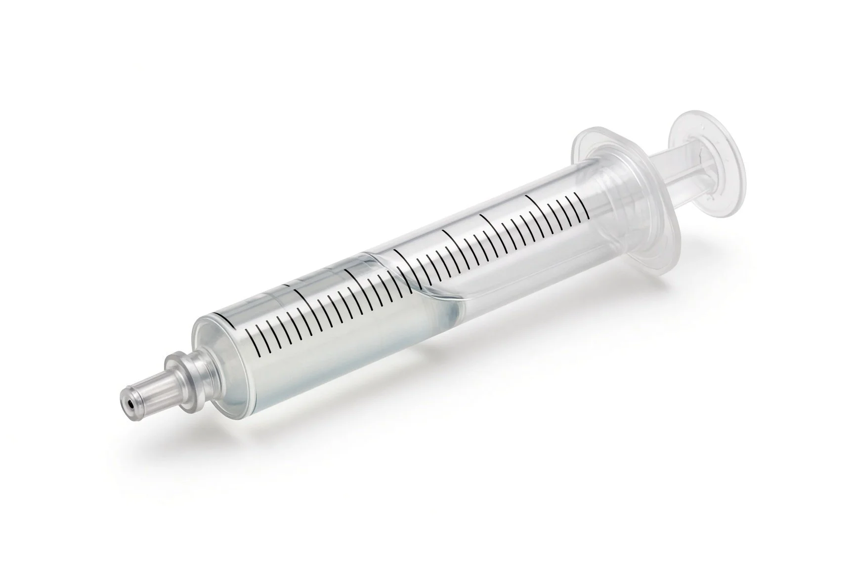
Contrast syringe.
Contrast syringe.
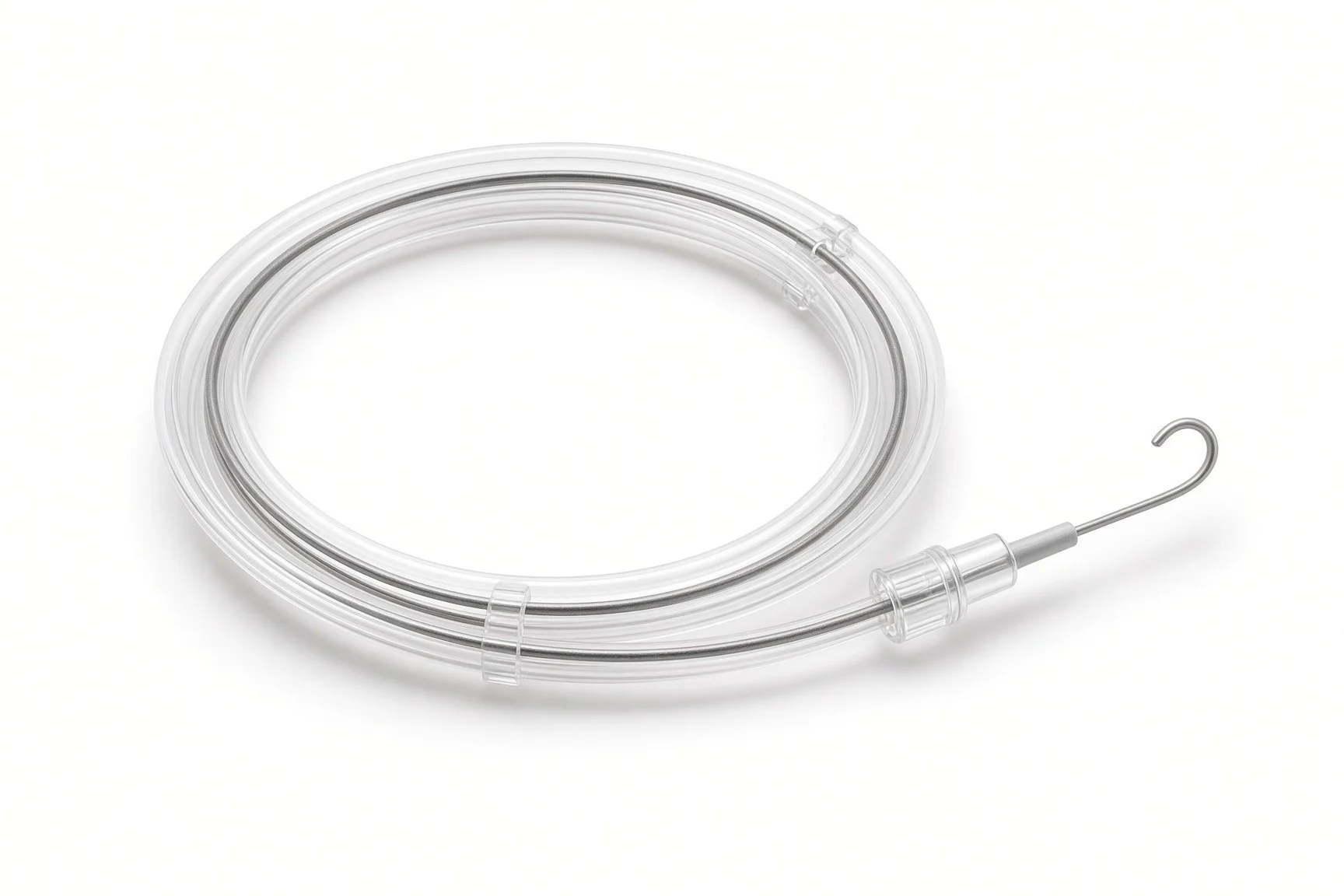
Guidewires.
Procedural steps (Barts example)
Getting ready
Access
Blocking the collateral
Oxygen levels and pressures
Finishing
First time in the cath lab?
Learn the circulation first. Ask whether this patient has a Glenn, a Fontan or MAPCAs; it changes what a good result looks like.
Track every device. Record each coil and plug size and lot number as it goes in.
Watch the oxygen saturation. It is the quickest sign that closing a vessel has helped or harmed.
Keep the snare handy. Non-target embolisation is uncommon, but retrieval is easier when the kit is ready.
Record the starting oxygen saturation and the inspired oxygen. The change after closure is part of the result.
Keep a snare open on the trolley. If a coil drifts, it is easier to retrieve early.
FAQ
Are all the collaterals closed?
No. Only vessels that are not needed, or that are causing harm, are closed. Some MAPCAs are the only supply to part of a lung and are kept.
Will my oxygen levels go up?
Closing venovenous collaterals often raises them. Closing collaterals to the lungs may not, and that can be expected.
Do the coils stay in for ever?
Yes. They stay in place and the vessel scars closed around them.
Can collaterals come back?
Yes, new ones can grow over time, especially after a Glenn or Fontan, so checks continue.
Related Cath Lab 101 guides
Coronary artery fistula closure (coming soon)
Sources
All checked on 12 November 2026. Each page's own date is shown where it gives one.
Baumgartner H, et al. 2020 ESC Guidelines for the management of adult congenital heart disease. Eur Heart J 2021;42(6):563–645
Feltes TF, et al. Indications for cardiac catheterization and intervention in pediatric cardiac disease: a scientific statement from the American Heart Association. Circulation 2011;123(22):2607–2652
StatPearls (NCBI Bookshelf), search: Fontan procedure. ncbi.nlm.nih.gov/books
Disclaimer
Education only. This page is for general education for healthcare students, cath lab staff and curious readers. It isn't medical advice and doesn't replace advice from your own doctor, heart team or local unit protocols. Equipment and medicines are described generically. No product or manufacturer is recommended. If you think someone is having a heart emergency, call 999.
Disclosure: I work for a company that makes a device used in this procedure. This page is independent education, isn't sponsored or reviewed by any company, and doesn't recommend any product.
All diagrams on this page are original illustrations created for rodnieoro.com.