Coronary artery fistula closure: a Cath Lab 101 guide
Reviewed by Rodnie Oro, cath lab charge nurse · Last reviewed: November 2026 · Education only, not medical advice
Coronary artery fistula closure in one minute
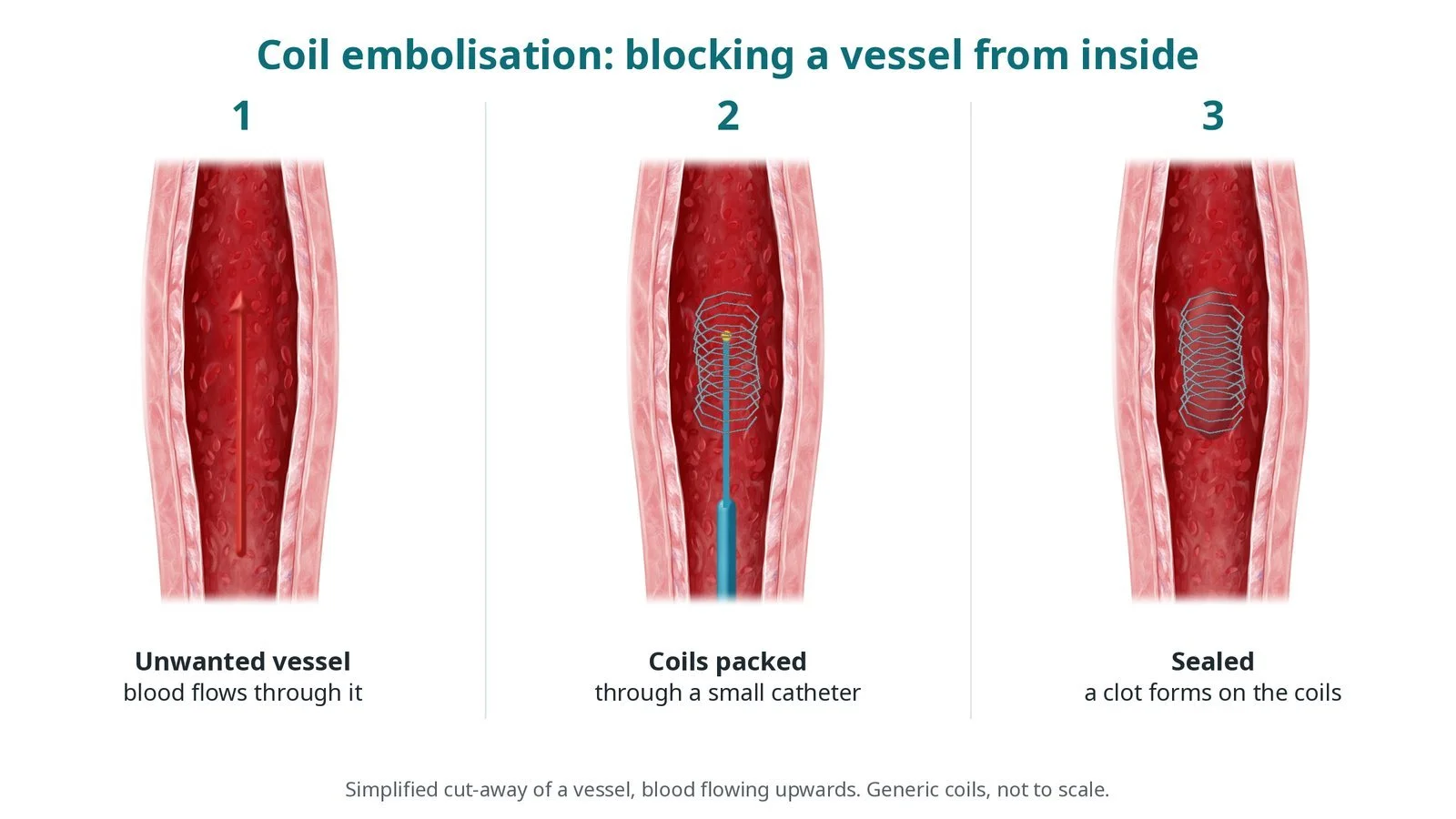
What it is: A coronary artery fistula is an abnormal connection between a coronary artery and a heart chamber or blood vessel, such as the right ventricle, right atrium or pulmonary artery. Blood takes this short cut instead of feeding the heart muscle. In catheter closure, coils, a vascular plug or a covered stent are placed inside the channel to block it.
Who it is for: Children and adults with a large fistula, or one causing symptoms, heart strain or complications, as decided by a heart team. Many small fistulas cause no problems and are simply watched.
Main risks: A heart attack from a clot in the widened artery or a blocked side branch, the device moving, a remaining leak, abnormal heart rhythms, damage to the artery, and problems at the access site. Your team will explain your own risk.
Usually: Under general anaesthetic for children, and local anaesthetic with sedation or a general anaesthetic for adults, about 1 to 3 hours, often home the next day.
On this page: What is coronary artery fistula closure? · Who is it for? · When is it done? · Step by step · Recovery · Risks · Kit list · First time in the lab? · FAQ
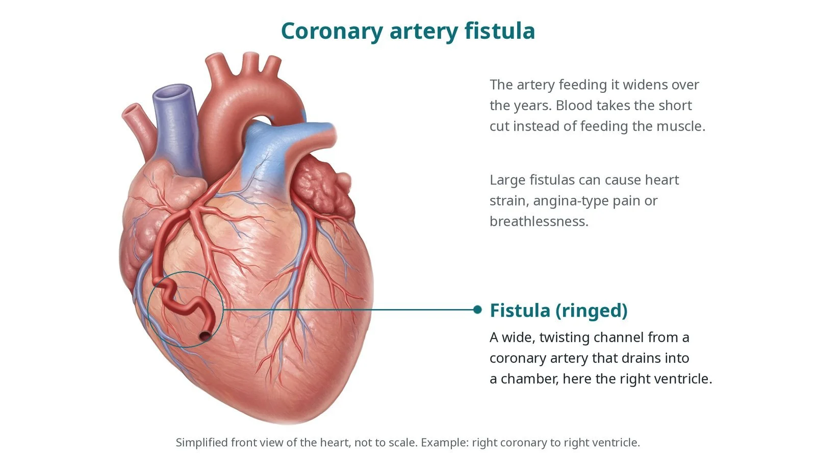
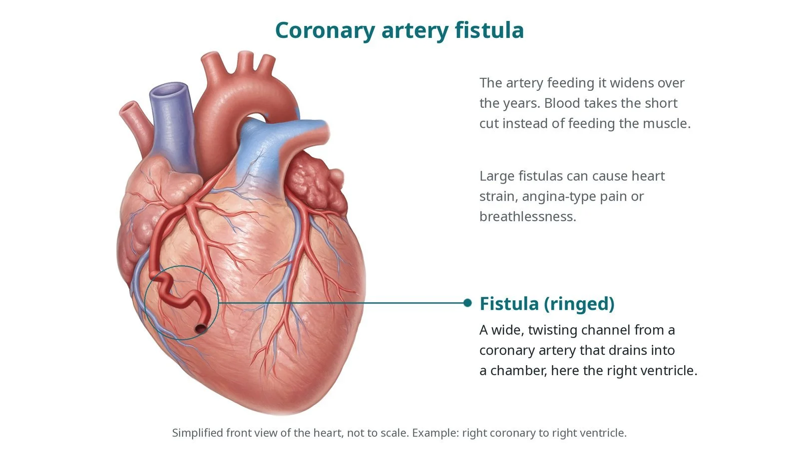
Coronary artery fistula (ringed): a wide, twisting channel from a coronary artery that drains into a heart chamber, here the right ventricle. The artery feeding it widens over time.
What is coronary artery fistula closure?
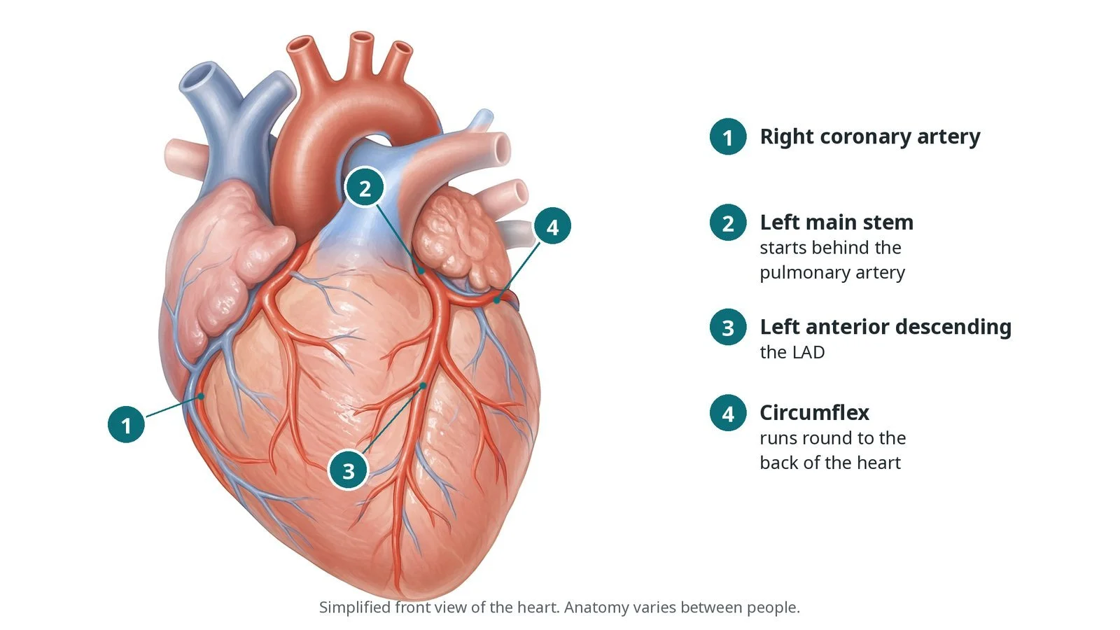
The coronary arteries normally branch into ever smaller vessels inside the heart muscle. In a coronary artery fistula, a branch connects directly to a low-pressure chamber or vessel, most often the right ventricle, right atrium, pulmonary artery or coronary sinus. Most are present from birth; a few follow surgery, trauma or heart biopsies.
Because the fistula is a low-resistance short cut, a lot of blood flows through it. Over the years the artery feeding it can widen and twist, sometimes forming an aneurysm. Blood may be "stolen" from the heart muscle, and the extra flow loads the heart.
Small fistulas are common and usually harmless. Large ones can cause breathlessness, angina-type chest pain, heart enlargement, rhythm problems, endocarditis (infection) and, rarely, rupture.
Closure aims to block the channel close to where it drains, so the normal branches before it keep their blood supply. After closure, blood in the widened artery flows slowly and can clot, which can cause a heart attack, so blood-thinning medicine is planned by the team.
New to the acronyms? See the Cath Lab Abbreviations A–Z.
Who is it for?
A heart team decides, using echo, CT coronary angiography and sometimes a stress test. Reasons to close include:
a large fistula, even without symptoms, especially in children
symptoms such as breathlessness or chest pain, or evidence of reduced blood supply to the muscle
heart enlargement, an aneurysm, or endocarditis
Who it may not suit: Very twisting anatomy, several fistulas, normal branches arising close to the exit, or a need for other heart surgery may make surgical closure the better choice.
The decision is made by your heart team, who will talk through the reasons, the alternatives and the risks with you.
When is it done?
It is a planned procedure. Before it, a CT scan usually maps the fistula's course, its exit point and the normal branches near it.
gives you a chance to ask questions and give consent
arranges blood tests, including kidney function and a blood count, and an ECG
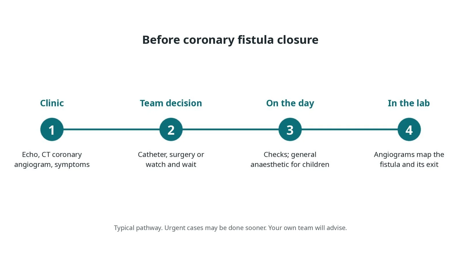
Before coronary fistula closure: echo and CT coronary angiogram; team decision between catheter, surgery or watching; checks on the day; angiograms map the fistula in the lab.
Step by step
1. Getting ready
Children have a general anaesthetic; adults may have sedation or a general anaesthetic. A 12-lead ECG is recorded at the start for comparison. In children all drugs and contrast are weight-based, as per centre protocol.
2. Access
A sheath is placed in the femoral artery or, in adults, sometimes the radial artery. A femoral vein sheath is added if the fistula will be closed from the drainage side using an artery-to-vein wire loop. Heparin is given, dose as per centre protocol.
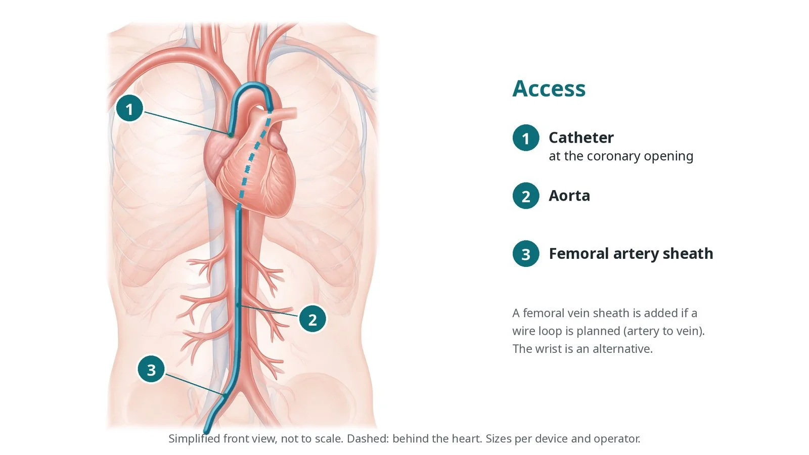
Access: (1) catheter at the coronary opening; (2) aorta; (3) femoral artery sheath. A femoral vein sheath is added if an artery-to-vein wire loop is planned.
3. Closing the fistula
A. Map. Angiograms of the coronary artery show the fistula's course, its exit and the normal branches.
B. Reach the exit. A microcatheter or guide is advanced as close to the drainage point as possible. If the vessel is very twisted, a wire can be passed through into the chamber, snared from the vein side and pulled out to form a loop, so the device can be delivered from the vein side (retrograde).
C. Close. Detachable coils, a vascular plug, or a covered stent in the feeding artery (when the fistula comes off its side) are placed, per operator. A balloon may first block the channel for a short test while the ECG is watched for ischaemia.
D. Check. An angiogram confirms the fistula is closed and the normal branches still fill, and the ECG is compared with the baseline.
Watch: Chest pain, ST changes or a new rhythm during the test occlusion or after closure may mean a normal branch is blocked: tell the operator straight away.
4. Preventing clot in the wide artery
After closure, blood in the widened feeding artery moves slowly and can clot. This can cause a heart attack, sometimes days later. The team decides on antiplatelet or anticoagulant medicine and how long to continue it.
Watch: New chest pain or ST changes after the case, including on the ward: record an ECG and call the team at once.
5. Finishing
The sheaths are removed and the access sites closed with pressure or a closure device, per operator. ECG monitoring continues on the ward.
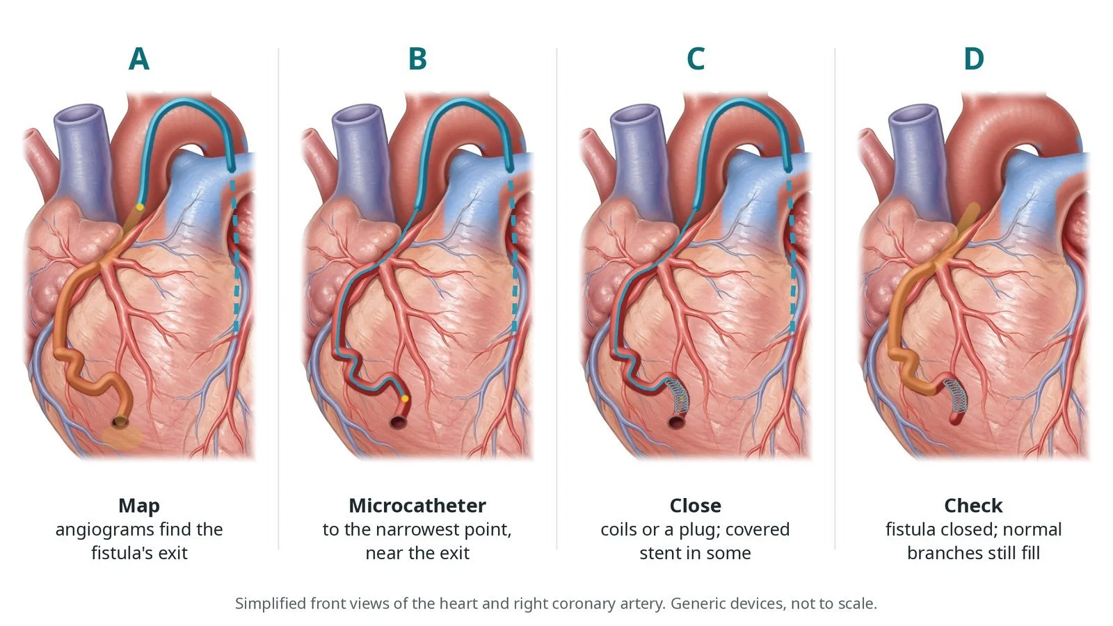
A: angiograms find the exit. B: microcatheter to the narrowest point near the exit. C: coils or a plug close it. D: fistula closed while normal branches still fill.
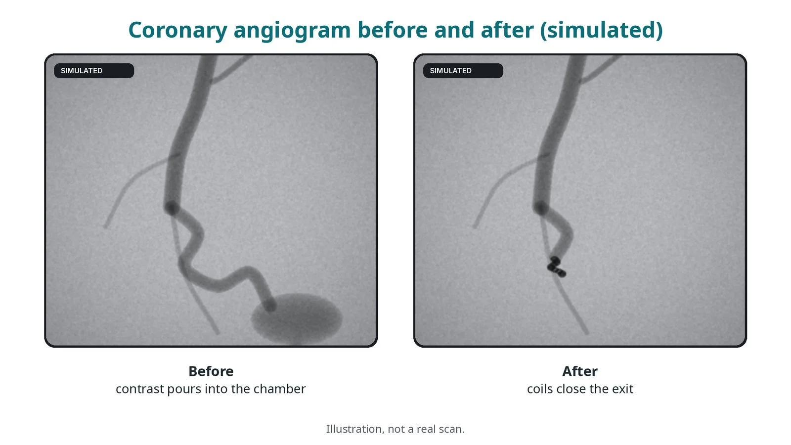
Illustration, not a real scan. Before: contrast pours from the fistula into the chamber. After: coils close the exit.
Recovery and aftercare
First hours: bed rest with checks of the access site, ECG and chest pain, as per centre protocol.
Going home: often the next day, after an echo or ECG.
Medicines: take antiplatelet or anticoagulant medicine exactly as prescribed; it lowers the risk of clot in the widened artery.
Activity: avoid heavy lifting for a few days while the access site heals, or as advised.
Driving (adults): ask your team; do not drive until the access site has healed and any sedation has worn off.
Follow-up: an echo and often a CT or angiogram later, to check the fistula stays closed and the artery is healthy.
Call 999 if you have chest pain, sudden breathlessness, fainting, signs of a stroke, or heavy bleeding from the access site.
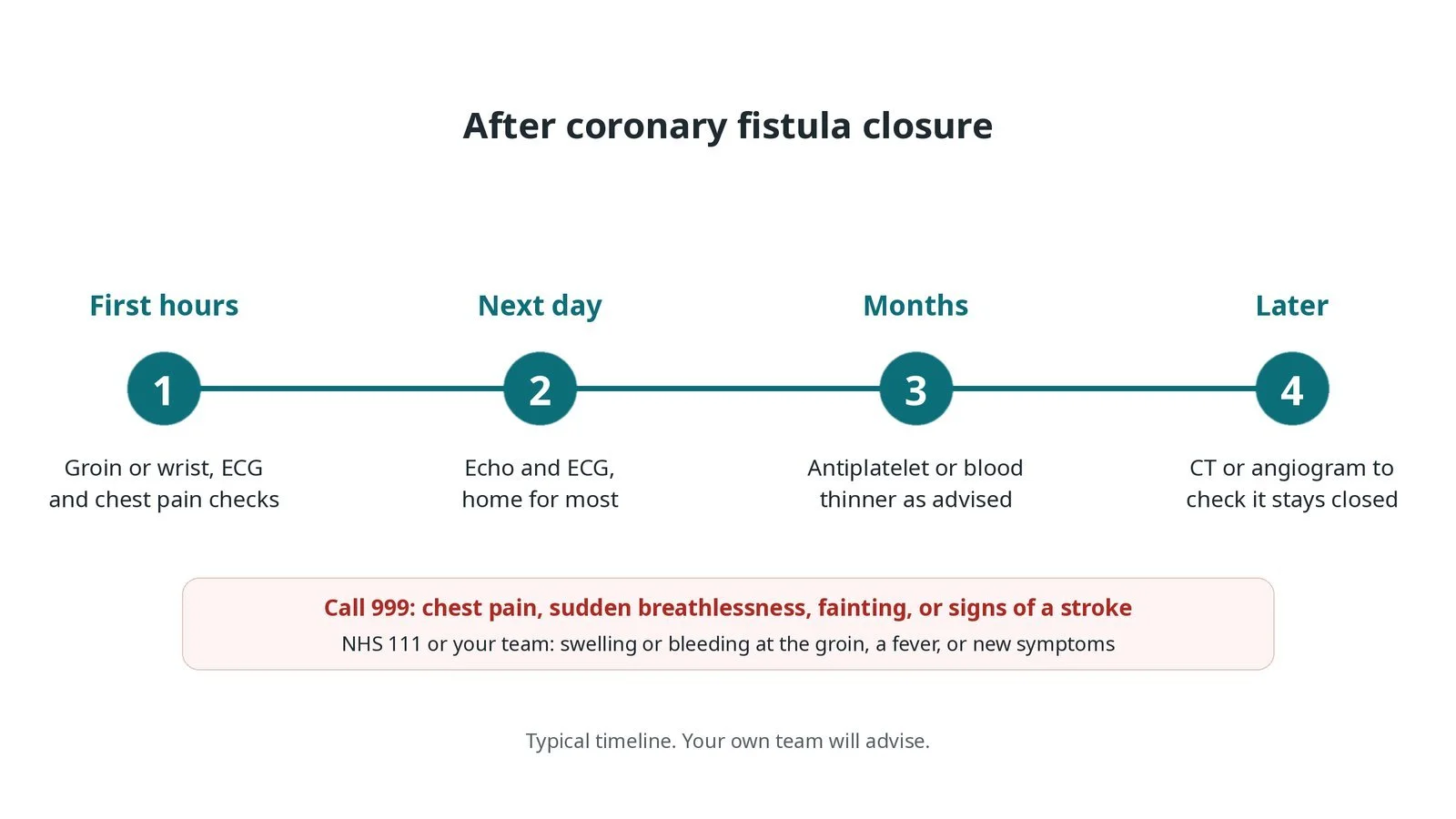
After fistula closure: (1) access site, ECG and chest pain checks; (2) echo and ECG, home the next day for most; (3) antiplatelet or blood thinner as advised; (4) CT or angiogram follow-up.
Risks and complications
Your team will explain the risks that apply to you. Possible problems include:
Heart attack from a clot in the widened artery or a blocked branch (uncommon)
A coil or plug moving to another place (uncommon)
A remaining leak through the fistula
Abnormal heart rhythms
Tear or perforation of the coronary artery (rare)
Bleeding or bruising at the access site
Cath lab kit list (printable)
Barts example kit list
An example from one UK centre, written generically. Every lab has its own protocol, and sizes and doses vary.
arterial sheath, femoral or radial; venous sheath if a wire loop is planned
coronary guide catheters, per operator
microcatheters and coronary guidewires
detachable embolisation coils in a range of sizes
vascular plugs, per operator
covered coronary stents, if needed
balloon for test occlusion
snare for an artery-to-vein loop or retrieval
12-lead ECG; heparin and antiplatelet medicines as per centre protocol
Procedural steps (Barts example)
Getting ready
Access
Closing the fistula
Preventing clot in the wide artery
Finishing
First time in the cath lab?
Know the map. Look at the CT or angiogram with the team so you know where the exit and the normal branches are.
Watch the ECG during test occlusion. ST changes mean the closure point may starve the heart muscle.
Count and record every device. Note each coil and plug size and its lot number for the record and follow-up.
Think clot afterwards. Chest pain after closure is not just bruising until an ECG says so.
Keep a baseline 12-lead ECG ready. ST changes during test occlusion or after closure are compared with it.
Have a snare and spare coils open before deployment in case a coil needs to be retrieved.
FAQ
I was told I have a small fistula. Does it need closing?
Often not. Many small fistulas never cause problems and are simply checked over time.
Will the widened artery shrink back?
It may shrink a little, but often stays wide. That is why follow-up scans and blood-thinning medicine matter.
Why do I need blood thinners afterwards?
Blood moves slowly in the widened artery after closure, so it can clot. Medicine lowers this risk.
Is surgery an alternative?
Yes. Surgery is chosen when the anatomy does not suit a catheter approach or other heart surgery is needed.
Related Cath Lab 101 guides
Collateral embolisation (MAPCAs and venovenous collaterals) (coming soon)
Sources
All checked on 12 November 2026. Each page's own date is shown where it gives one.
Stout KK, et al. 2018 AHA/ACC Guideline for the Management of Adults With Congenital Heart Disease. J Am Coll Cardiol 2019;73(12):e81–e192
Baumgartner H, et al. 2020 ESC Guidelines for the management of adult congenital heart disease. Eur Heart J 2021;42(6):563–645
StatPearls (NCBI Bookshelf), search: coronary artery fistula. ncbi.nlm.nih.gov/books
Disclaimer
Education only. This page is for general education for healthcare students, cath lab staff and curious readers. It isn't medical advice and doesn't replace advice from your own doctor, heart team or local unit protocols. Equipment and medicines are described generically. No product or manufacturer is recommended. If you think someone is having a heart emergency, call 999.
Disclosure: I work for a company that makes a device used in this procedure. This page is independent education, isn't sponsored or reviewed by any company, and doesn't recommend any product.
All diagrams on this page are original illustrations created for rodnieoro.com.