RVOT stenting in tetralogy of Fallot: a Cath Lab 101 guide
Reviewed by Rodnie Oro, cath lab charge nurse · Last reviewed: November 2026 · Education only, not medical advice
RVOT stenting in tetralogy of Fallot in one minute
What it is: In tetralogy of Fallot the pathway from the right ventricle to the lungs (the right ventricular outflow tract, RVOT) is narrowed by thick muscle and a small pulmonary valve. In RVOT stenting a metal stent is placed across this narrowing by catheter, from a vein, to let more blood reach the lungs.
Who it is for: Selected babies with tetralogy of Fallot who have low oxygen levels or cyanotic ("tet") spells and are too small, premature or unwell for early full repair, as decided by the congenital heart team. It is an alternative to a surgical shunt.
Main risks: A cyanotic spell during the procedure, abnormal heart rhythms, the stent moving, too much lung blood flow, and problems at the access site. The stent can also affect the type of repair later. Your team will explain your baby's own risk.
Usually: Under general anaesthetic, about 1 to 2 hours, then care on the cardiac or neonatal unit. Full surgical repair follows months later.
On this page: What is RVOT stenting in tetralogy of Fallot? · Who is it for? · When is it done? · Step by step · Recovery · Risks · Kit list · First time in the lab? · FAQ
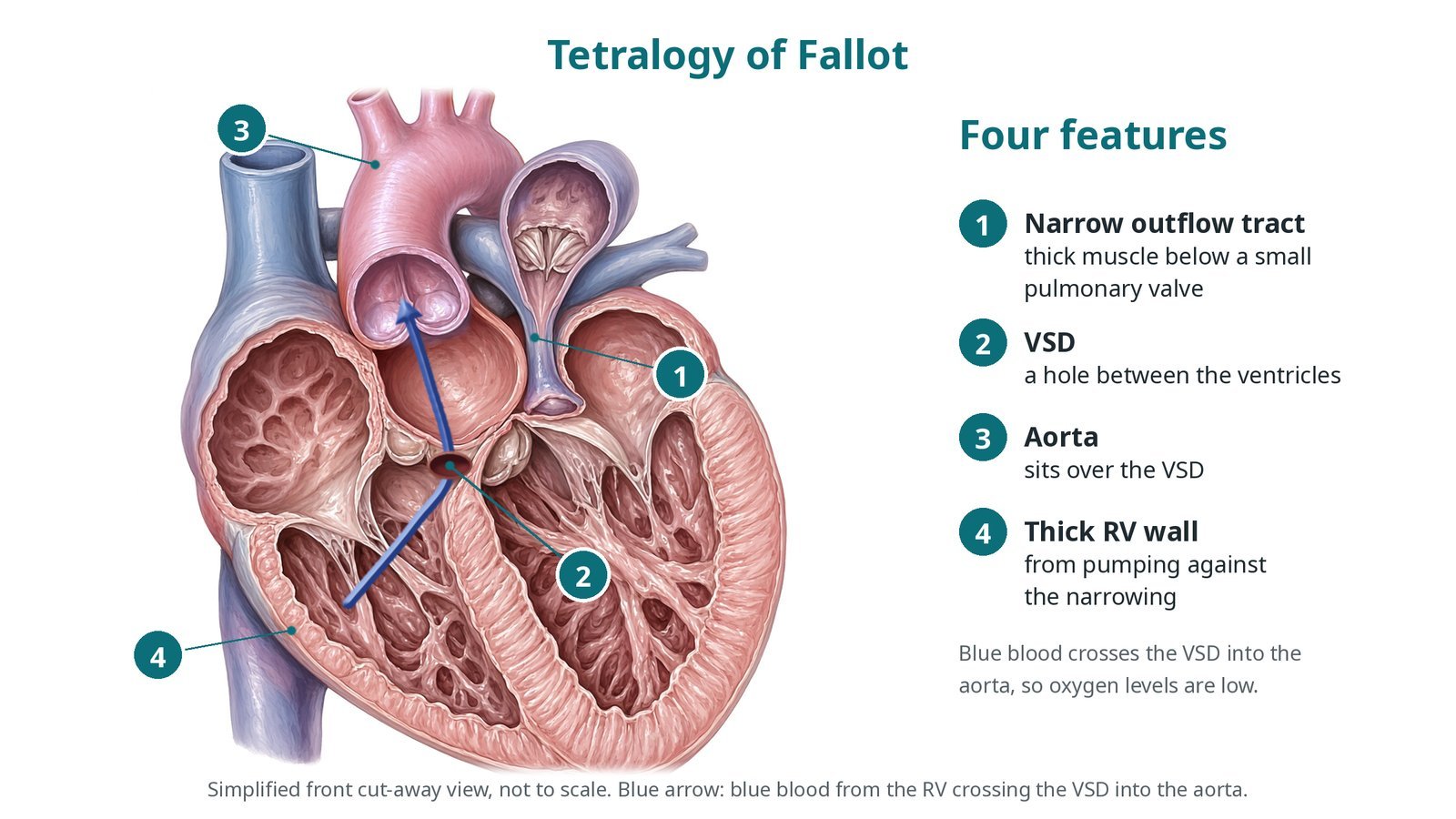
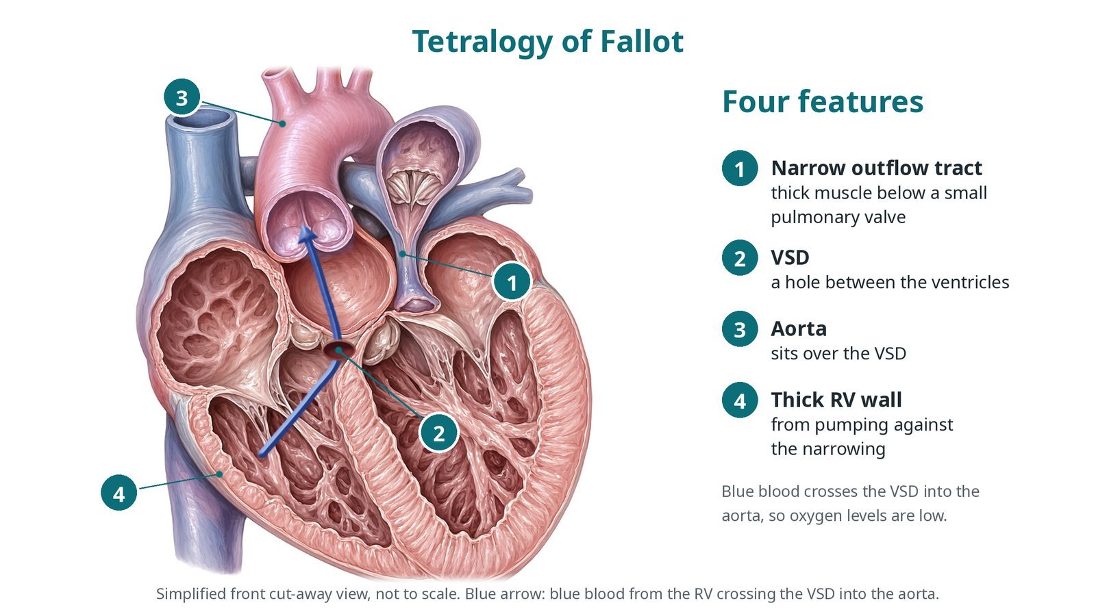
Tetralogy of Fallot: (1) narrow right ventricular outflow tract, thick muscle below a small pulmonary valve; (2) VSD; (3) the aorta sits over the VSD; (4) thick right ventricular wall.
What is RVOT stenting in tetralogy of Fallot?
Tetralogy of Fallot has four features: a narrowed right ventricular outflow tract (thick muscle below a small pulmonary valve, sometimes with small pulmonary arteries), a large hole between the ventricles (VSD), an aorta that sits over the VSD, and a thick right ventricular wall.
Because the outflow to the lungs is narrow, blue blood crosses the VSD into the aorta, so oxygen levels are low. The outflow muscle can suddenly tighten, for example with crying, pain, fever or dehydration, causing a hypercyanotic or "tet" spell: the baby becomes very blue, breathless and distressed, and may lose consciousness.
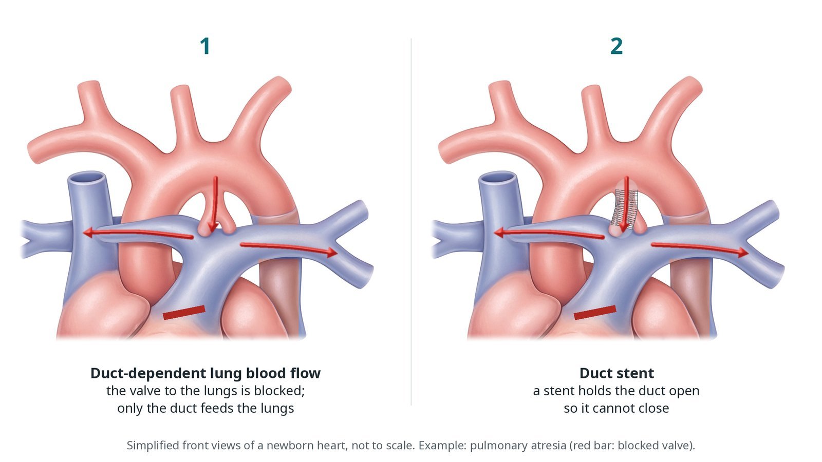
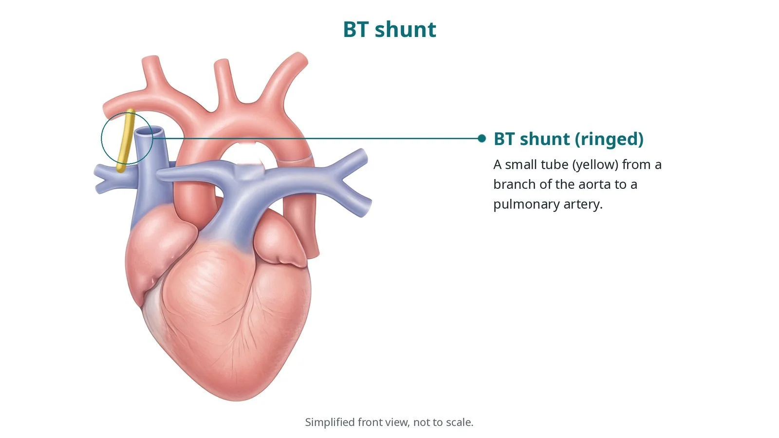
Most babies have full surgical repair in the first year of life. If a baby is too small or unwell for early repair and is very blue or having spells, a temporary way to boost lung blood flow is needed: a surgical shunt, a duct stent, or a stent across the outflow tract.
The RVOT stent holds the narrowed muscle (and often the small valve) open. More blood reaches the lungs, oxygen levels rise, and the pulmonary arteries can grow before surgery.
New to the acronyms? See the Cath Lab Abbreviations A–Z.
Who is it for?
Babies with tetralogy of Fallot, or similar anatomy, chosen by the congenital heart team when:
oxygen levels are low or spells are happening
the baby is too small, premature or unwell for early full repair
the anatomy suits a stent
Who it may not suit: Babies who are big enough for early full repair, or whose anatomy makes stenting risky, may go straight to surgery or have a surgical shunt instead.
The decision is made by your heart team, who will talk through the reasons, the alternatives and the risks with you.
When is it done?
Usually in the first weeks or months of life, as a planned or urgent procedure depending on how blue the baby is. Full repair is planned for later, once the baby has grown.
gives you a chance to ask questions and give consent
arranges blood tests, including kidney function and a blood count, and an ECG
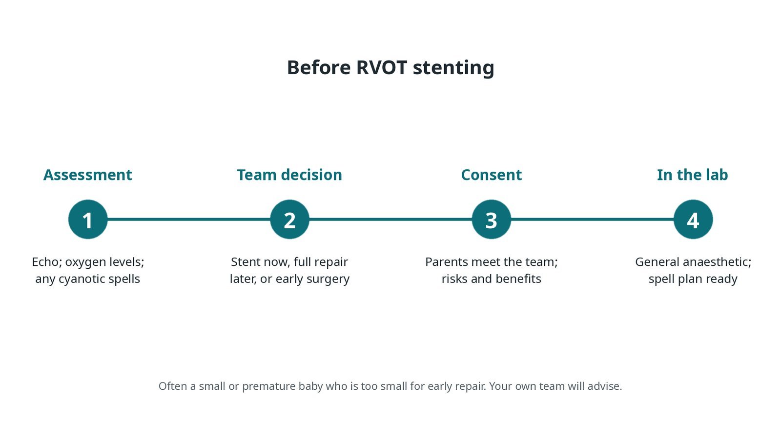
Before RVOT stenting: echo and oxygen levels; team decision on stent now and repair later; parents meet the team; general anaesthetic with a plan ready for cyanotic spells.
Step by step
1. Getting ready, with a spell plan
The baby has a general anaesthetic, a warming mattress and temperature monitoring. All drugs, fluids and contrast are weight-based, as per centre protocol. Before starting, the team agrees how a tet spell will be treated: oxygen, deeper anaesthesia and pain relief, fluid, and medicines that raise the body's blood pressure or relax the outflow muscle, all as per centre protocol.
2. Access
A sheath is placed in the femoral vein under ultrasound; the neck vein is sometimes used. An arterial line monitors blood pressure. Heparin is given, dose as per centre protocol.
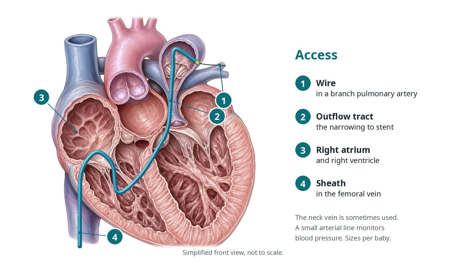
Access: (1) wire in a branch pulmonary artery; (2) the outflow tract narrowing to stent; (3) right atrium and ventricle; (4) sheath in the femoral vein.
3. Stenting the outflow tract
A. Angiogram. Contrast in the right ventricle outlines the narrowing, the valve and the pulmonary arteries, and the length to cover is measured.
B. Wire across. A catheter and soft wire cross the narrowing into a branch pulmonary artery. A long sheath or guide may be passed for support.
C. Stent. A stent on a balloon is positioned across the narrowed muscle, often also across the small valve, and opened. Size and length are per operator.
D. Check. Oxygen levels, pressures and an angiogram check the result, and that the stent does not reach into the pulmonary artery branches or the tricuspid valve.
Watch: Catheters in the outflow tract can trigger a spell: oxygen levels and blood pressure fall. Tell the operator at once and start the agreed spell plan.
4. Finishing
The sheath is removed and pressure applied. Oxygen levels often rise a lot, and the team watches for too much blood flow to the lungs. The baby returns to the cardiac or neonatal unit with a handover covering the target oxygen range, medicines and the leg.
5. Planning the later repair
Full surgical repair is done once the baby has grown. The surgeon removes the stent and closes the VSD. If the stent crossed the pulmonary valve, the valve usually cannot be kept, and a patch across the valve ring is often needed. This leaves a leaking pulmonary valve, which is watched over life.
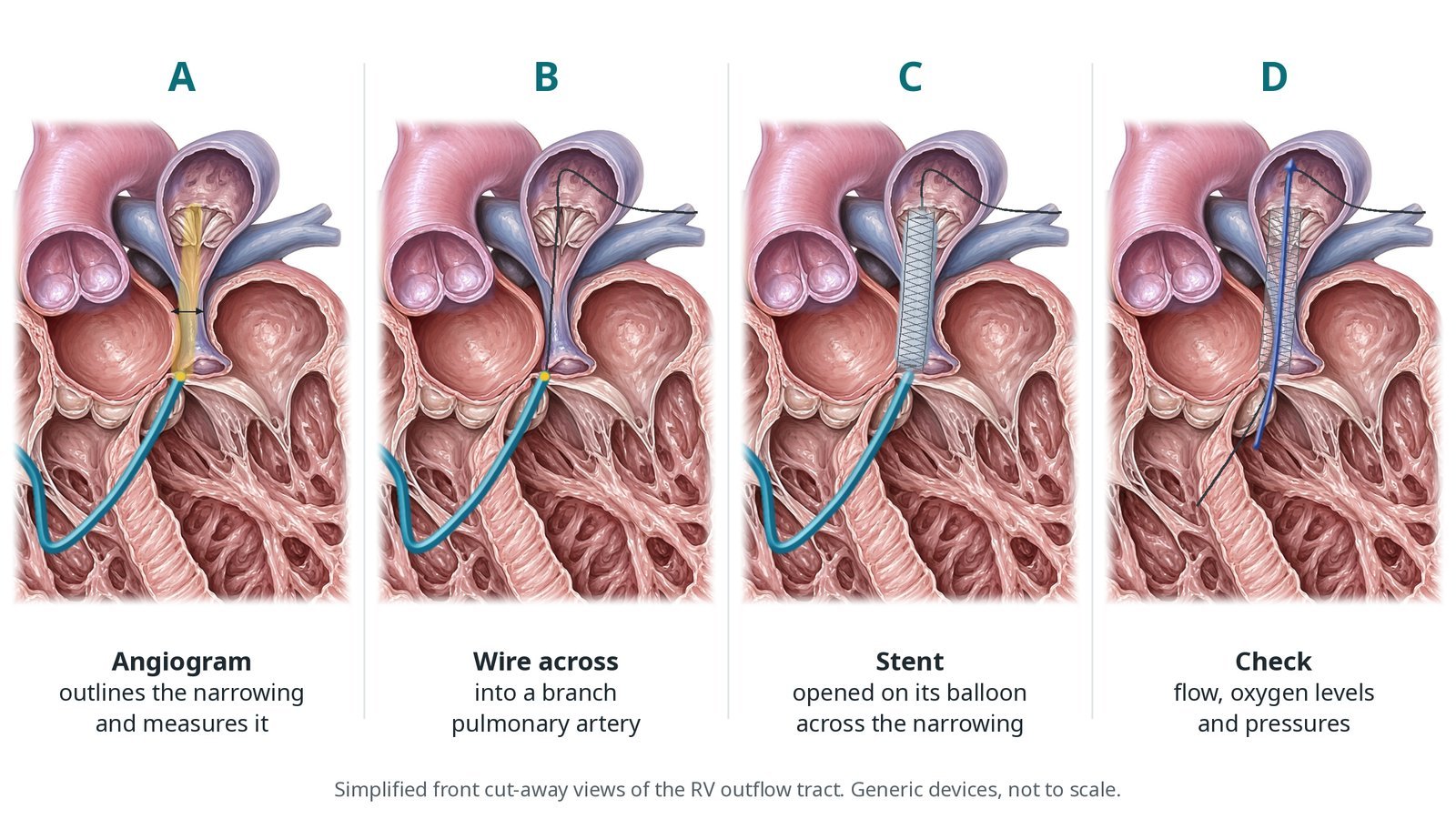
A: angiogram outlines and measures the narrowing. B: wire across into a branch pulmonary artery. C: stent opened on its balloon. D: flow, oxygen levels and pressures checked.
Recovery and aftercare
First hours: oxygen levels, heart rhythm, blood pressure, temperature and the leg are checked closely, as per centre protocol.
Echo: checks flow through the stent and the pulmonary arteries.
Going home: after a few days for many babies, once feeding and oxygen levels are settled.
Medicines: some babies have an antiplatelet medicine; follow the team's advice.
Growth: feeding and weight gain are checked in clinic, because a bigger baby has a safer repair.
Follow-up: echo and clinic checks until full surgical repair, usually months later.
Call 999 if your baby is more blue than usual, floppy, breathing fast or struggling, or very sleepy and not feeding.
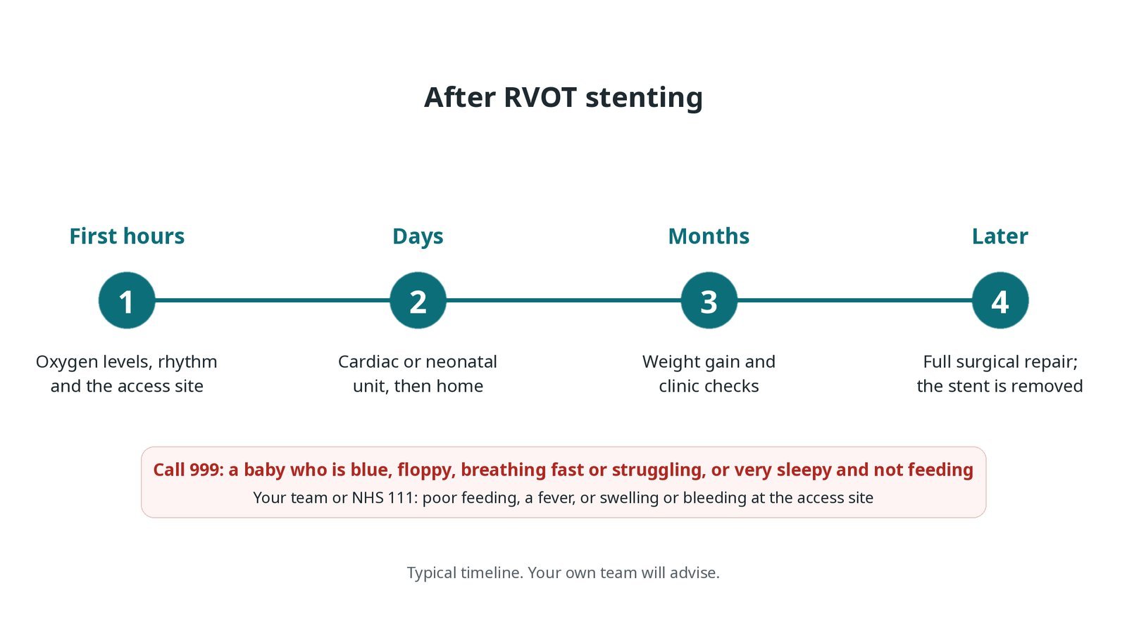
After RVOT stenting: (1) oxygen levels, rhythm and access site; (2) cardiac or neonatal unit, then home; (3) weight gain and clinic checks; (4) full surgical repair later, when the stent is removed.
Risks and complications
Your team will explain the risks that apply to you. Possible problems include:
A cyanotic spell during the procedure
Abnormal heart rhythms
The stent moving, needing retrieval or surgery (uncommon)
Too much blood flow to the lungs
Narrowing again in or around the stent, needing a further procedure
Bleeding or a blocked vessel at the access site (uncommon)
Less chance of keeping the pulmonary valve at repair if the stent crosses it
Cath lab kit list (printable)
Barts example kit list
An example from one UK centre, written generically. Every lab has its own protocol, and sizes and doses vary.
small venous sheath; arterial line
ultrasound for vascular access
angiographic and shaped catheters
long sheath or guide catheter, per operator
soft and supportive guidewires
stents on balloons in several sizes and lengths, per operator
inflation device
spell medicines and fluid bolus, weight-based, as per centre protocol
warming mattress and temperature probe
snare for a stent that moves
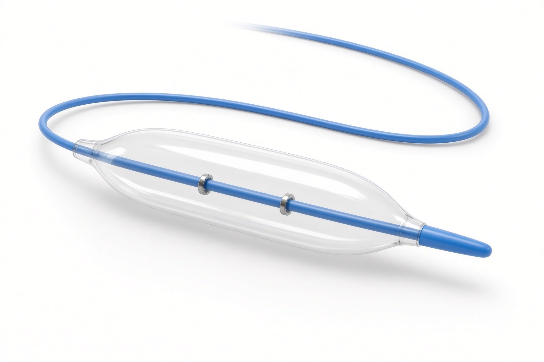
Stents on balloons.
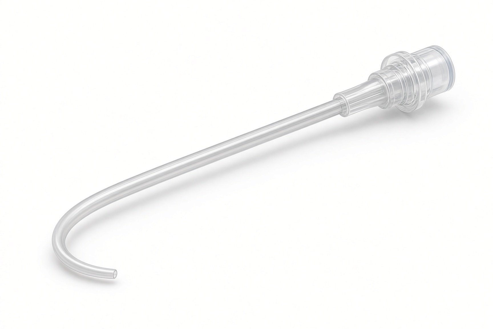
Angiography catheter.
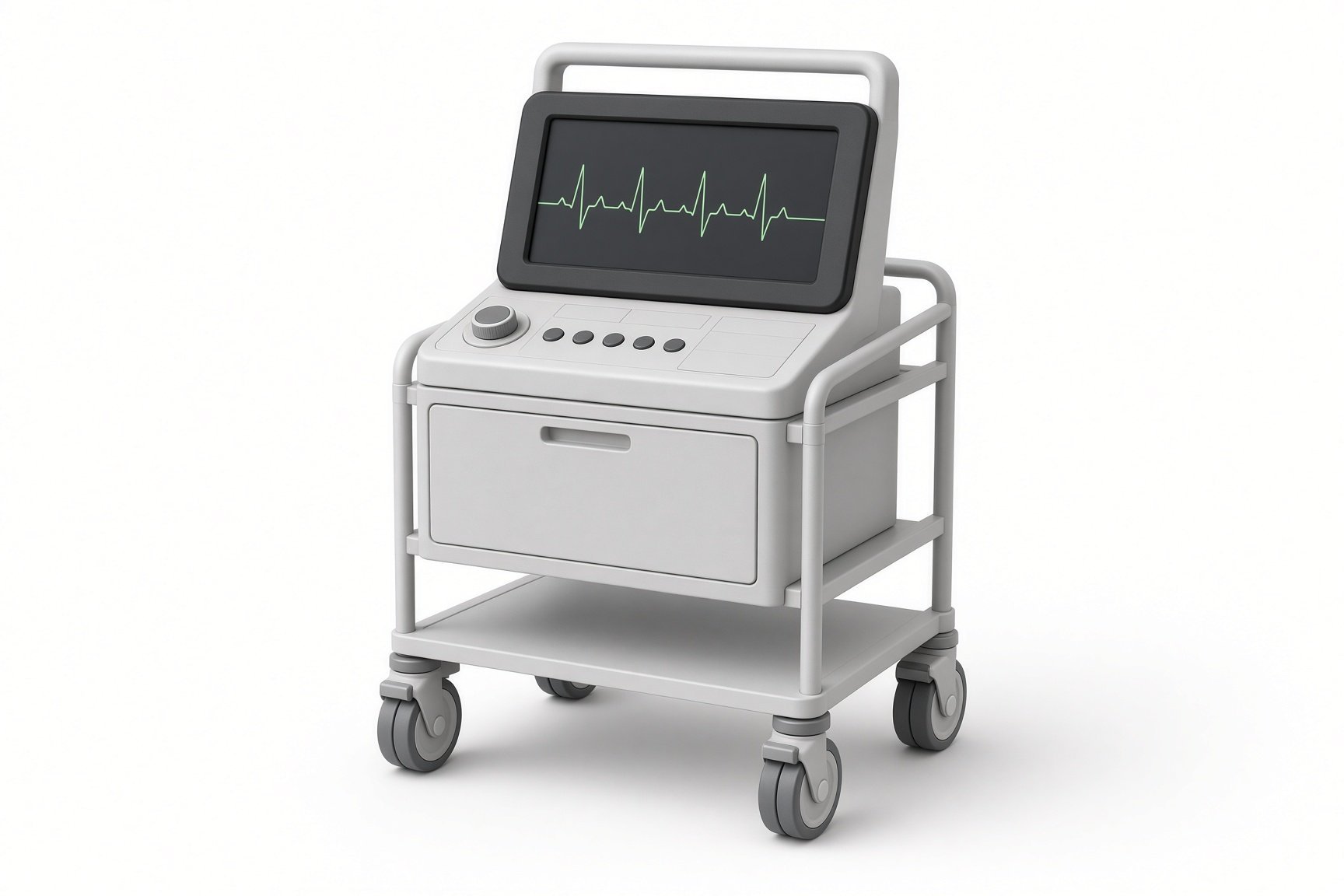
Echo machine.
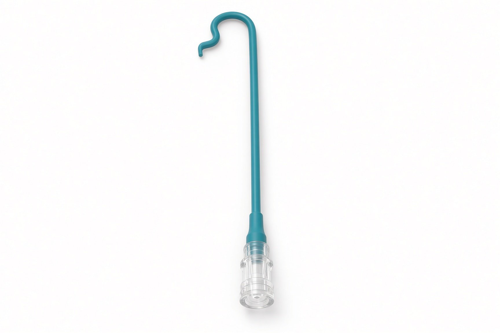
Long sheath or guide catheter.
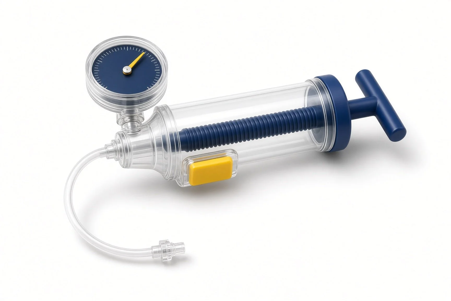
Inflation device.
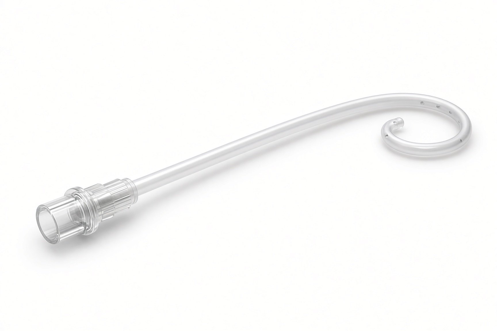
Angiography catheter.
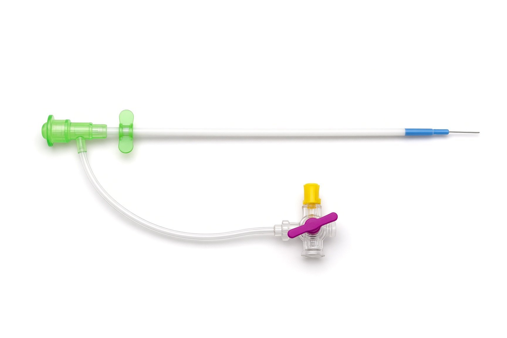
Small venous sheath.
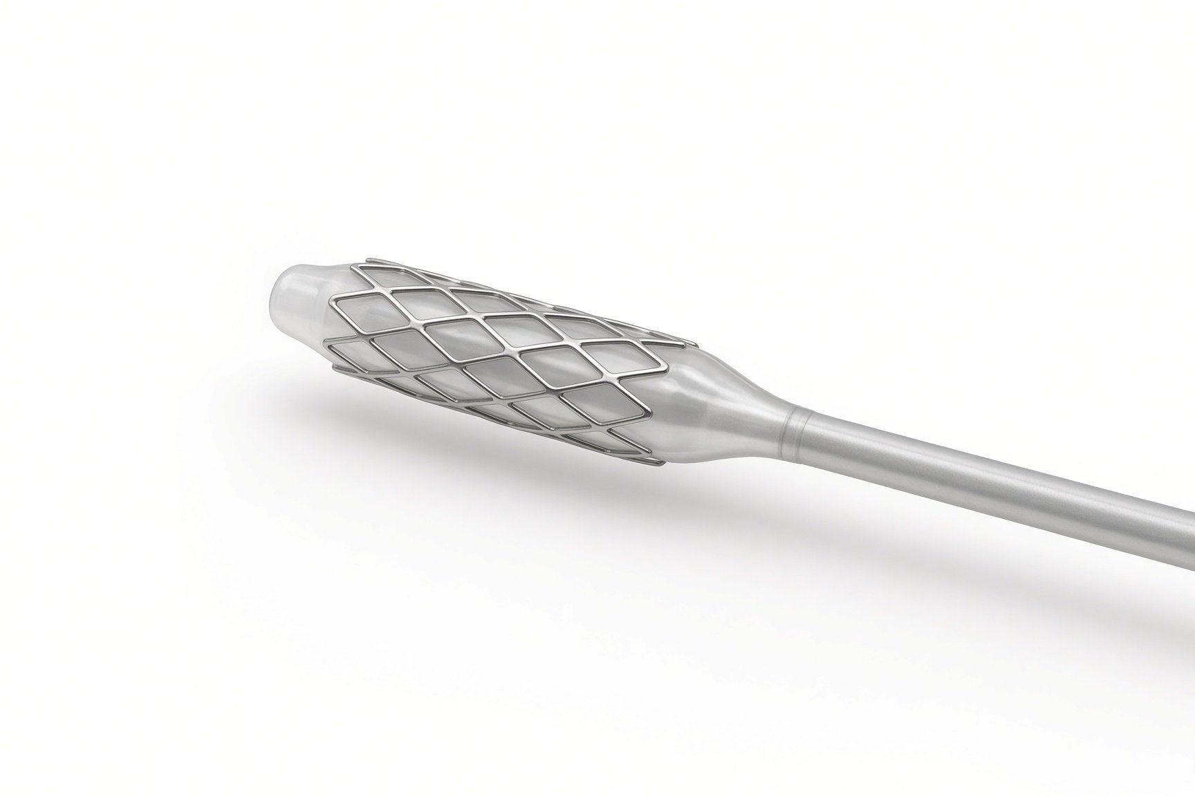
Stents on balloons.

Spell medicines per protocol.
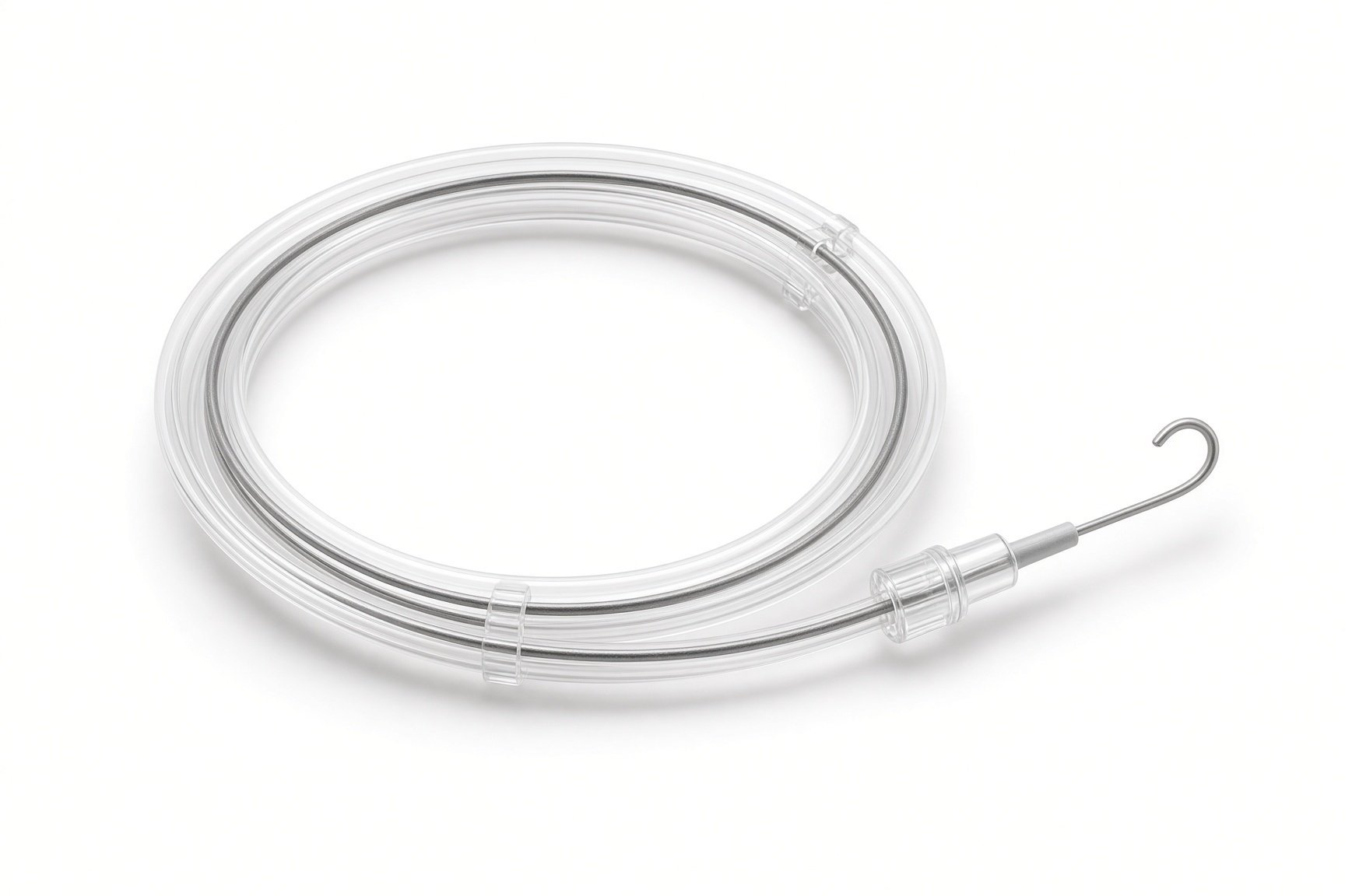
Guidewires.
Procedural steps (Barts example)
Getting ready, with a spell plan
Access
Stenting the outflow tract
Finishing
Planning the later repair
First time in the cath lab?
Know the spell plan. Before the case starts, know who does what and where the drugs and fluid are if a spell happens.
Watch the saturation trace. A spell shows first as a falling oxygen level, often with catheter movement in the outflow tract.
Keep the baby warm and still. Cold, pain and light anaesthesia can all trigger a spell.
Explain the plan to parents. This is a bridge to surgery, not the final repair. Make sure they know that.
Draw up the spell medicines and fluid bolus before the drapes go on, labelled with the weight-based doses.
Check the leg's colour, warmth and pulse on both sides before leaving the lab.
FAQ
Is this instead of the heart operation?
No. It is a bridge. Your baby still needs full surgical repair, usually months later.
What is a tet spell?
A sudden worsening of blueness, often with crying or upset, when the outflow muscle tightens. Your team will show you what to do and when to call 999.
Will the stent be removed?
Yes. The surgeon usually removes it during the full repair.
Why a stent rather than a shunt operation?
A stent avoids opening the chest and sends blood forwards into the lung arteries, which can help them grow. The team chooses based on your baby's anatomy.
Related Cath Lab 101 guides
Ductal (PDA) stenting in newborns (coming soon)
Balloon pulmonary valvuloplasty (coming soon)
Sources
All checked on 12 November 2026. Each page's own date is shown where it gives one.
Feltes TF, et al. Indications for cardiac catheterization and intervention in pediatric cardiac disease: a scientific statement from the American Heart Association. Circulation 2011;123(22):2607–2652
StatPearls (NCBI Bookshelf), search: tetralogy of Fallot. ncbi.nlm.nih.gov/books
Disclaimer
Education only. This page is for general education for healthcare students, cath lab staff and curious readers. It isn't medical advice and doesn't replace advice from your own doctor, heart team or local unit protocols. Equipment and medicines are described generically. No product or manufacturer is recommended. If you think someone is having a heart emergency, call 999.
Disclosure: I work for a company that makes a device used in this procedure. This page is independent education, isn't sponsored or reviewed by any company, and doesn't recommend any product.
All diagrams on this page are original illustrations created for rodnieoro.com.