PPVI (Percutaneous Pulmonary Valve Implantation): Cath Lab Guide
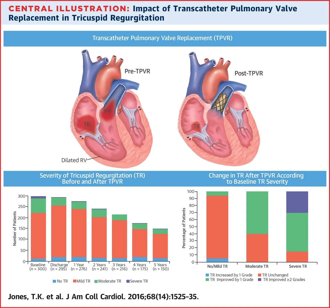
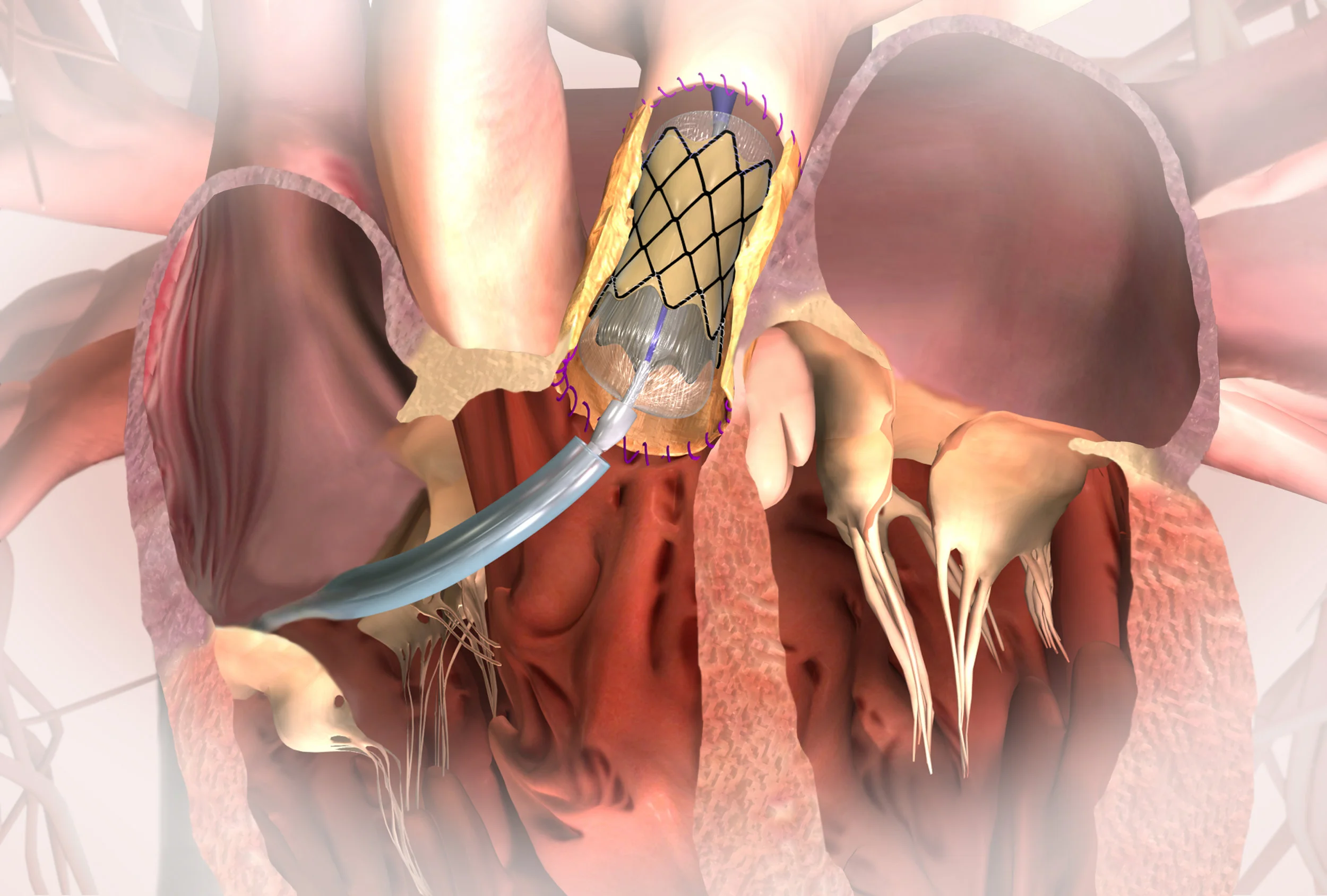
PPVI stands for percutaneous pulmonary valve implantation: a keyhole (catheter-based) procedure that places a new pulmonary valve inside a narrowed or leaking right ventricular outflow tract (RVOT), without open heart surgery. A folded-down valve is passed up through a vein, usually in the groin, and opened inside the old valve, conduit or outflow tract under X-ray and echo guidance.
You may also see it called TPVI (transcatheter pulmonary valve implantation) or TPVR (transcatheter pulmonary valve replacement). They all mean the same procedure. This guide explains PPVI from the cath lab side: who it is for, the kit, the steps, the team and what happens afterwards. See also the cath lab abbreviations glossary.
Who is PPVI for?
PPVI is mostly used for children (usually older children) and adults with congenital heart disease whose RVOT is not working well. The RVOT is the pathway that carries blood from the right ventricle (RV) to the pulmonary artery (PA). Typical patients include:
People with repaired tetralogy of Fallot (ToF). Many have had an earlier surgical repair and later develop severe pulmonary regurgitation (a leaking valve), stenosis (narrowing), or both.
Patients with an RV-to-PA conduit. This is a surgically placed tube, homograft or bioprosthetic valve (for example after a Ross operation, or repair of truncus arteriosus or pulmonary atresia) that has worn out over time.
Selected patients with a native or patched RVOT. Newer, larger valves and pre-stents mean some of these patients can now be treated too.
Whether PPVI or surgery is better is decided by the adult congenital heart disease (ACHD) multidisciplinary team. They look at symptoms, RV size and function on MRI/echo, arrhythmias and anatomy. NICE says PPVI should only be done in specialist units with cardiac surgical support available.
Valve types (general overview)
Several valve families are used. Which one is chosen depends on anatomy, size and the team's experience:
Melody transcatheter pulmonary valve: a bovine (cow) jugular vein valve sewn into a balloon-expandable stent. This was the first valve used for PPVI and was designed for conduits.
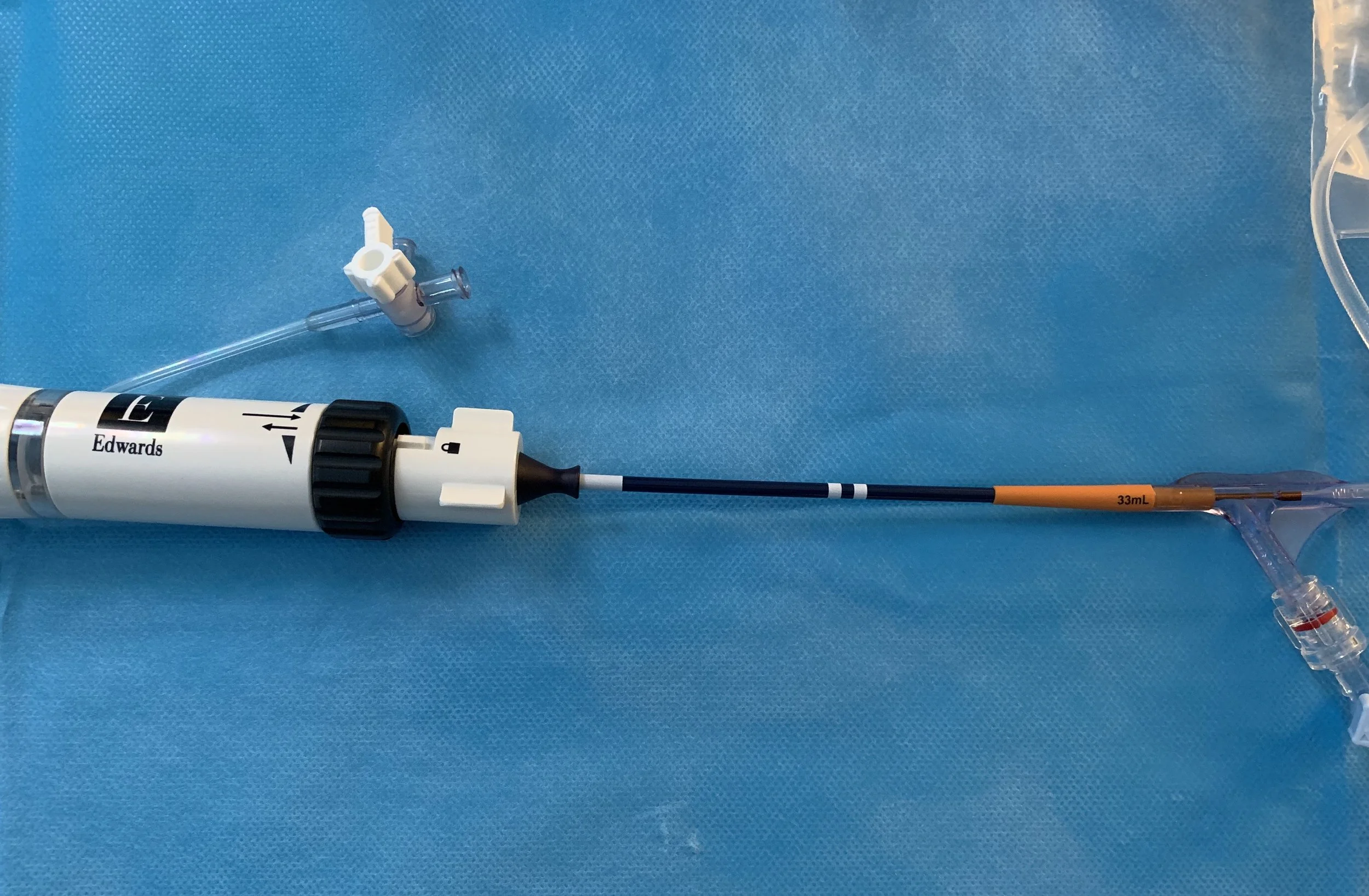
Edwards SAPIEN valves in the pulmonary position: balloon-expandable bovine pericardial valves first designed for the aortic position (TAVI), and also used in the RVOT.
Harmony transcatheter pulmonary valve: a self-expanding valve designed for larger native or patched RVOTs.
Others include self-expanding valves and dedicated pre-stents for large outflow tracts. Not all of these are available in every country.
(Description only, not a recommendation of any device. Device choice is a clinical decision.)
The procedure, step by step (cath lab view)
Every centre has its own protocol. This is a general outline.
Preparation.
The case is planned in advance from CT/MRI and echo.
Consent and WHO checklist; blood group and cross-match available.
Usually done under general anaesthetic with biplane imaging.
Prophylactic antibiotics and heparin per protocol (ACT monitored).
Defibrillator pads on. Surgical and perfusion backup confirmed.
Access.
Femoral venous access is most common. The jugular vein is an alternative.
Arterial access is used for pressure monitoring and aortic/coronary angiography.
The large venous sheath may be "pre-closed" with suture-based closure devices.
Haemodynamics.
A right heart study records RA, RV and PA pressures and the RVOT gradient.
Angiography of the RVOT shows its size, shape and how much the valve leaks.
Sizing. A compliant sizing balloon is inflated in the RVOT to measure the "waist" and check for a safe landing zone.
Coronary compression testing.
While the balloon is inflated in the RVOT, the team runs an aortic root or selective coronary angiogram.
This checks that expanding the outflow tract will not squash a coronary artery. In ToF and some other conditions the coronaries can run very close to the RVOT.
If there is compression, the valve is not implanted. Aortic root distortion is checked at the same time.
Pre-stenting.
One or more stents (bare or covered) are often placed first. They create a stable landing zone and help protect the valve frame.
Covered stents are kept ready in case the conduit tears.
Deployment.
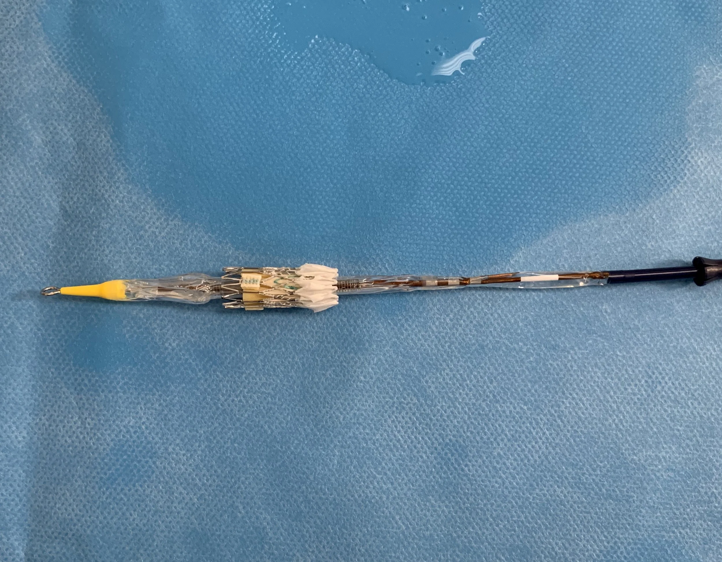
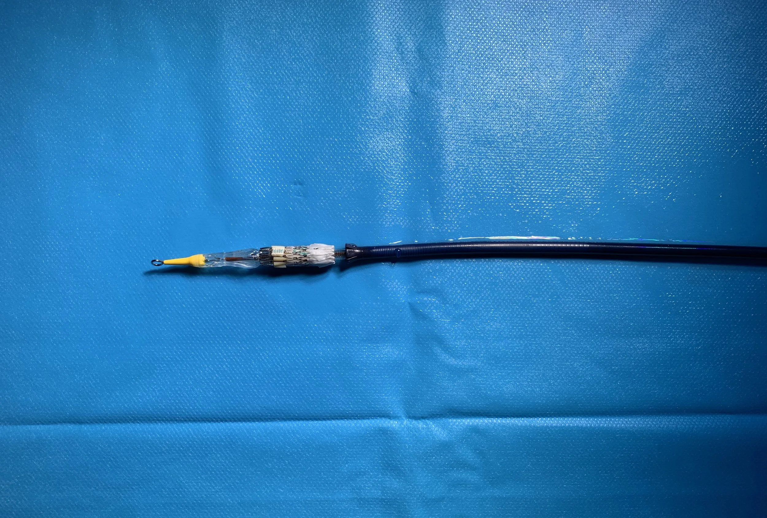
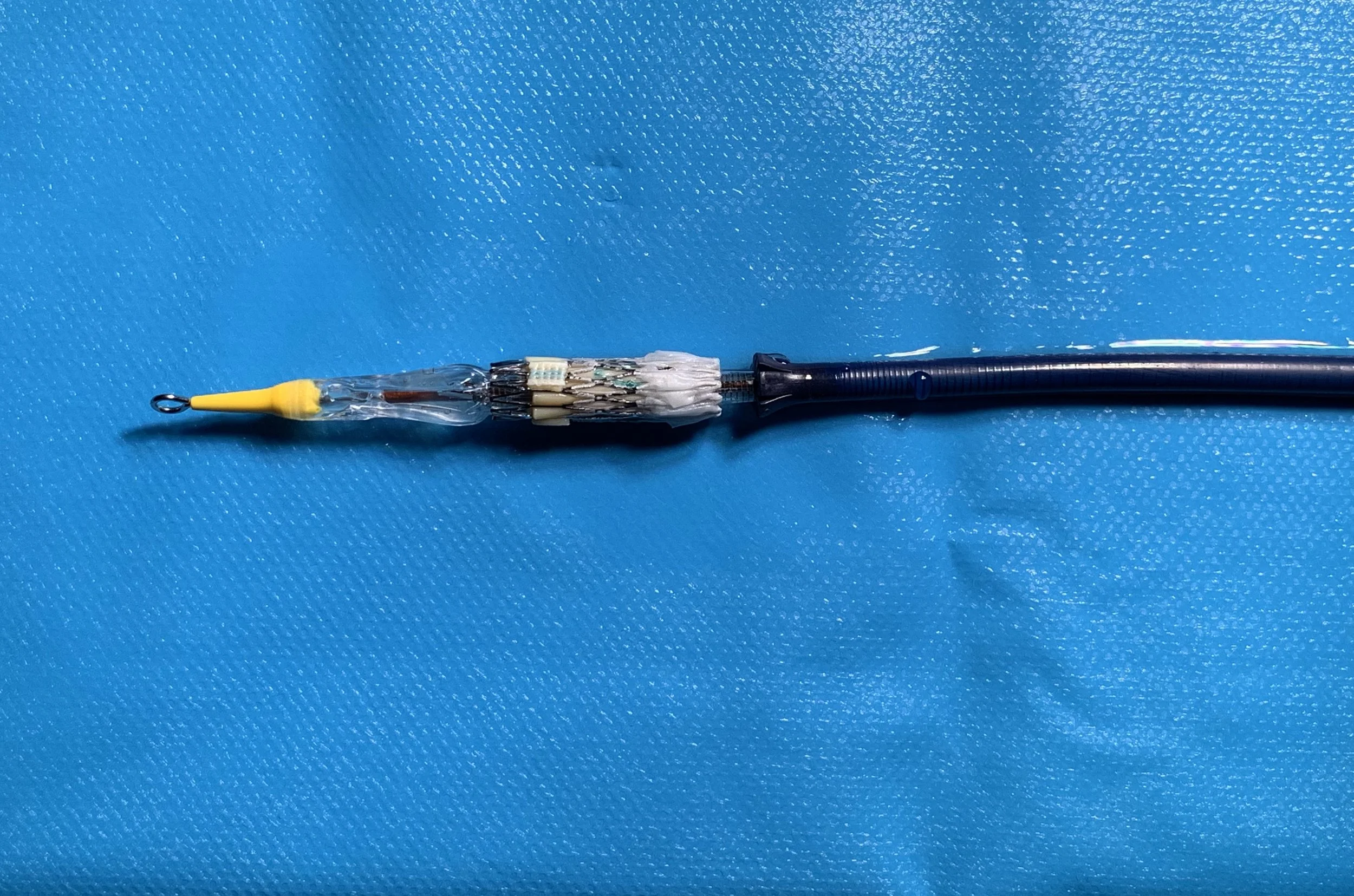
The valve is crimped onto its delivery system. Check orientation carefully on the back table: the valve sits "upside down" compared with TAVI.
The valve is passed over a stiff wire, usually through a large sheath.
It is then positioned and deployed, by balloon inflation or self-expansion.
Some teams use rapid ventricular pacing during balloon-expandable deployment.
Post checks.
Pressures are measured again to confirm the gradient has improved.
Angiography checks valve position and leak, and looks for conduit injury or contrast leaking out.
Coronary flow is checked again.
Then haemostasis with closure devices or a suture, and recovery.
Who does what: team roles
Interventional cardiologists (congenital/structural): plan and perform the implant.
Anaesthetist and ODP: general anaesthetic and airway management, and TOE if used.
Scrub nurse: sets up the trolley, preps balloons and stents, and helps crimp the valve.
Circulating/running nurse: gets kit, gives drugs, keeps track of time-out checks, and watches the patient.
Cardiac physiologist: records pressures and ECG, and supports pacing.
Radiographer: biplane imaging, projections and radiation safety.
Imaging cardiologist: CT/MRI planning and echo.
Cardiac surgeon and perfusionist: on standby for emergencies.
Risks and possible complications
PPVI is less invasive than surgery, but it is a specialist procedure with known risks. These include:
coronary artery compression
conduit tear or rupture (bleeding)
valve or stent malposition or embolisation
arrhythmias
damage to the tricuspid valve or pulmonary arteries
vascular access complications
reactions to contrast or anaesthetic
radiation exposure
In the longer term, NICE highlights stent fracture. Reviews also stress infective endocarditis (infection of the valve). Valves may eventually need replacing, sometimes with another valve placed inside the first one ("valve-in-valve"). Patients should discuss their own risks with their team.
Aftercare
Monitoring. Usually at least an overnight stay on a cardiac ward, with access site checks, ECG monitoring and an echo before going home.
Medicines. Antiplatelet or other medicines as prescribed by the team.
Endocarditis awareness. Good dental hygiene, regular dental check-ups, and getting fever looked at promptly.
Follow-up. Long-term review in an ACHD or congenital cardiology clinic.
PPVI FAQ
What does PPVI stand for?
Percutaneous pulmonary valve implantation. "Percutaneous" means "through the skin": the valve goes in through a vein rather than through an open chest.
Is PPVI open heart surgery?
No. It is a catheter procedure done in the cath lab, usually through a vein in the groin. The breastbone is not opened. A surgical team is always on standby.
How long does PPVI take?
Usually a few hours in the lab. It depends on the anatomy, whether pre-stenting is needed and how much testing is done. Anaesthetic and recovery time come on top of this.
PPVI vs surgical pulmonary valve replacement (PVR): which is better?
Neither is best for everyone. PPVI avoids another sternotomy and usually means a shorter recovery. Surgery may be needed if the anatomy is unsuitable (too large, or there is a coronary compression risk), or if other heart repairs are needed at the same time. The ACHD team decides with the patient.
Related guides
Sources
NICE. Percutaneous pulmonary valve implantation for right ventricular outflow tract dysfunction. HealthTech guidance HTG297 (formerly IPG436). https://www.nice.org.uk/guidance/htg297
ESC. 2020 ESC Guidelines for the management of adult congenital heart disease. https://www.escardio.org/guidelines/clinical-practice-guidelines/all-esc-practice-guidelines/adult-congenital-heart-disease/
Writing Committee. 2025 ACC/AHA/HRS/ISACHD/SCAI Guideline for the Management of Adults With Congenital Heart Disease. Circulation 2026. doi:10.1161/CIR.0000000000001402. https://pubmed.ncbi.nlm.nih.gov/41411375/
Bonhoeffer P, et al. Percutaneous replacement of pulmonary valve in a right-ventricle to pulmonary-artery prosthetic conduit with valve dysfunction. Lancet 2000. doi:10.1016/S0140-6736(00)02844-0. https://pubmed.ncbi.nlm.nih.gov/11052583/
Patel ND, Levi DS, Cheatham JP, Qureshi SA, Shahanavaz S, Zahn EM. Transcatheter Pulmonary Valve Replacement: A Review of Current Valve Technologies. JSCAI 2022. doi:10.1016/j.jscai.2022.100452. https://pubmed.ncbi.nlm.nih.gov/39132347/
Giugno L, Faccini A, Carminati M. Percutaneous Pulmonary Valve Implantation. Korean Circ J 2020. doi:10.4070/kcj.2019.0291. https://pubmed.ncbi.nlm.nih.gov/32157831/
Morray BH, et al. Risk of coronary artery compression among patients referred for transcatheter pulmonary valve implantation: a multicenter experience. Circ Cardiovasc Interv 2013. doi:10.1161/CIRCINTERVENTIONS.113.000202. https://pubmed.ncbi.nlm.nih.gov/24065444/
British Heart Foundation. Tetralogy of Fallot. https://www.bhf.org.uk/informationsupport/conditions/tetralogy-of-fallot
NHS. Congenital heart disease. https://www.nhs.uk/conditions/congenital-heart-disease/
Educational disclaimer: This page is for general education for healthcare professionals, students and interested readers. It is not medical advice and does not replace advice from your own doctor or specialist team. Procedures, kit and protocols vary between hospitals. Device names are mentioned for education only and are not an endorsement. If you are a patient, please speak to your cardiology team about your own care.
Barts PPVI kit list and steps
PPVI (PERCUTANEOUS PULMONARY VALVE IMPLANTATION)
under General Anaesthetic
BiPlane views
Scrub (SMALL) TROLLEY:
1 Barts TAVI pack
1 10F Femoral Sheath
1 5F JR4 Catheter
1 6F Pigtail catheter
1 Multi-track catheter
1 PTS-X sizing balloon (ask for size)
1 12Fr Mullins Sheath 75cm length or 12F Mullins 85cm
1 BIB balloon (ask for size)
1 AndraStentXL or Covered CP stent (ask for size)
1 26Fr Gore Dryseal sheath (65cm length)
2 Proglide
1 J-tip Terumo 260cm guidewire
1 Lunderquist wire
20ml 1% Lidocaine
20ml 0.5% Bupivacaine
10ml Heparin
1:1000 Adrenaline
If needs stenting,
1 BIB balloon (75ml:25ml saline:contrast mixture)
Orange- outer balloon - 20mL
Indigo-inner balloon - 10mL
Cyan - catheter - flushing
2 3-way tap
3 10ml luerlock syringe
1 20ml luerlock syringe
BIG TROLLEY (VALVE CRIMPING): (inverted orientation. Skirt towards RV)
*valve to be crimped on the balloon directly, with only medium force applied on the crimper
3-way tap
60mL luerlock syringe (for de-airing)
30mL luerlock syringe (for flushing)
Contrast 15mL/Saline 85mL
A pair of Scissor and forceps (borrow from small trolley)
2 Bowls of Saline for soaking valve (minimum of 1 minute each bowl)
Procedure:
Right femoral venous puncture
6F -> 10F femoral sheath with Proglide
6F MPA1 diagnostic catheter with 260cm guidewire
Insert wire up to distal pulmonary artery
6F Multitrack catheter
Exchange 10F femoral sheath with 12F 75cm Mullins sheath
Insert sizing balloon up to pulmonary stenosis and dilate
Mount AndraStentXL to BIB balloon and deploy in the stenotic area
Exchange Mullins sheath with Gore Dryseal sheath
Insert Edwards valve and deploy slowly
Insert Multitrack catheter to measure changes
Close access using Proglide