PDA closure (patent ductus arteriosus): a Cath Lab 101 guide
Reviewed by Rodnie Oro, cath lab charge nurse · Last reviewed: October 2026 · Education only, not medical advice
PDA closure in one minute
What it is: the ductus arteriosus is a blood vessel that joins the aorta to the pulmonary artery before birth. It normally closes in the first days of life. If it stays open, it is a patent ductus arteriosus (PDA). Many PDAs can be closed with a keyhole procedure: a small plug or coil is passed through a vein or artery in the groin and placed inside the duct.
Who it is for: children and adults whose PDA is large enough to overload the heart or lungs, or to cause symptoms, and selected smaller PDAs, as decided by a congenital heart team. Premature babies are managed differently.
Main risks: bleeding or a clot at the groin, the device moving, and partial narrowing of a nearby artery. Your team will explain your own risk.
Usually: under general anaesthetic (children) or local anaesthetic with sedation (adults), about 1 hour, often home the next day.
On this page: What is a PDA? · Who is it for? · When is it done? · Step by step · Recovery · Risks · Kit list · First time in the lab? · FAQ
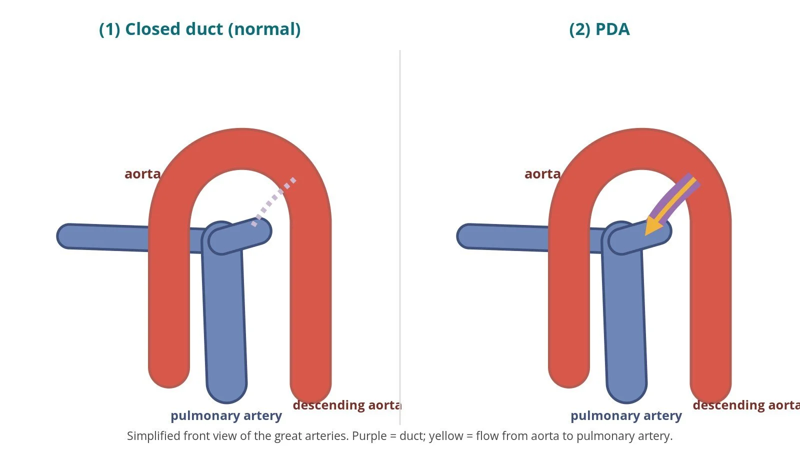
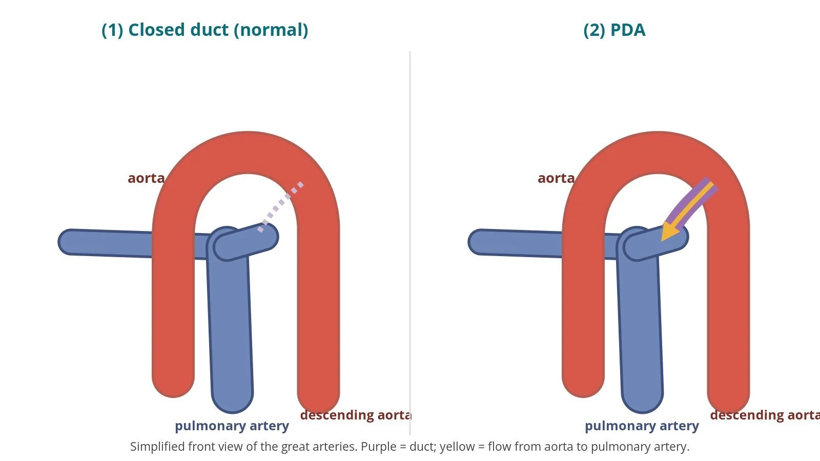
(1) After birth the duct normally closes. (2) PDA: blood flows from the aorta into the pulmonary artery, overloading the lungs.
What is a PDA?
Before birth a baby's lungs are not used, because oxygen comes from the placenta. The ductus arteriosus lets blood leaving the right side of the heart bypass the lungs by flowing from the pulmonary artery into the aorta.
After birth, when the baby starts breathing, the duct normally narrows and closes within a few days and becomes a small band of tissue. If it stays open, it is called a patent ("open") ductus arteriosus (PDA). It is more common in babies born early.
After birth, pressure in the aorta is higher than in the pulmonary artery, so blood flows the other way: from the aorta into the pulmonary artery. This sends extra blood to the lungs and back to the left side of the heart. A small PDA may cause only a heart murmur. A large PDA can enlarge the left heart, cause breathlessness, poor feeding and weight gain in babies, and over time raise the pressure in the lungs (pulmonary hypertension). A PDA also carries a small risk of infection (endarteritis).
In some babies with other heart problems, the duct is vital and is deliberately kept open with medicine until surgery.
New to the acronyms? See the Cath Lab Abbreviations A–Z.
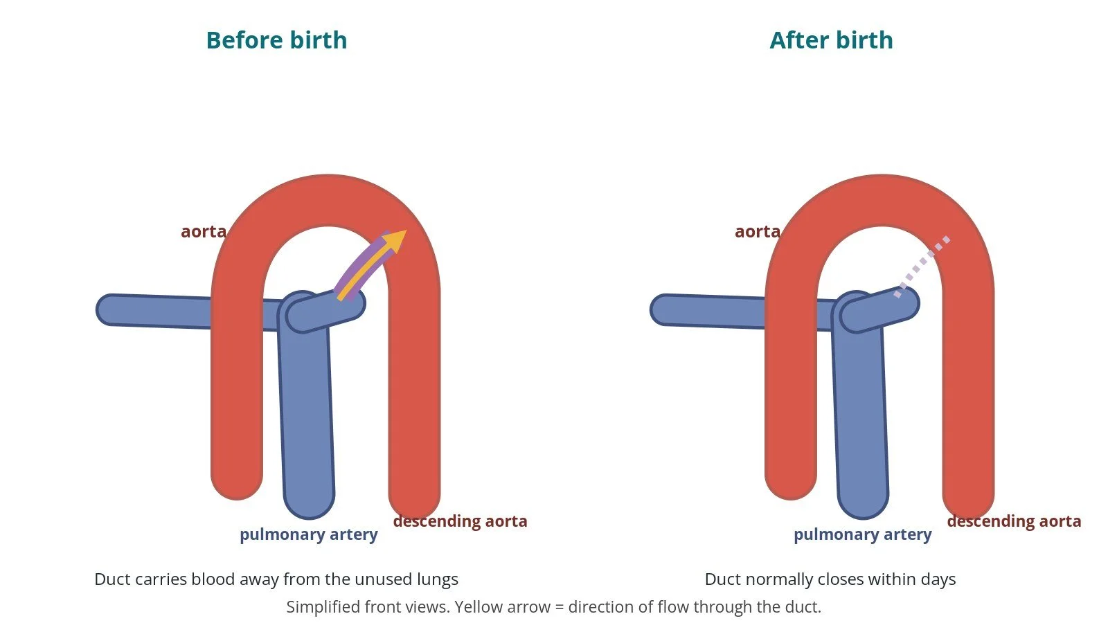
Before birth the duct lets blood bypass the lungs (pulmonary artery to aorta). After birth it normally closes within days.
Who is it for?
The decision is made by a paediatric or adult congenital heart team. In line with the ESC 2020 guidelines for adult congenital heart disease, closure is generally recommended when a PDA causes left heart enlargement without severe pulmonary hypertension, and it may be considered in other selected cases. Lung pressures are checked carefully, because closing a PDA when lung pressures are very high can be harmful.
NICE guidance on endovascular PDA closure (IPG97, now HTG59) supports the procedure with normal arrangements, in units with cardiac surgical support. Very small, "silent" PDAs found by chance often need no treatment. Premature babies are usually treated first with medicines or, if needed, surgery or specialist catheter techniques, which are outside this page.
When is it done?
It is usually a planned procedure. Before it, the team usually:
uses echo (and sometimes MRI or CT) to measure the duct and its shape
checks the pulses in the groins, blood tests and allergies
plans the anaesthetic: general anaesthetic for children, local anaesthetic and sedation for many adults
explains the procedure and alternatives, including surgery
Step by step
1. Anaesthetic and set-up
X-ray, often biplane (two cameras), is used throughout, sometimes with echo. Antibiotics are given as per centre protocol.
2. Access
A sheath is placed in the femoral vein and often a small sheath in the femoral artery, under ultrasound. Sizes are per device and operator.
Heparin is given, dose as per centre protocol.
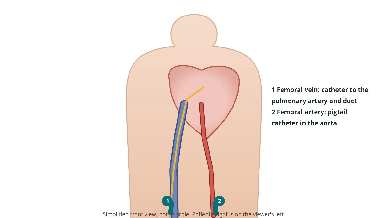
Access: (1) femoral vein, catheter up to the pulmonary artery and across the duct; (2) femoral artery for a pigtail catheter in the aorta.
3. Measuring and closing the duct
A. Picture and measure. A pigtail catheter in the aorta injects contrast (an aortogram) to show the duct's shape and size. Pressures in the pulmonary artery may be measured.
B. Cross the duct. A catheter and wire are passed across the duct, usually from the pulmonary artery side (via the vein and right heart) into the descending aorta, or from the aortic side, per operator. The wire is exchanged for a stiff exchange wire if needed and the delivery sheath is placed.
C. Deploy. The occluder (a self-expanding mesh plug) or a coil is pushed out so that it fills the duct, with its larger end on the aortic side. Device choice and size are per device and operator.
D. Check and release. A repeat aortogram checks the position, any leak, and that the device does not narrow the aorta or the left pulmonary artery. The device is then released.
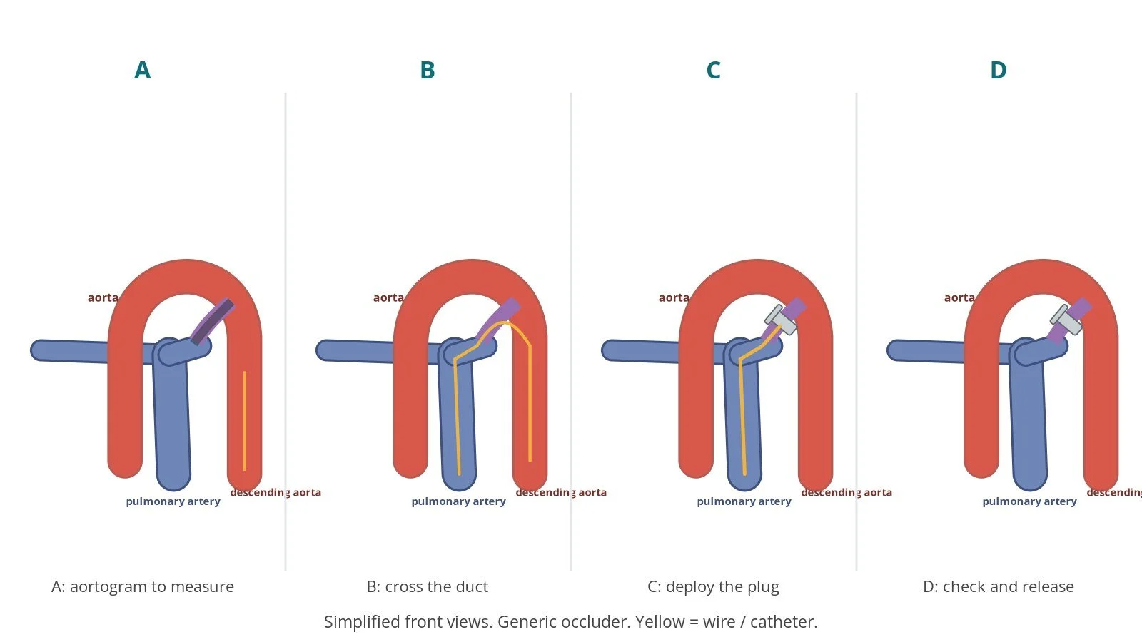
A: measure with an aortogram. B: cross the duct. C: deploy the plug. D: check and release.
4. Finishing
The sheaths are removed and pressure is applied to the groin. In small children the leg pulses are checked carefully afterwards.
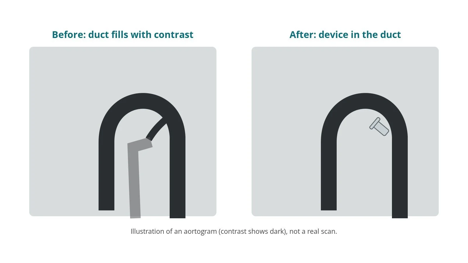
Illustration, not a real scan. Before: contrast crosses the duct. After: the device blocks it.
Recovery and aftercare
First hours: bed rest with checks of the groin, leg pulses, blood pressure and heart rate, as per centre protocol.
Going home: usually the next day after an echo and sometimes a chest X-ray.
Activity: avoid heavy lifting or rough play for about a week, or as advised.
Follow-up: echo checks with the congenital heart team.
Driving (adults): ask your team; follow the DVLA guidance for congenital heart disease.
Call 999 if there is heavy bleeding from the groin that does not stop with firm pressure, or the leg becomes cold, pale or painful.
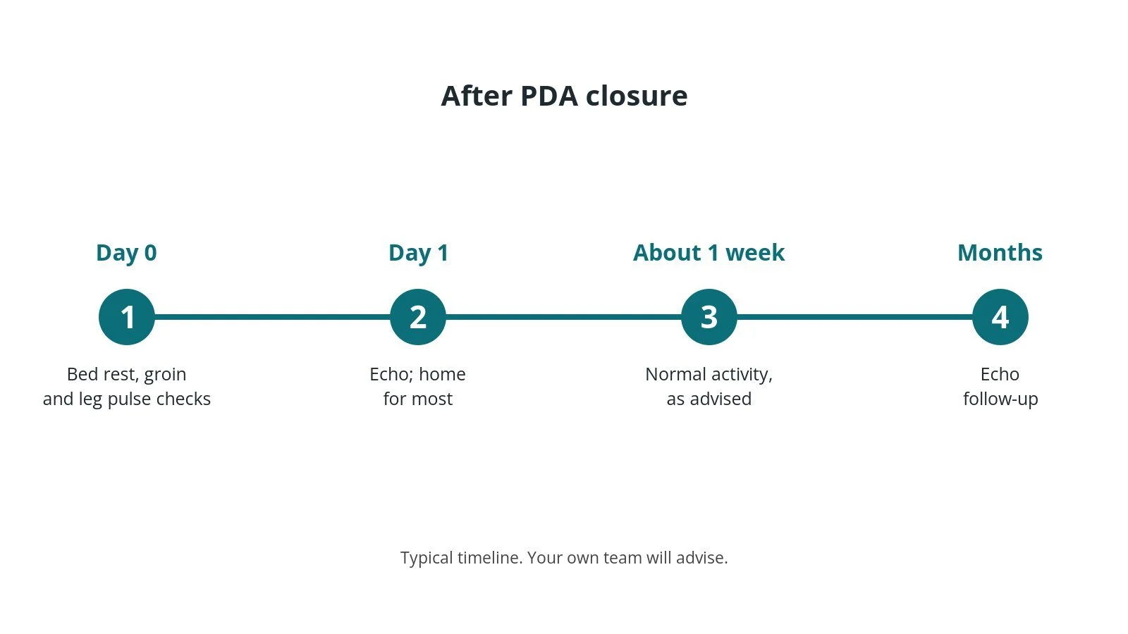
After PDA closure: (1) bed rest and checks; (2) echo, home; (3) normal activity after about a week; (4) echo follow-up.
Risks and complications
Your team will discuss your own risks. Possible problems include:
bleeding, bruising or a clot at the groin, including loss of the leg pulse in small children
the device or coil moving (embolisation), which usually can be retrieved by catheter
a small remaining leak
narrowing of the aorta or left pulmonary artery by the device
damage to red blood cells (haemolysis) if a leak is fast, which is rare
reaction to contrast or anaesthetic
Cath lab kit list (printable)
Barts example kit list
An example from one UK centre, written generically. Every lab has its own protocol, and sizes and doses vary.
angiography pack and femoral access needle
femoral venous sheath and femoral arterial sheath, sizes per device and operator
stiff exchange guidewire, J-tip, length per operator
right coronary-shaped and multipurpose-shaped catheters (venous side), per operator
pigtail catheter (arterial side) and power injector for aortograms
flush set and saline; heparin and local anaesthetic, as per centre protocol
PDA occluder or coil (size per device and operator) and delivery system
snare and retrieval kit available
Procedural steps (Barts example)
General anaesthetic (or sedation); biplane X-ray
Femoral venous and arterial access; heparin as per centre protocol
Aortogram via pigtail to size the duct
Duct crossed (usually from the pulmonary artery side); delivery sheath placed
Device deployed with its larger end in the aortic end of the duct
Repeat aortogram; device released
Sheaths removed; haemostasis; leg pulses checked
First time in the cath lab?
💡 First-time tip
Children are not small adults. Contrast, fluid and heparin are worked out by weight; double-check every syringe with the team. Keep them warm. Babies and small children lose heat quickly in the lab. Have the snare ready. Know where retrieval kit is before the device opens. Check the leg. After the case, the foot on the access side should be pink and warm with a pulse. Talk to the family. Parents are often waiting anxiously; keep them updated as per your unit's practice.
FAQ
Will the PDA come back?
Once closed with a device and healed over, it is very unlikely to reopen. Echo checks confirm this.
Does my child need surgery instead?
Most PDAs in children and adults can be closed by catheter. Surgery is used for very large ducts, unusual shapes or some premature babies.
Is there a scar?
No chest scar; just a small puncture in the groin.
Can I have an MRI scan afterwards?
Most PDA devices and coils are labelled MR conditional. Always tell the scanning team.
Related Cath Lab 101 guides
Sources
All checked on 10 October 2026. Each page's own date is shown where it gives one.
NICE. Endovascular closure of patent ductus arteriosus (IPG97, now HTG59). nice.org.uk/guidance/htg59 (published 27 Oct 2004)
Baumgartner H, et al. 2020 ESC Guidelines for the management of adult congenital heart disease. Eur Heart J 2021;42(6):563–645. PubMed 32860028
NHS. Congenital heart disease: types. nhs.uk
British Heart Foundation. Congenital heart conditions. bhf.org.uk
DVLA / GOV.UK. Cardiovascular disorders: assessing fitness to drive (congenital heart disease). gov.uk
Disclaimer
Education only. This page is for general education for healthcare students, cath lab staff and curious readers. It isn't medical advice and doesn't replace advice from your own doctor, heart team or local unit protocols. Equipment and medicines are described generically. No product or manufacturer is recommended. If you think someone is having a heart emergency, call 999.
Disclosure: I work for a company that makes a device used in this procedure. This page is independent education, isn't sponsored or reviewed by any company, and doesn't recommend any product.
All diagrams on this page are original illustrations created for rodnieoro.com.