Balloon mitral valvuloplasty (BMV) for mitral stenosis: a Cath Lab 101 guide
Reviewed by Rodnie Oro, cath lab charge nurse · Last reviewed: October 2026 · Education only, not medical advice
Balloon mitral valvuloplasty in one minute
What it is: in mitral stenosis the mitral valve, between the left atrium and left ventricle, becomes narrowed and stiff, most often after rheumatic fever. Balloon mitral valvuloplasty (BMV), also called percutaneous mitral commissurotomy, opens the valve by inflating a special balloon inside it, passed up a vein from the groin and across the wall between the atria.
Who it is for: people with significant mitral stenosis causing symptoms, whose valve shape is suitable, as decided by a heart team. It is also used in pregnancy in selected cases.
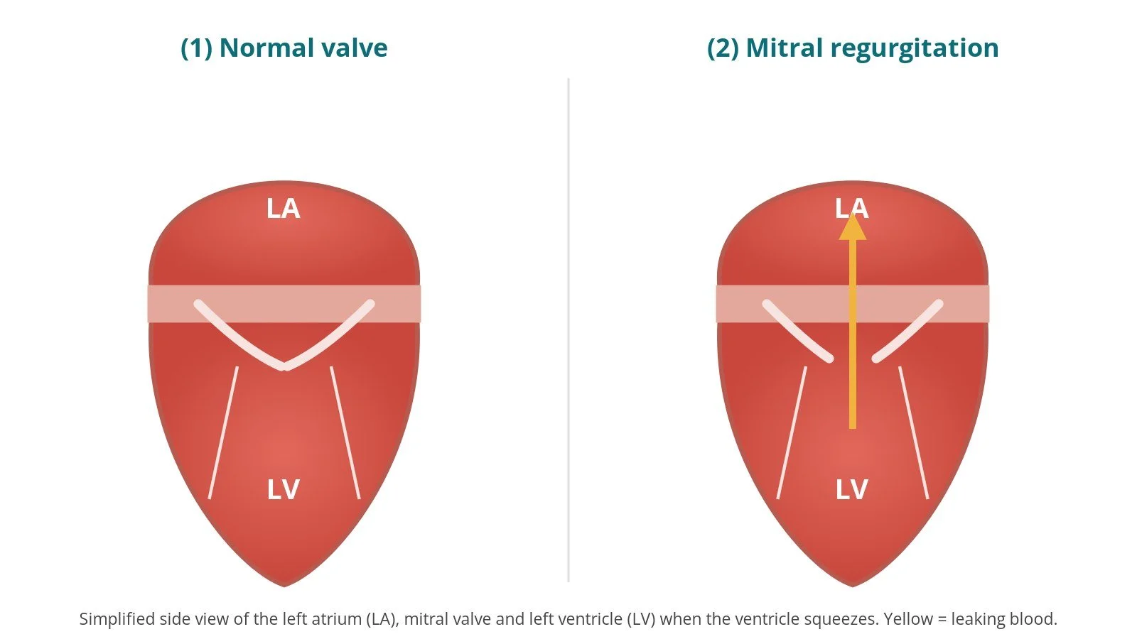
Main risks: a leak in the valve after stretching (mitral regurgitation), fluid around the heart, stroke, and a small hole left in the septum. Your team will explain your own risk.
Usually: under local anaesthetic with sedation or general anaesthetic, guided by echo, about 1 to 2 hours, often home the next day.
On this page: What is balloon mitral valvuloplasty? · Who is it for? · When is it done? · Step by step · Recovery · Risks · Kit list · First time in the lab? · FAQ
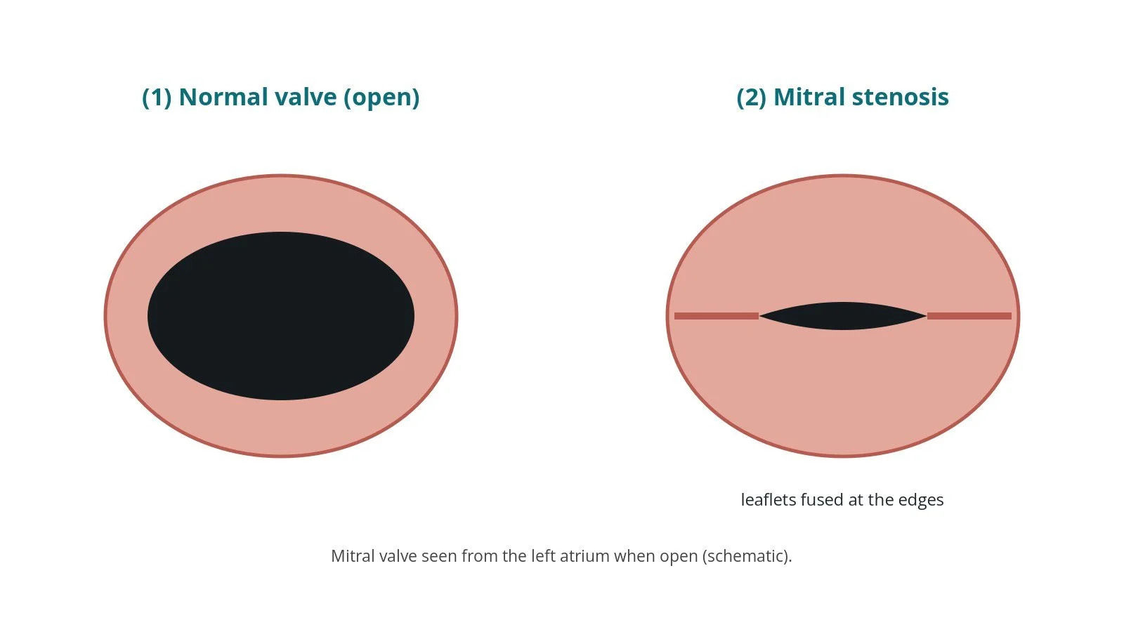
(1) Normal mitral valve, wide open. (2) Mitral stenosis: the leaflets are fused at the edges, leaving a narrow opening.
What is balloon mitral valvuloplasty?
The mitral valve lets blood flow from the left atrium (LA) into the left ventricle (LV). In mitral stenosis, the two leaflets become thickened and stuck together at their edges (the commissures), so the opening narrows. The most common cause worldwide is rheumatic heart disease, after rheumatic fever in childhood; in older people, calcium build-up can also narrow the valve.
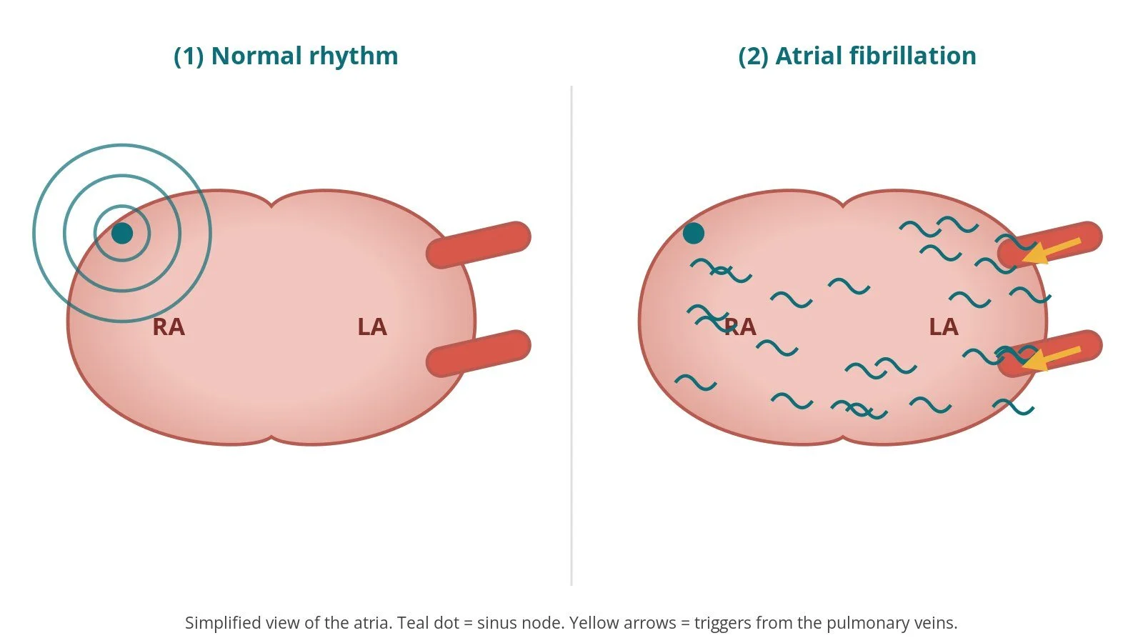
Because blood cannot easily leave the LA, pressure builds up behind the valve. The LA enlarges, which can lead to atrial fibrillation (AF), clots and stroke, and pressure rises in the lungs, causing breathlessness, tiredness and sometimes coughing up blood.
Balloon mitral valvuloplasty (BMV) splits the fused commissures by inflating a balloon across the valve. A waisted, self-positioning balloon is commonly used: it inflates in stages so it sits in the valve opening before the middle expands. The valve opening becomes wider, and pressure in the LA and lungs falls.
New to the acronyms? See the Cath Lab Abbreviations A–Z.
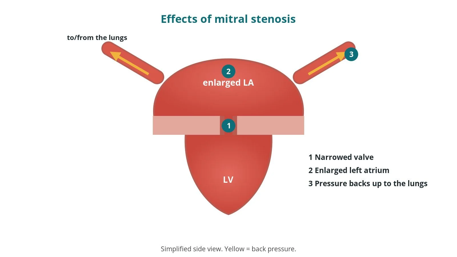
Effects of mitral stenosis: (1) narrowed valve; (2) enlarged left atrium; (3) pressure backs up into the lungs.
Who is it for?
NICE guidance on heart valve disease (NG208) and the ESC/EACTS valvular heart disease guidelines describe BMV as a treatment for people with significant (moderate to severe) rheumatic mitral stenosis and symptoms, when the valve anatomy is favourable. The team uses echo to look at how thick, mobile and calcified the leaflets and supporting cords are, and how much leak there is already.
BMV is generally not suitable if there is a clot in the left atrium, more than mild mitral regurgitation, or heavy calcium. Then valve surgery may be better. Calcified mitral stenosis in older people usually responds poorly to a balloon.
When is it done?
It is usually planned. Before it, the team usually arranges:
echo, including TOE on or before the day, to exclude clot in the left atrium and its appendage and to score the valve
an ECG and blood tests
a review of anticoagulants (often taken for AF), as per centre protocol
in pregnancy, careful planning with the obstetric team to limit X-ray
Step by step
1. Anaesthetic and set-up
BMV is done under local anaesthetic with sedation or general anaesthetic, depending on whether TOE guidance is used. X-ray is used throughout. Some centres also use intracardiac echo.
2. Access and transseptal puncture
A sheath is placed in the right femoral vein, under ultrasound; sizes are per device and operator. Some operators place a suture-based closure device.
A transseptal needle and sheath cross the atrial septum into the LA, guided by X-ray and echo. Heparin is given after the crossing, dose as per centre protocol.
Pressures in the LA (and often the LV, via an artery) are measured to calculate the gradient across the valve.
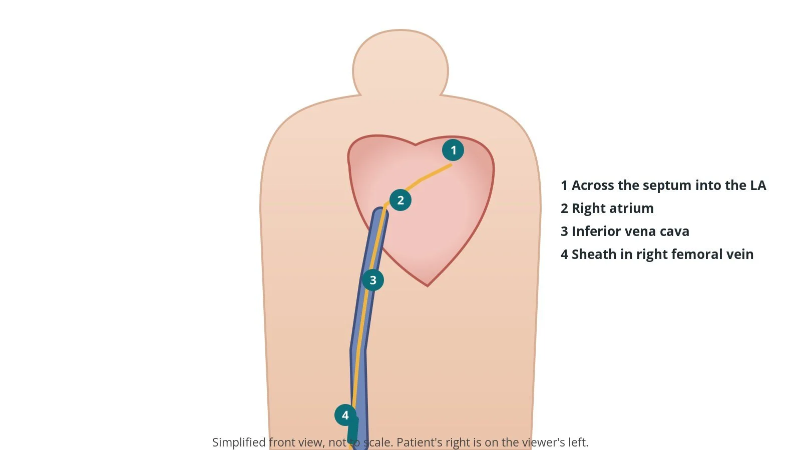
Access: (1) across the septum into the left atrium; (2) right atrium; (3) inferior vena cava; (4) sheath in the right femoral vein.
3. Balloon dilatation
A. Wire into the LA. A coiled wire is placed in the LA and the septum is gently dilated so the balloon can pass.
B. Cross the valve. The balloon is passed over the wire into the LA, then steered through the mitral valve into the LV with a stylet.
C. Inflate. The far part of the balloon is inflated first and pulled back against the valve, then the whole balloon is inflated so its waist splits the fused edges. The balloon is sized from the patient's height and valve, and the volume increased in steps, per device and operator.
D. Check. After each inflation, the gradient and the echo are checked for the valve opening and any new leak. Further inflations are made in small steps until the result is good, or stopped if the leak increases.
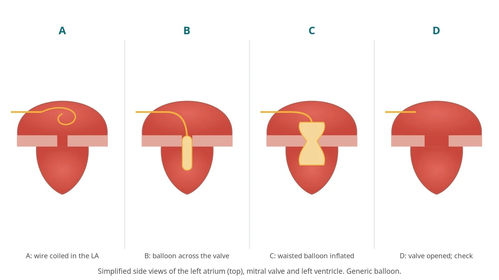
A: wire in the left atrium. B: balloon across the valve. C: waisted balloon inflated. D: valve opened; check.
4. Finishing
The balloon is removed. The size of the hole in the septum is checked on echo. The sheath is removed and the vein is closed with pressure or the closure device, per operator.
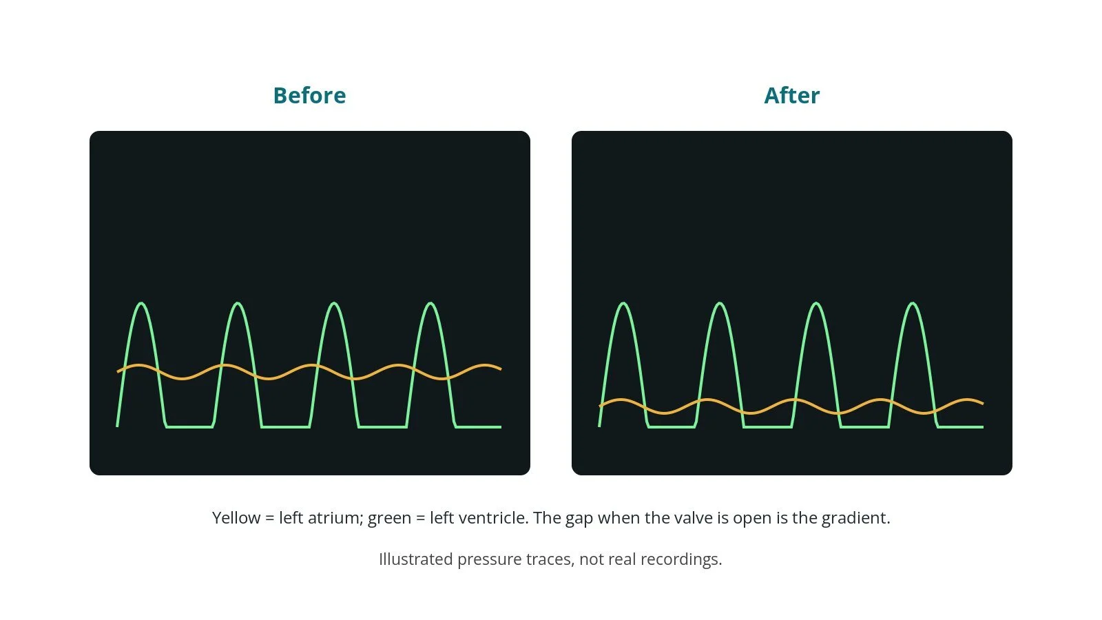
Illustrated, not a real recording. Before: large pressure gap across the valve. After: much smaller gap.
Recovery and aftercare
First hours: bed rest with checks of the groin, blood pressure and heart rhythm, as per centre protocol.
Going home: usually the next day after an echo.
Medicines: anticoagulants restarted if needed. Antibiotics to prevent rheumatic fever returning may continue, as advised.
Activity: avoid heavy lifting for about a week, or as advised.
Follow-up: regular echo checks, as the valve can narrow again over years.
Driving: DVLA guidance after a percutaneous valve procedure for a car licence is not to drive for 2 weeks; bus and lorry drivers must tell the DVLA. Ask your team.
Call 999 if you have chest pain, sudden breathlessness, fainting, signs of a stroke, or heavy groin bleeding.
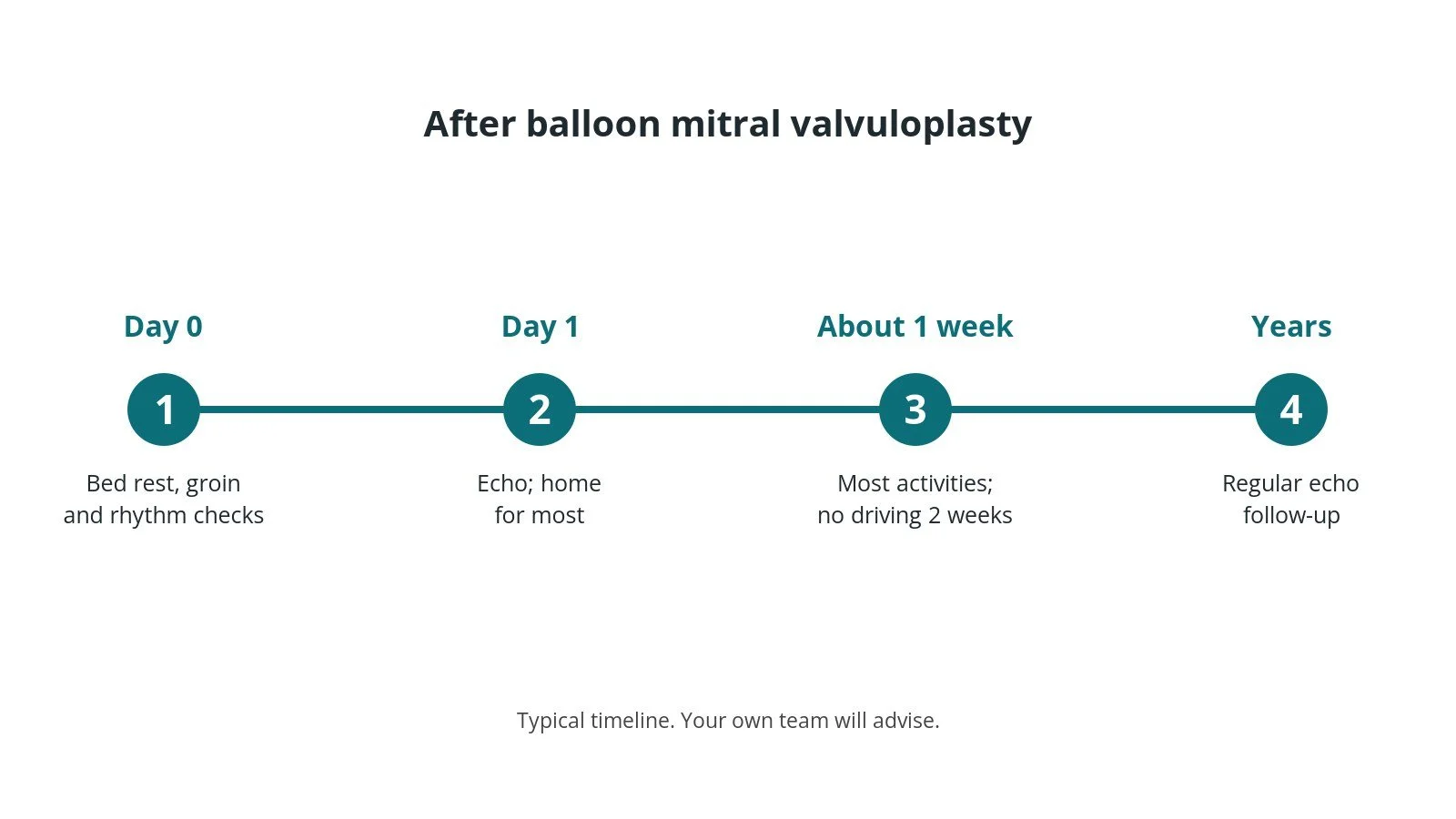
After BMV: (1) checks; (2) echo, home; (3) most activities after about a week; (4) regular echo follow-up.
Risks and complications
Your team will discuss your own risks. Possible problems include:
a new or worse mitral regurgitation (leak), sometimes needing surgery
fluid around the heart (tamponade) from the transseptal puncture or wire
stroke from a clot or air
a small remaining hole in the atrial septum, which usually closes or causes no problems
bleeding at the groin
the valve narrowing again in later years
Cath lab kit list (printable)
Barts example kit list
An example from one UK centre, written generically. Every lab has its own protocol, and sizes and doses vary.
angiography pack and femoral access needle
femoral venous sheath, size per device and operator
suture-based vascular closure device, per operator
waisted mitral valvuloplasty balloon set (balloon, stylet, coiled wire, septal dilator), size per device and operator
large and medium Luer-lock syringes for inflation
contrast and saline mix for the balloon, as per centre protocol
transseptal needle and transseptal sheath, per operator
flush set; heparin and local anaesthetic, as per centre protocol
TOE or intracardiac echo
Procedural steps (Barts example)
Anaesthetic or sedation; TOE confirms no LA clot
Right femoral venous access; closure suture if used
Transseptal puncture; heparin as per centre protocol; pressures measured
Coiled wire into the LA; septum dilated
Balloon across the valve; staged inflations, checking gradient and leak each time
Balloon removed; septum checked on echo
Sheath removed; haemostasis
First time in the cath lab?
💡 First-time tip
Prepare the balloon carefully. Use the correct contrast mix and measured syringe volumes; the operator will call out each inflation volume. Record the gradients. Pressures before and after each inflation guide the next step. Watch the echo for a leak. A new severe leak means stopping. Respect the air. The balloon is in the left heart; keep everything bubble-free. Watch the pressure. A falling blood pressure can mean fluid around the heart.
FAQ
Is this the same as valve replacement?
No. BMV opens your own valve; nothing is left behind.
Will the valve narrow again?
It can, over years. Some people can have a repeat BMV; others later need surgery.
Can BMV be done in pregnancy?
Yes, in selected cases where symptoms cannot be controlled with medicines, with careful planning to limit X-ray exposure.
Will I need antibiotics long term?
Some people with rheumatic heart disease take antibiotics to prevent rheumatic fever returning. Your team will advise.
Related Cath Lab 101 guides
Sources
All checked on 10 October 2026. Each page's own date is shown where it gives one.
NICE. Heart valve disease presenting in adults: investigation and management (NG208). nice.org.uk/guidance/ng208
Vahanian A, et al. 2021 ESC/EACTS Guidelines for the management of valvular heart disease. Eur Heart J 2022;43(7):561–632. PubMed 34453165
NHS. Heart valve disease. nhs.uk/conditions/heart-valve-disease
British Heart Foundation. Heart valve disease. bhf.org.uk
DVLA / GOV.UK. Cardiovascular disorders: assessing fitness to drive (heart valve surgery, including percutaneous valve intervention). gov.uk
Disclaimer
Education only. This page is for general education for healthcare students, cath lab staff and curious readers. It isn't medical advice and doesn't replace advice from your own doctor, heart team or local unit protocols. Equipment and medicines are described generically. No product or manufacturer is recommended. If you think someone is having a heart emergency, call 999.
Disclosure: I work for a company that makes a device used in this procedure. This page is independent education, isn't sponsored or reviewed by any company, and doesn't recommend any product.
All diagrams on this page are original illustrations created for rodnieoro.com.