LAMPOON (mitral leaflet laceration before TMVR): a Cath Lab 101 guide
Reviewed by Rodnie Oro, cath lab charge nurse · Last reviewed: October 2026 · Education only, not medical advice
LAMPOON in one minute
What it is: LAMPOON (Laceration of the Anterior Mitral leaflet to Prevent left ventricular Outflow ObstructioN) is a catheter technique done just before transcatheter mitral valve replacement (TMVR). An electrified wire splits the front (anterior) mitral leaflet down the middle, so that when the new valve opens, the leaflet does not get pushed into the outflow tract and block blood leaving the heart.
Who it is for: people having TMVR (for example inside a surgical ring, or in a valve with heavy calcium) whose CT predicts a high risk of left ventricular outflow tract (LVOT) obstruction, as decided by a heart team. It is a specialist technique done in experienced centres.
Main risks: stroke, damage to the heart or valve structures, a severe leak before the new valve is placed, LVOT obstruction despite the procedure, and the risks of TMVR. Your team will explain your own risk.
Usually: under general anaesthetic with TOE, with TMVR in the same procedure, about 3 to 4 hours in total, often home after a few nights.
On this page: What is LAMPOON? · Who is it for? · When is it done? · Step by step · Recovery · Risks · Kit list · First time in the lab? · FAQ
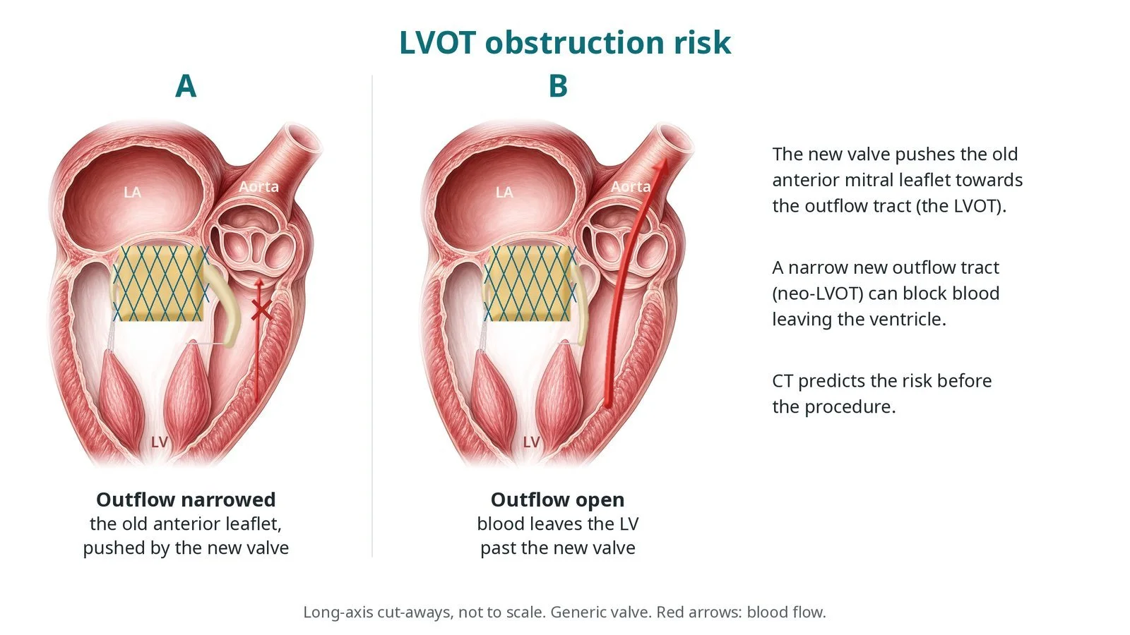
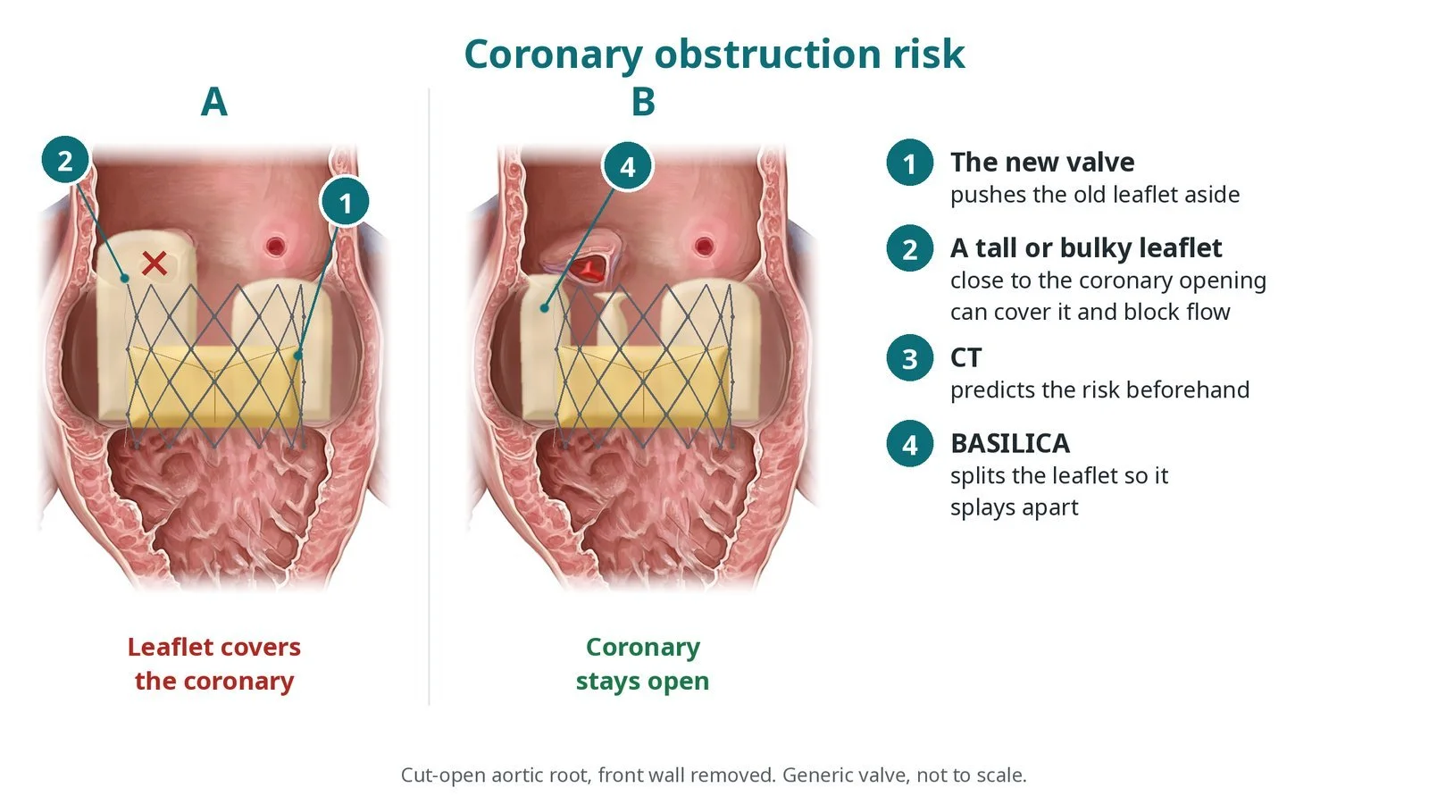
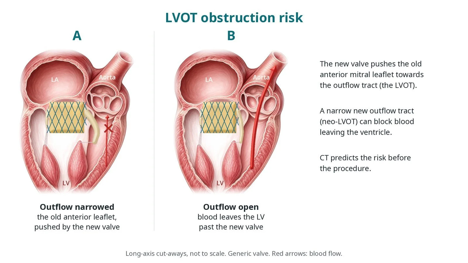
LVOT obstruction risk: (A) the anterior mitral leaflet, pushed by the new valve, narrows the outflow tract; (B) outflow open.
What is LAMPOON?
The anterior mitral leaflet sits right next to the left ventricular outflow tract (LVOT), the channel through which blood leaves the left ventricle for the aorta. When a transcatheter valve is expanded in the mitral position, it pushes the anterior leaflet towards the LVOT. In some people, the leaflet and new valve together can narrow or block the LVOT, which is dangerous.
LAMPOON splits the anterior leaflet before the new valve goes in:
a wire crosses the base of the leaflet using a brief burst of electrosurgical energy
it is caught by a snare in the left atrium and brought out to form a loop
pulling the energised wire splits the leaflet from base to tip
when the new valve opens, the two halves of the leaflet splay aside, and blood can pass through the frame of the new valve into the LVOT
See the TMVI guide for transcatheter mitral valve implantation.
New to the acronyms? See the Cath Lab Abbreviations A–Z.
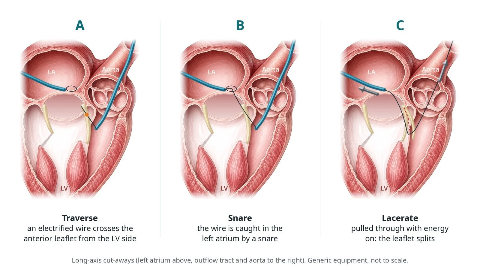
LAMPOON in outline: (A) an electrified wire crosses the anterior leaflet from the LV side; (B) it is caught by a snare in the left atrium; (C) pulled through with energy on, it splits the leaflet.
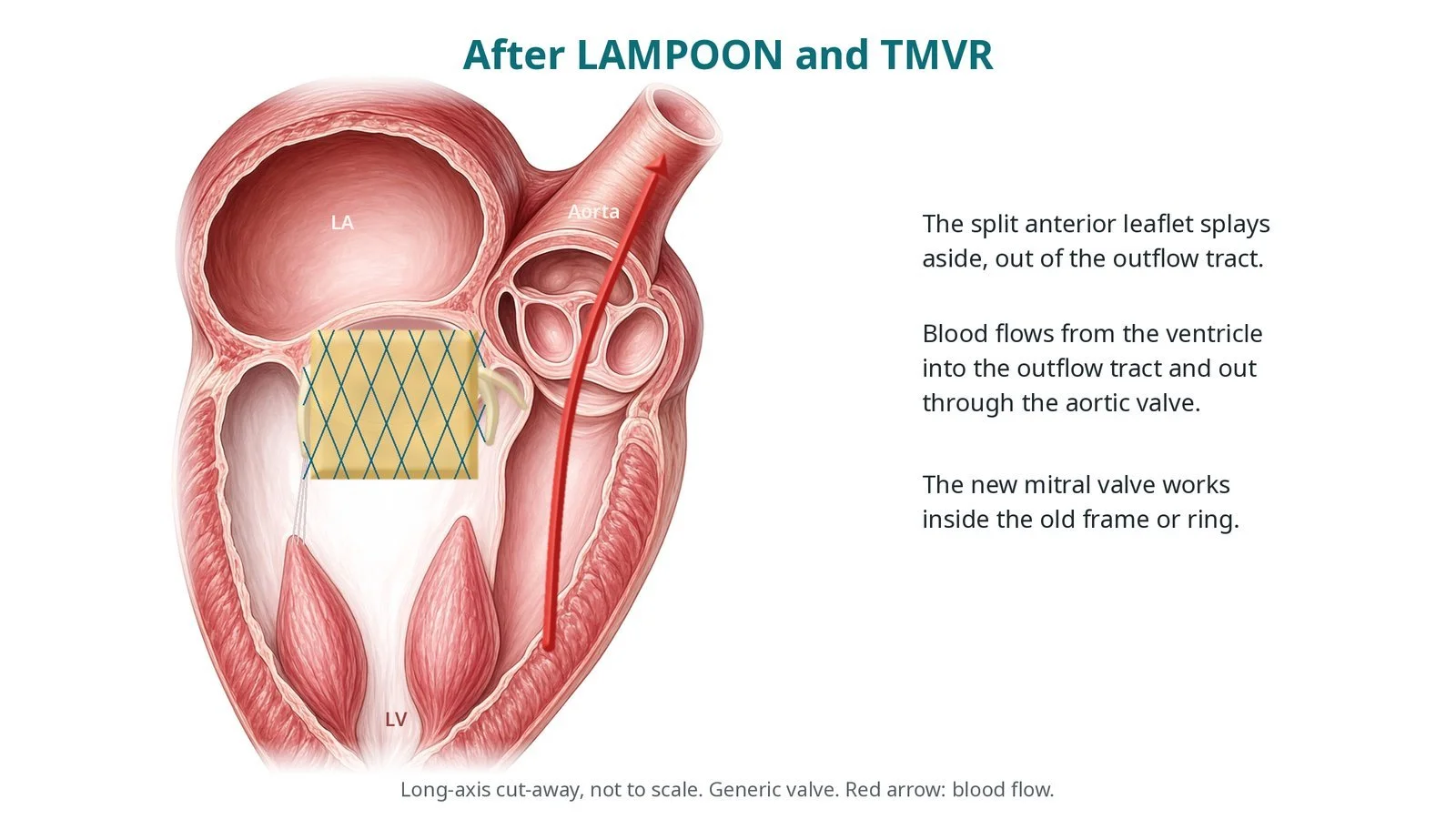
After LAMPOON and TMVR, the split leaflet splays aside and blood can flow into the outflow tract.
Who is it for?
The ESC/EACTS valvular heart disease guidelines and NICE guidance on mitral valve-in-valve (HTG592) and valve-in-ring (HTG593) implantation recognise LVOT obstruction as a key risk of transcatheter mitral procedures. CT predicts the size of the remaining outflow channel (the neo-LVOT); if it is too small, the heart team may consider LAMPOON, other techniques or a different plan. LAMPOON is a specialist, evolving technique, and consent includes discussion of this.
When is it done?
It is planned with the TMVR. Before it, the team usually arranges:
detailed CT planning, including the predicted neo-LVOT
TOE, ECG, blood tests and a group and save
a review of blood thinners, as per centre protocol
Step by step
1. Anaesthetic and access
Done under general anaesthetic with TOE. Sheaths are placed in the femoral veins (for transseptal access, pacing or monitoring) and a femoral artery (often with a long sheath); sizes per device and operator. A transseptal puncture is made and a steerable sheath placed in the left atrium. Heparin is given, dose as per centre protocol.
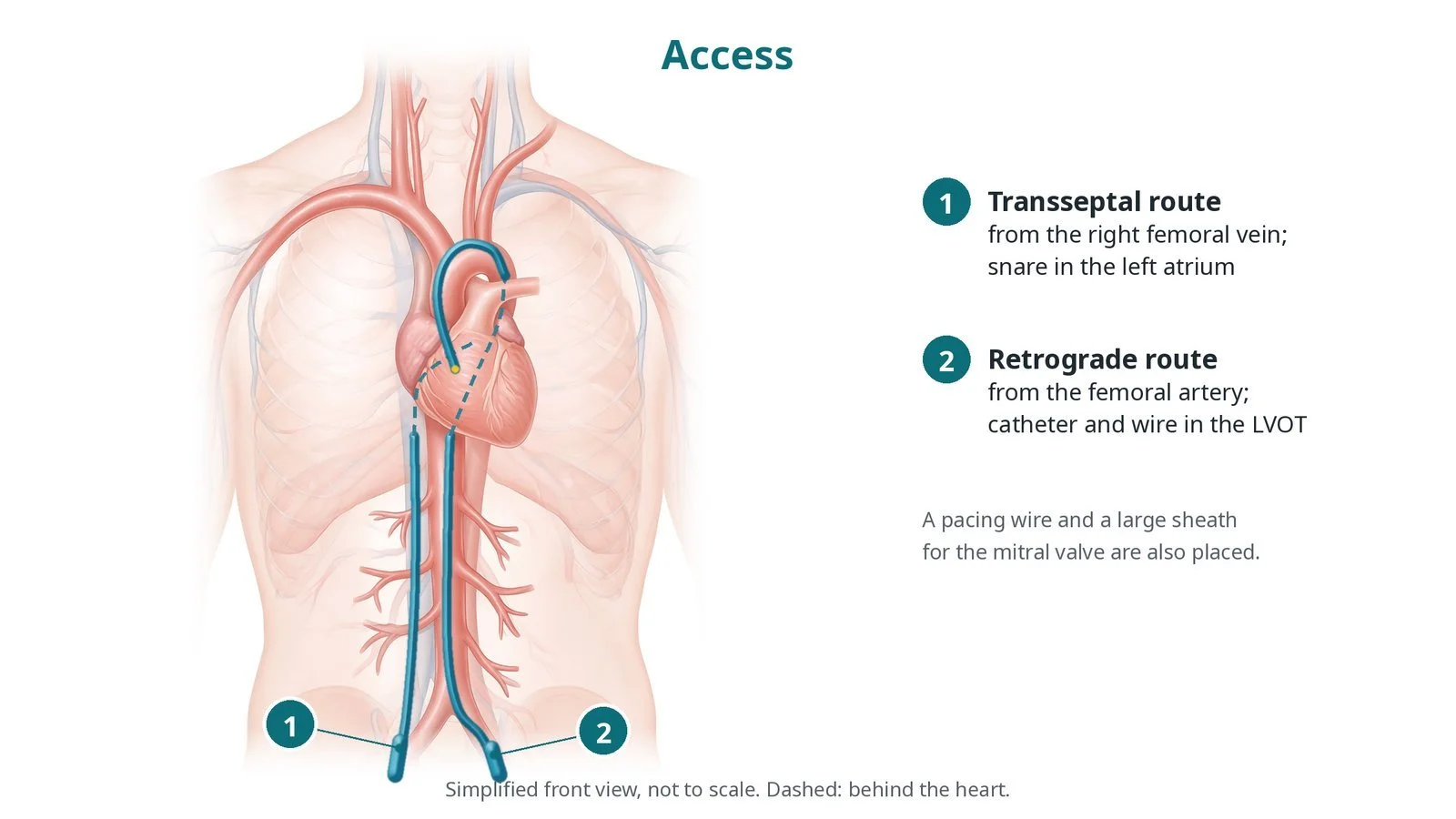
Access: (1) transseptal route from the femoral vein; (2) retrograde route from the femoral artery.
2. Leaflet laceration
A. Set up. From the femoral artery, guide catheters pass retrogradely across the aortic valve and are aimed at the base of the anterior mitral leaflet. From the transseptal side, a snare waits in the left atrium.
B. Traverse. An insulated wire, with only its tip exposed and connected to an electrosurgery pen, crosses the leaflet base with a brief burst of energy (settings per operator, with non-conducting fluid flushing as per centre protocol). It is caught by the snare in the left atrium.
C. Lacerate. The wire is brought out to form a loop with a V-shaped bend at the leaflet. Pulling the energised wire splits the leaflet from base to free edge, guided by TOE.
D. TMVR. The transcatheter mitral valve is implanted promptly, because the split leaflet can cause a leak until then.
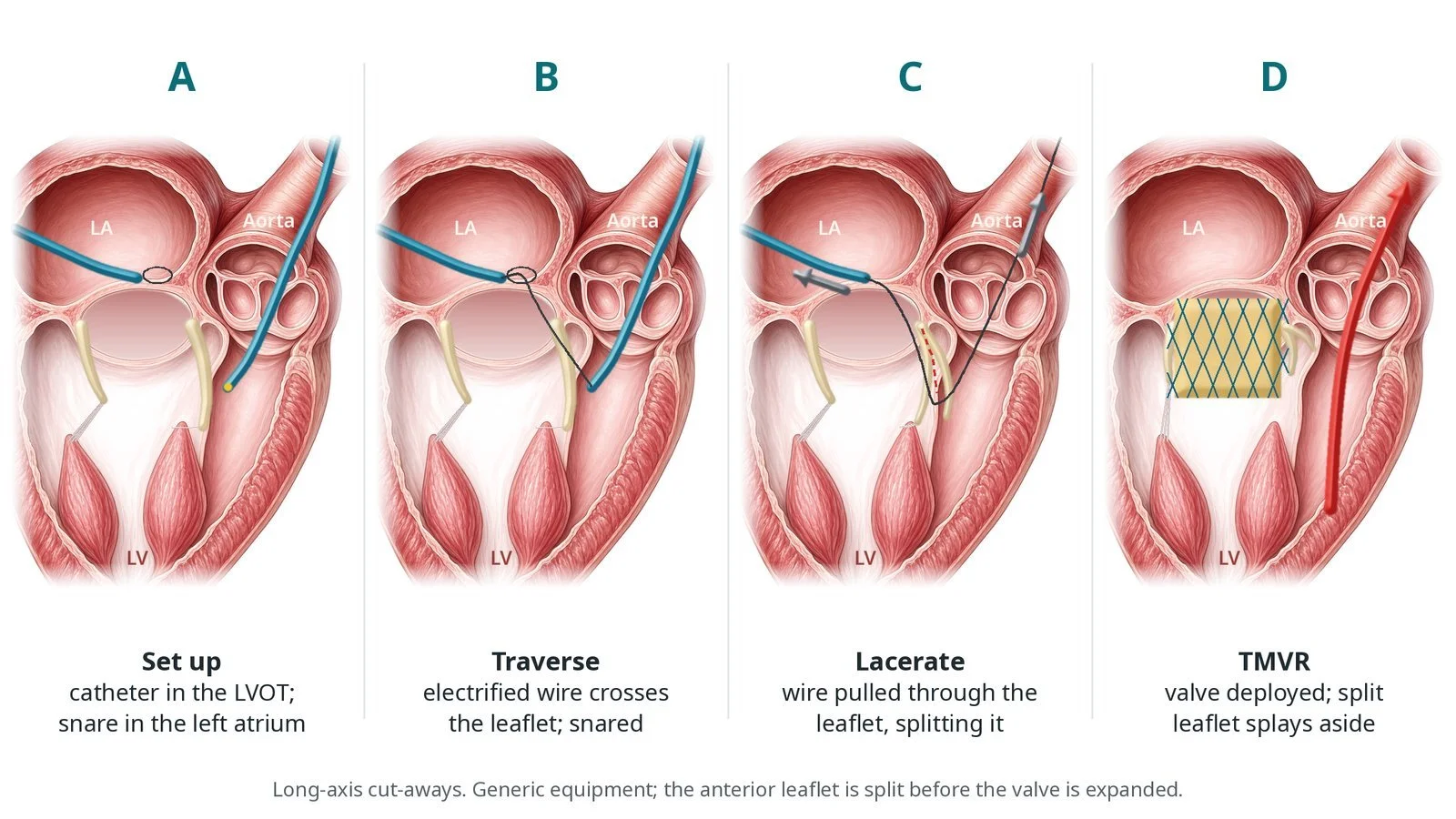
A: catheter in the LVOT and snare in the left atrium. B: the wire crosses the leaflet and is snared. C: the leaflet is lacerated. D: TMVR, with the split leaflet splayed aside.
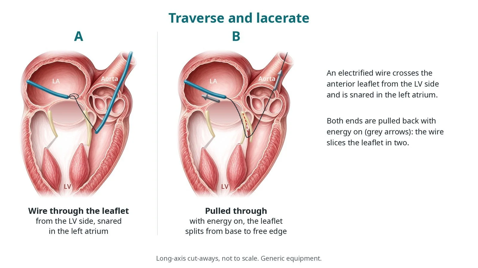
A: the wire crosses the anterior leaflet. B: pulled through with energy on, it splits the leaflet from its base towards the free edge.
3. Checking
TOE and pressure measurements check the LVOT, the new valve and any leak.
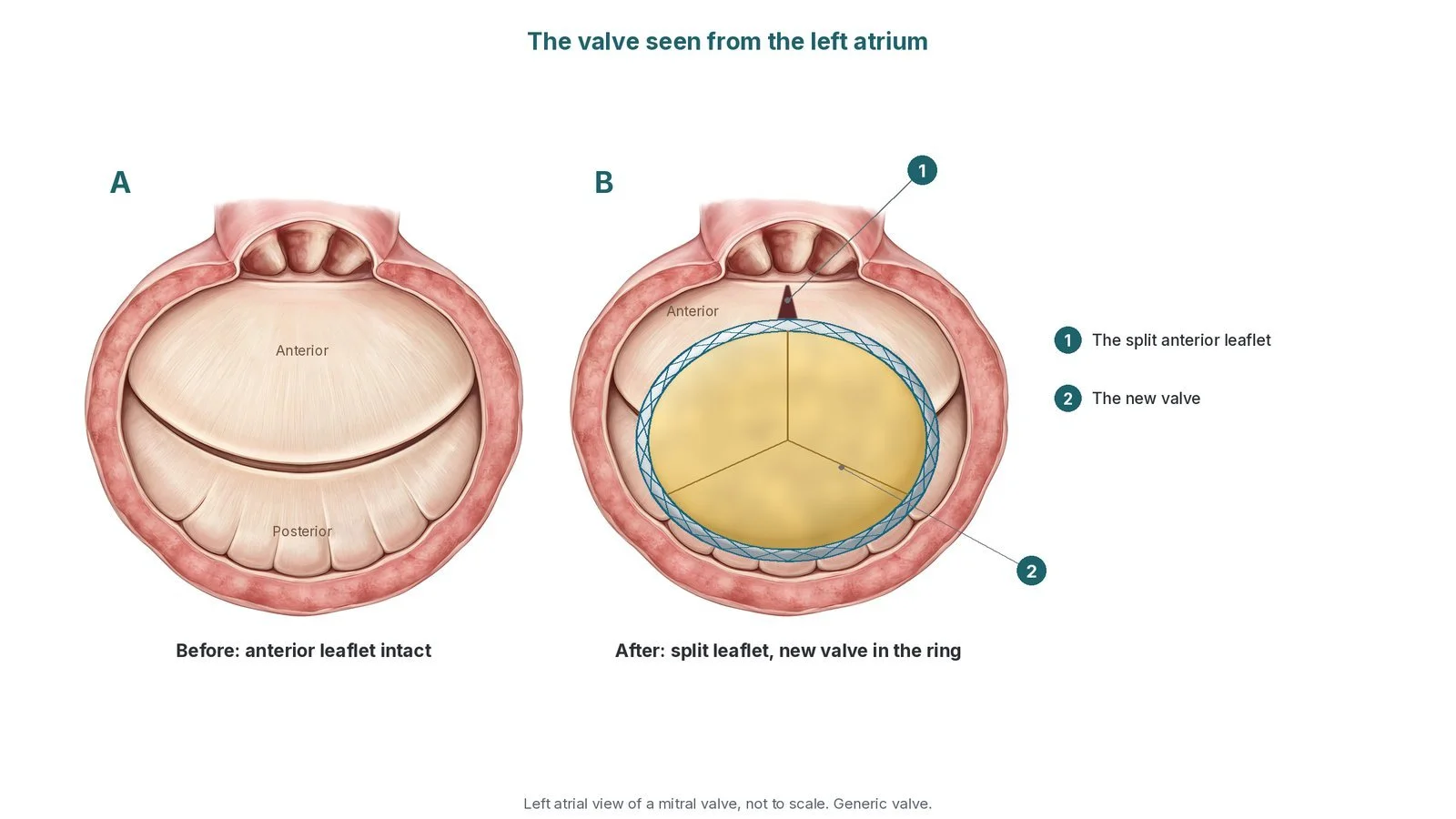
The valve seen from the left atrium. (A) Before: the anterior leaflet is intact. (B) After LAMPOON and TMVR: (1) the split anterior leaflet; (2) the new valve in the ring.
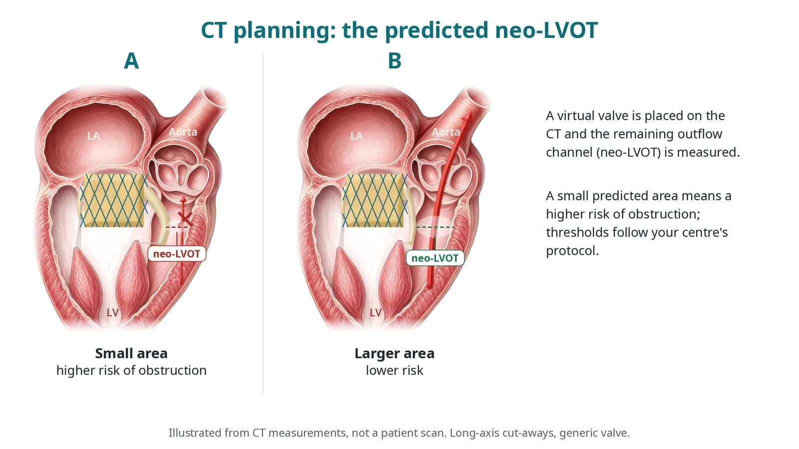
Illustrated CT planning, not a patient scan: the predicted neo-LVOT. A small area means a higher risk of obstruction; thresholds follow your centre's protocol.
Recovery and aftercare
First hours: recovery, often in a high-dependency area, with checks of the groins, heart rhythm and blood pressure, as per centre protocol.
Echo and blood tests: before discharge.
Going home: often after a few nights.
Medicines: anticoagulant medicine after a mitral valve, as per your team's plan.
Driving: DVLA guidance after a percutaneous valve procedure for a car licence is not to drive for 2 weeks; bus and lorry drivers must tell the DVLA. Ask your team.
Call 999 if you have chest pain, sudden breathlessness, fainting, signs of a stroke, or bleeding from the groin.
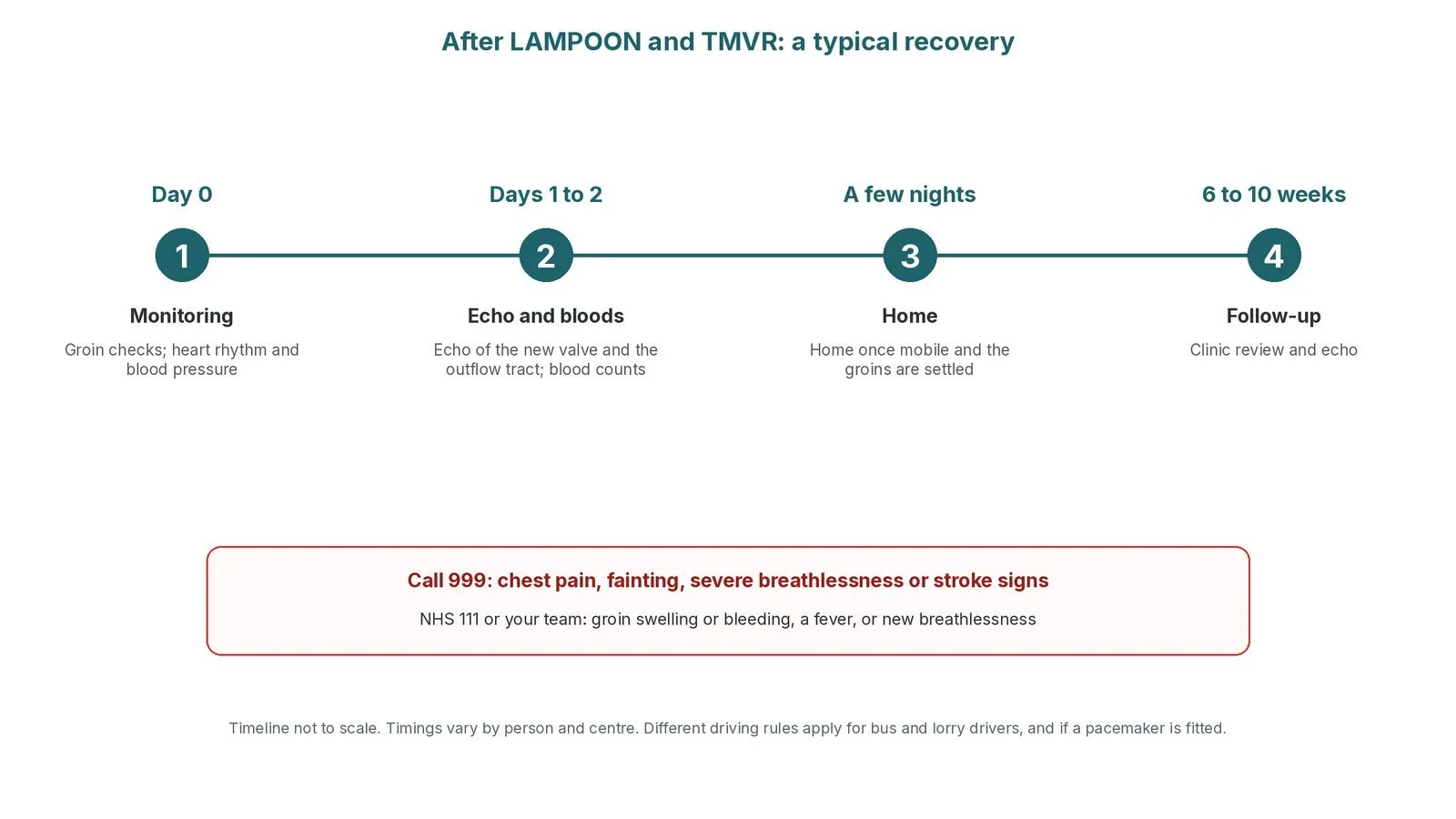
After LAMPOON and TMVR: (1) monitoring; (2) echo and bloods; (3) home after a few nights; (4) follow-up at 6 to 10 weeks.
Risks and complications
Your team will discuss your own risks. Possible problems include:
stroke
injury to the mitral valve apparatus, the aortic valve or the heart wall
a severe leak between laceration and TMVR, which is why the valve follows promptly
LVOT obstruction despite the laceration
fluid around the heart (tamponade)
bleeding or injury at the access sites
the risks of TMVR itself
Cath lab kit list (printable)
Barts example kit list
An example from one UK centre, written generically. Every lab has its own protocol, and sizes and doses vary.
Access
femoral venous sheaths (one to be upsized), femoral arterial sheath (long), sizes per device and operator
transseptal needle and transseptal sheath; steerable sheath, per operator
balloon-tipped pulmonary artery catheter, per operator
Catheters
guide catheters (left coronary shapes), per operator
pigtail catheter
microcatheter or wire converter and a braided microcatheter, per operator
Wires, snares, balloon
stiff-tipped 0.014" traversal wire (insulated except the tip), per operator; 0.014" buddy wire; long exchange wire
stiff pre-shaped LV wire and extra-stiff wire, per operator
snares (large single-loop and multi-loop), per operator; haemostasis valves; 3-way tap
balloon for septostomy, size per device and operator
Electrosurgery
diathermy pen and generator, settings per operator; non-conducting flush, as per centre protocol
TMVR equipment, per device and operator
Procedural steps (Barts example)
General anaesthetic and TOE; venous and arterial access; transseptal puncture; steerable sheath
Snare in the left atrium via the transseptal sheath
Retrograde guide catheters aimed at the anterior leaflet base
Traversal wire energised and advanced; snared in the LA; externalised
V-shaped bend at the leaflet; energised pull splits the leaflet under TOE
Septostomy as needed; TMVR implanted promptly; LVOT and valve checked
First time in the cath lab?
💡 First-time tip
Have the valve ready. TMVR follows the laceration promptly; prepare it beforehand. Diathermy only on command. Activate only when the operator asks. Know the flush. Non-conducting flush is used during energy delivery, as per centre protocol; label syringes. Many catheters, two directions. Label which catheter came from the vein and which from the artery. Watch the pressure. A big leak after laceration can drop the blood pressure.
FAQ
Why split my leaflet?
Because CT predicts the leaflet and new valve could block blood leaving the heart. Splitting it reduces that risk.
Will my valve leak afterwards?
Briefly, until the new valve is placed, which follows straight away.
Is LAMPOON widely done?
It is offered in experienced centres for selected patients.
Are there alternatives?
Yes, depending on the case, such as other ways to reduce the leaflet's effect, or surgery. Your team will discuss them.
Related Cath Lab 101 guides
Sources
All checked on 10 October 2026. Each page's own date is shown where it gives one.
NICE. Transapical transcatheter mitral valve-in-valve implantation for a failed surgically implanted mitral valve bioprosthesis (HTG592). nice.org.uk/guidance/htg592
NICE. Transapical transcatheter mitral valve-in-ring implantation after failed annuloplasty for mitral valve repair (HTG593). nice.org.uk/guidance/htg593
Vahanian A, et al. 2021 ESC/EACTS Guidelines for the management of valvular heart disease. Eur Heart J 2022;43(7):561–632. PubMed 34453165
NHS. Heart valve disease. nhs.uk/conditions/heart-valve-disease
DVLA / GOV.UK. Cardiovascular disorders: assessing fitness to drive. gov.uk
Disclaimer
Education only. This page is for general education for healthcare students, cath lab staff and curious readers. It isn't medical advice and doesn't replace advice from your own doctor, heart team or local unit protocols. Equipment and medicines are described generically. No product or manufacturer is recommended. If you think someone is having a heart emergency, call 999.
Disclosure: I work for a company that makes a device used in this procedure. This page is independent education, isn't sponsored or reviewed by any company, and doesn't recommend any product.
Diagrams are original illustrations created for rodnieoro.com, except procedure images credited in their captions.