UNICORN (valve through the leaflet in TAVI): a Cath Lab 101 guide
Reviewed by Rodnie Oro, cath lab charge nurse · Last reviewed: October 2026 · Education only, not medical advice
UNICORN in one minute
What it is: UNICORN stands for Undermining Iatrogenic Coronary Obstruction with Radiofrequency Needle. An electrified wire makes a hole near the base of the old valve leaflet in front of an at-risk coronary artery. Balloons widen the hole, and the new TAVI valve is then placed through the hole and expanded, so the leaflet is opened and pushed away from the coronary opening.
Who it is for: selected people having TAVI, mostly valve-in-valve TAVI in a failed surgical or transcatheter valve, whose CT shows a high risk of coronary obstruction and where simpler options are unlikely to work, as decided by an experienced heart team.
Evidence: first described in 2022. Evidence so far is case reports, technique papers and small retrospective registries. There are no randomised trials, long-term results are unknown, and no guideline specifically recommends it.
Main risks: a sharp drop in blood pressure during the procedure, stroke, coronary obstruction despite the procedure (including rare late blockage), a loose leaflet flap, and the usual TAVI risks. Your team will explain your own risk.
On this page: What is UNICORN? · Who is it for? · When is it done? · Step by step · Recovery · Risks · Kit list · First time in the lab? · FAQ
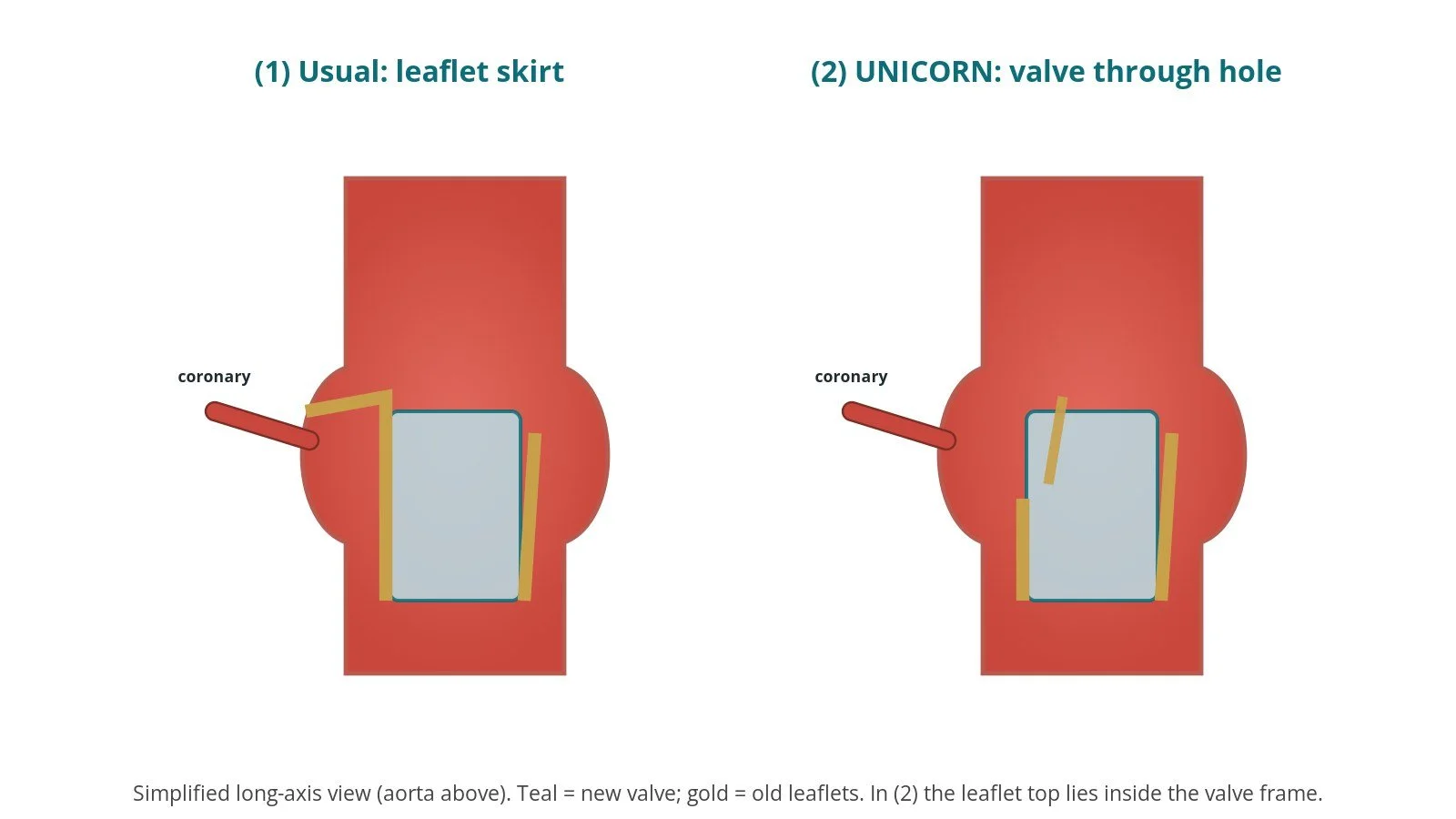
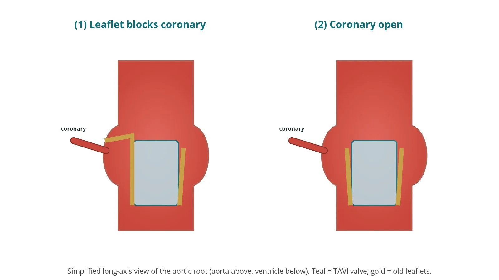
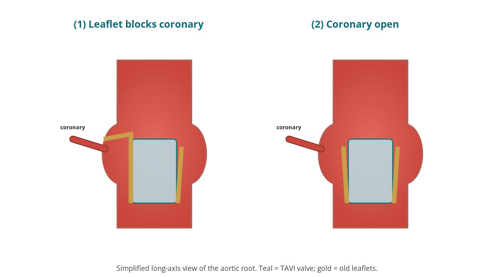
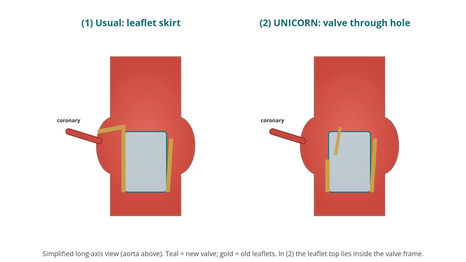
(1) Usual valve-in-valve: the old leaflet forms a skirt that can cover the coronary. (2) UNICORN: the valve goes through a hole in the leaflet, leaving a small skirt on that side.
What is UNICORN?
When a new TAVI valve is expanded inside an old valve, the old leaflets are pushed outwards and form a tube around it, sometimes called a "neo-skirt". If that tube reaches above a coronary artery opening, or seals the space around it, blood flow to the heart muscle can be blocked. This coronary obstruction is rare but life-threatening, and the risk is assessed on CT before TAVI.
UNICORN changes what happens to one leaflet. Instead of cutting it from top to bottom, the team makes a hole near its base and widens it with balloons. The new valve is then placed through that hole ("intraleaflet" deployment). As the valve expands, the leaflet on that side ends up opened and displaced, leaving only a small skirt on that side and keeping the coronary opening clear.
How it differs from related techniques:
BASILICA splits the leaflet from base to tip with an electrified wire loop before the valve goes in. It needs a snare and more catheters, but the cut follows the wire. See the BASILICA guide.
UNICORN uses only a hole and balloons, and in its original form the valve itself does the work of opening the leaflet. No snare is needed.
LLAMACORN is a later variation that uses a large balloon to tear the leaflet fully before the valve, rather than placing the valve through the hole. See the LLAMACORN guide.
The name UNICORN is now used loosely for several balloon-based variations, including ones with a full tear and "recrossing" methods for self-expanding valves. This page describes the original idea. All belong to the same family of electrosurgical leaflet techniques as BASILICA and LAMPOON.
New to the acronyms? See the Cath Lab Abbreviations A–Z.
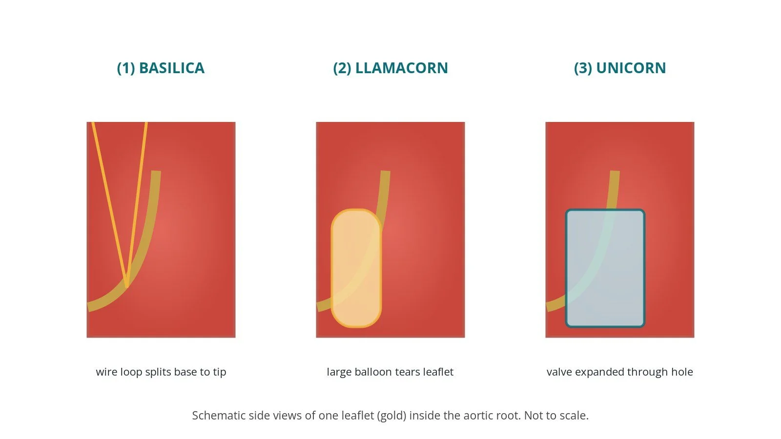
(1) BASILICA: wire loop splits the leaflet. (2) LLAMACORN: balloon tears it. (3) UNICORN: valve expanded through a hole.
Who is it for?
UNICORN may be considered by an experienced heart team for people having TAVI whose CT predicts a high risk of coronary obstruction, for example a short distance between the expected leaflet position and the coronary opening, or a narrow aortic root. Most reports involve valve-in-valve TAVI in failed surgical or transcatheter valves; a few describe use in native (including bicuspid) valves, which can be more challenging.
It was first described with a balloon-expandable valve, which expands from the inside out through the hole. Modified versions for self-expanding valves have since been reported.
Evidence, stated plainly: UNICORN is newer than BASILICA. The published literature (2022 to 2026) includes a first-in-human report, many case reports and technique papers, bench testing, a single-centre registry and a small multicentre retrospective study. These suggest it is feasible in selected patients at experienced centres, but there are no randomised trials, no direct comparison with BASILICA in people, and no long-term data. Published reports also describe failures and problems, including a late blockage of a coronary artery and a loose leaflet flap. The ESC/EACTS valvular guidelines and NICE guidance on valve-in-valve TAVI (HTG518) recognise coronary obstruction as a risk to assess; the choice of technique rests with the heart team. Consent should include this uncertainty.
When is it done?
It is planned with the TAVI, after:
detailed CT planning of the aortic root, the old valve and the coronary arteries, including the X-ray angles for the target leaflet and the planned hole position
echo, ECG, blood tests and a group and save
a review of blood thinners, as per centre protocol
Step by step
1. Anaesthetic and access
Usually under general anaesthetic with TOE. Sheaths go in both femoral arteries and a femoral vein for temporary pacing; sizes per device and operator. Other routes (for example via the neck) have been reported when the groin cannot be used. Cerebral embolic protection may be used, per operator. Heparin is given, dose as per centre protocol.
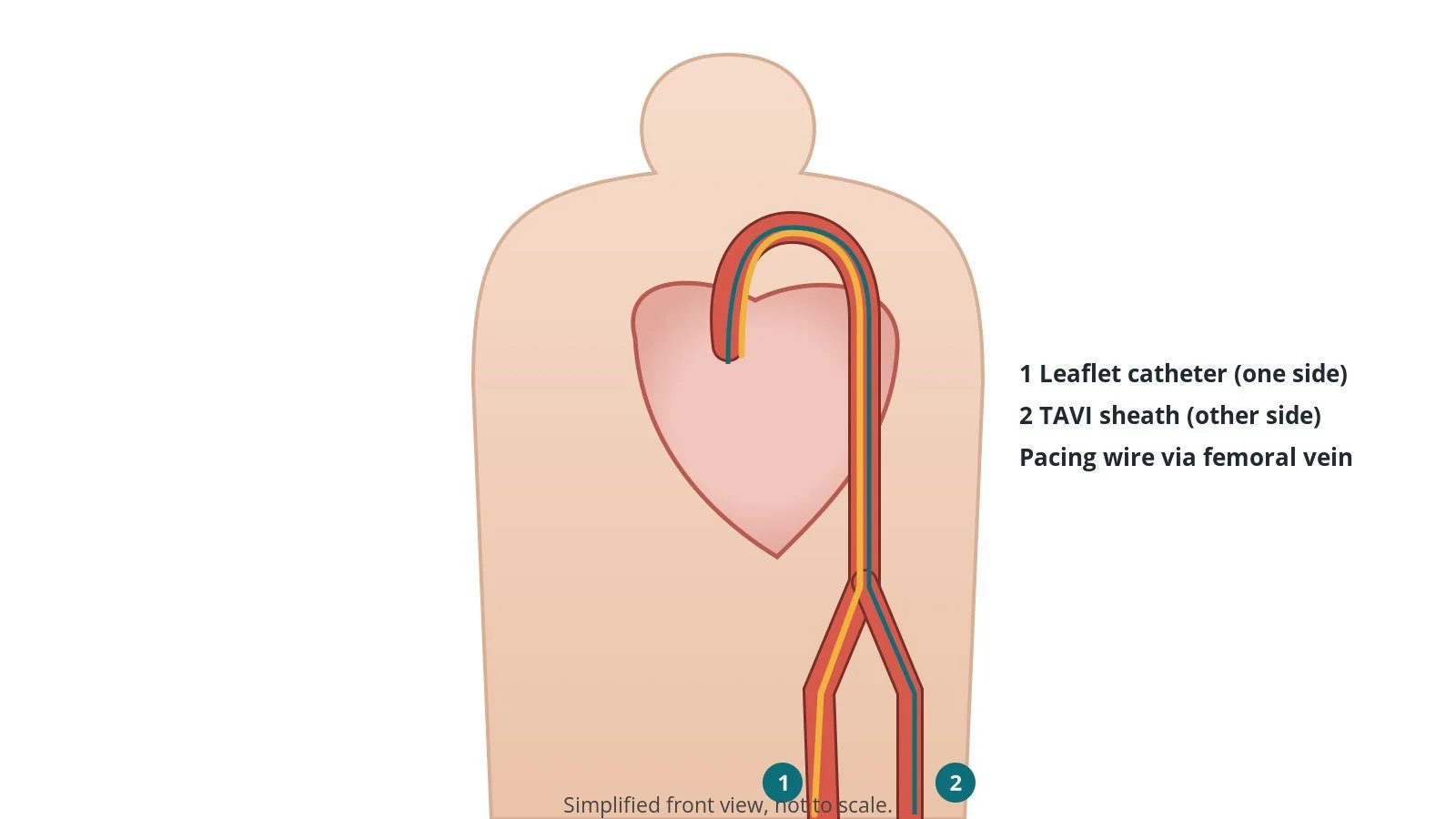
Access: (1) leaflet catheter from one femoral artery; (2) TAVI sheath from the other; pacing via a femoral vein.
2. Leaflet hole and valve
A. Traverse. A guide catheter and microcatheter are aimed at the lower part of the target leaflet, with the position confirmed on TOE and X-ray. A wire connected to an electrosurgery pen crosses the leaflet with a brief burst of energy (settings per operator) into the left ventricle.
B. Widen the hole. A supportive wire is placed and balloons of increasing size, coronary then larger, widen the hole into a window ("fenestration"), per device and operator. A stiff wire goes through the hole into the left ventricle.
C. Valve through the hole. The new valve is advanced over the stiff wire through the window in the leaflet.
D. Expand. The valve is expanded, usually under rapid pacing. As it opens, the target leaflet is split open and pushed aside, away from the coronary.
The blood pressure can fall during these steps, so the anaesthetic team is ready with support, as per centre protocol.
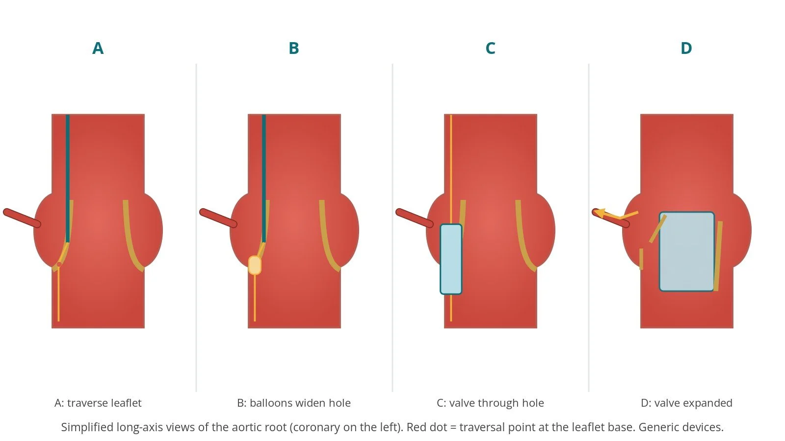
A: traverse. B: balloons widen the window. C: valve through the window. D: valve expanded; leaflet pushed aside.
3. Checking
Coronary angiography, echo and pressures check that both coronary arteries fill and the new valve works well.
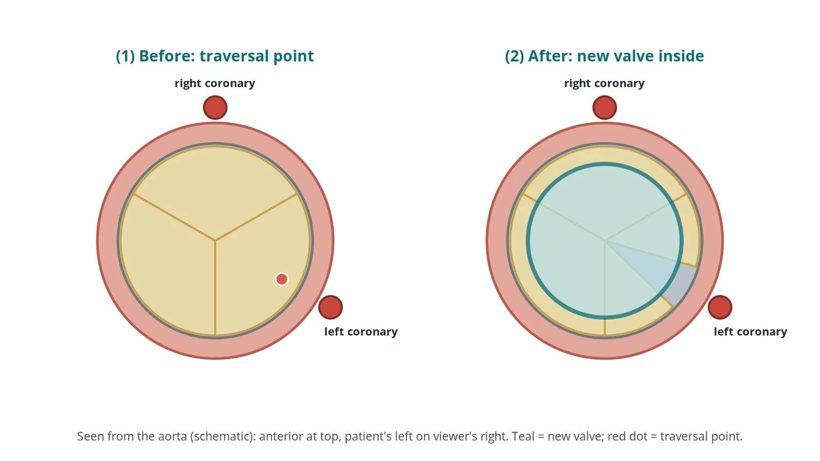
Seen from above: (1) traversal point on the at-risk leaflet; (2) the new valve inside, with that leaflet opened away from the coronary.
Recovery and aftercare
First hours: recovery, often in coronary care, high-dependency or intensive care, with checks of the groins, heart rhythm (including for heart block) and blood pressure, as per centre protocol.
Echo and blood tests: before discharge.
Going home: often after a few nights.
Medicines: antiplatelet or anticoagulant medicines, as per your team's plan.
Driving: DVLA guidance after a percutaneous valve procedure for a car licence is not to drive for 2 weeks; bus and lorry drivers must tell the DVLA. Ask your team.
Call 999 if you have chest pain, sudden breathlessness, fainting, signs of a stroke, or bleeding from the groin. Chest pain in the weeks afterwards should also be checked urgently.
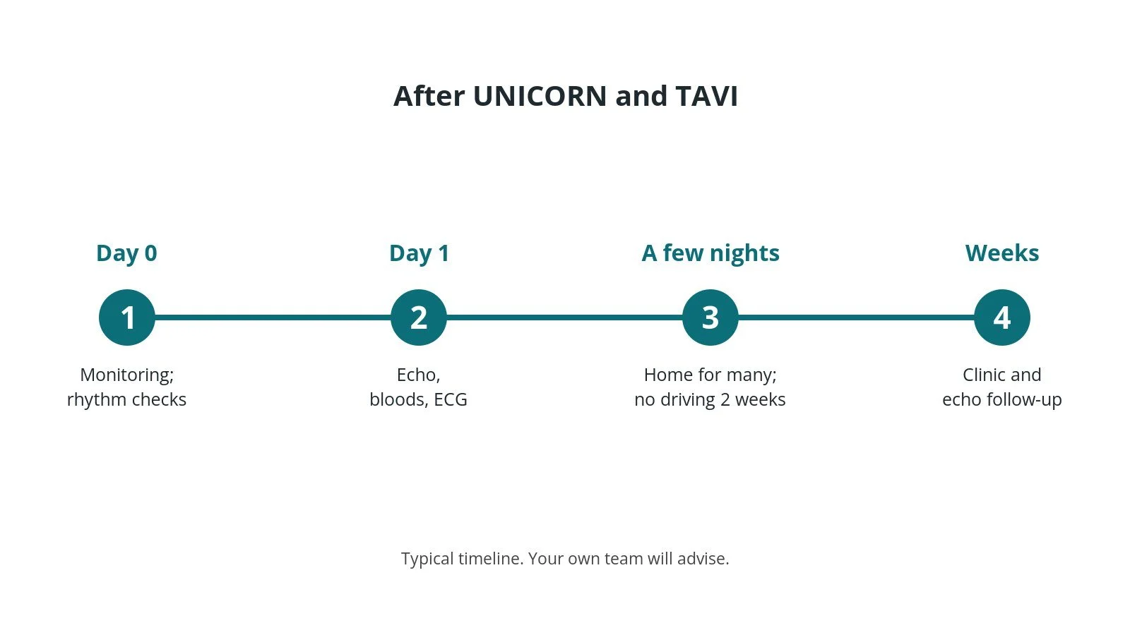
After UNICORN and TAVI: (1) monitoring; (2) echo and bloods; (3) home after a few nights; (4) follow-up.
Risks and complications
Your team will discuss your own risks. Reported and expected problems include:
low blood pressure during the procedure, sometimes needing medicines to support it
stroke, from debris or air
coronary obstruction despite UNICORN, including rare reports of blockage some time after the procedure
a loose or torn leaflet flap that could itself move towards a coronary opening
difficulty crossing a heavily calcified leaflet, or the valve not passing through the hole
injury to the aortic root or other structures
bleeding or injury at the access arteries
the usual TAVI risks, including heart block needing a pacemaker
Cath lab kit list (printable)
Example kit list
Written generically from published technique descriptions. Every lab has its own protocol, and sizes and doses vary.
Access
femoral arterial sheaths on both sides (large-bore for TAVI), femoral venous sheath for pacing; sizes per device and operator
suture-based closure devices, per operator
cerebral embolic protection device, if used
Leaflet traversal
guide catheter (shape for the target cusp) with inner catheter or microcatheter, per operator
traversal wire suitable for electrosurgery (0.014" or 0.035" radiofrequency wire, per operator), back end prepared for the pen if needed
diathermy pen and generator or radiofrequency generator, settings per operator; non-conducting flush, as per centre protocol
Fenestration
non-compliant coronary and larger peripheral balloons in increasing sizes, per device and operator
extra-stiff pre-shaped LV wire, per operator
Usual TAVI equipment, valve per device and operator
Procedural steps (example)
General anaesthetic and TOE; both femoral arteries and a femoral vein; pacing wire; heparin
CT-derived X-ray angles; angiogram of the target cusp
Guide at the lower part of the leaflet; TOE confirms position; energised traversal into the LV
Exchange wire; serial balloons to make the window; stiff wire through the window
Vasopressor support ready, as per centre protocol
Valve advanced through the window and expanded under rapid pacing
Coronary angiography and echo
First time in the cath lab?
💡 First-time tip
Watch the blood pressure. Drops are common during the balloon and valve steps; call it out early. Diathermy only on command. Activate the pen only when the operator asks. Know which wire is which. The stiff wire through the leaflet is the one the valve follows; label it. Pacing ready. Rapid pacing is used for balloons and the valve. Keep the valve close. Once the window is made, the valve should follow without delay.
FAQ
Is UNICORN the same as LLAMACORN?
They are closely related. In UNICORN the new valve is expanded through a hole in the leaflet. In LLAMACORN a large balloon tears the leaflet first, then the valve is placed.
Why is it called UNICORN?
It is an acronym: Undermining Iatrogenic Coronary Obstruction with Radiofrequency Needle.
Is it proven?
Not fully. Small studies suggest it is feasible in selected people, but there are no randomised trials or long-term results.
What are the alternatives?
BASILICA, LLAMACORN, a stent placed in the coronary artery during TAVI, or surgery, depending on your situation.
Related Cath Lab 101 guides
Sources
All checked on 10 October 2026. Each page's own date is shown where it gives one.
Chan KYE, et al. First-in-human undermining iatrogenic coronary obstruction with radiofrequency needle (UNICORN) procedure during valve-in-valve TAVR. Circ Cardiovasc Interv 2022;15(11):928–931. PubMed 36268707
Chan KYE, et al. Leaflet modification technique with UNICORN in failed aortic bioprosthesis: procedural step-by-step, best practice, and troubleshooting. JACC Cardiovasc Interv 2026;19(12):1565–1578. PubMed 42331405
Wong CK, et al. UNICORN for valve-in-valve TAVR: UNICORN Hong Kong Registry. JACC Asia 2026;6(1):52–63. PMC12833605
Hebbo E, et al. UNICORN during TAVR in native and bioprosthetic aortic valves: a multicenter study. JACC Cardiovasc Interv 2026;19(12):1595–1603. PubMed 42331407
Khokhar AA, et al. Risk of leaflet avulsion after UNICORN leaflet modification. EuroIntervention 2026;22(6):e370–e371. PMC12978347
Ghazal R, et al. Transcatheter valve-in-valve and "Unicorn" leaflet laceration resulting in late left main occlusion. JACC Cardiovasc Interv 2025;18(12):1595–1598. PubMed 40338789
Veillette JB, et al. Feasibility and challenges of the UNICORN technique in native aortic valve TAVI. EuroIntervention 2026;22(11):e645–e647. PMC13214564
NICE. Valve-in-valve TAVI for aortic bioprosthetic valve dysfunction (HTG518). nice.org.uk/guidance/htg518 (published 26 Jun 2019)
Vahanian A, et al. 2021 ESC/EACTS Guidelines for the management of valvular heart disease. Eur Heart J 2022;43(7):561–632. PubMed 34453165
DVLA / GOV.UK. Cardiovascular disorders: assessing fitness to drive. gov.uk
Disclaimer
Education only. This page is for general education for healthcare students, cath lab staff and curious readers. It isn't medical advice and doesn't replace advice from your own doctor, heart team or local unit protocols. Equipment and medicines are described generically. No product or manufacturer is recommended. If you think someone is having a heart emergency, call 999.
Disclosure: I work for a company that makes a device used in this procedure. This page is independent education, isn't sponsored or reviewed by any company, and doesn't recommend any product.
All diagrams on this page are original illustrations created for rodnieoro.com.