Native mitral valve replacement (TMVR): a Cath Lab 101 guide
Reviewed by Rodnie Oro, cath lab charge nurse · Last reviewed: November 2026 · Education only, not medical advice
Native mitral valve replacement in one minute
What it is: Transcatheter mitral valve replacement (TMVR) places a new valve inside the native mitral valve, through the heart's apex (transapical) or from a vein in the groin across the atrial septum (transseptal). It aims to stop the leak completely.
Who it is for: Adults with severe mitral regurgitation and symptoms who are not suitable for surgery and whose valve cannot be treated well with TEER, as decided by a heart team. It is mostly offered in trials and specialist centres.
Main risks: Blocked outflow from the left ventricle (LVOT obstruction), bleeding, valve moving or leaking around its edge, stroke, and problems at the apex or septum. Your team will explain your own risk.
Usually: Under general anaesthetic with TOE, about 2 to 3 hours, often a few days in hospital.
On this page: What is native mitral valve replacement? · Who is it for? · When is it done? · Step by step · Recovery · Risks · Kit list · First time in the lab? · FAQ
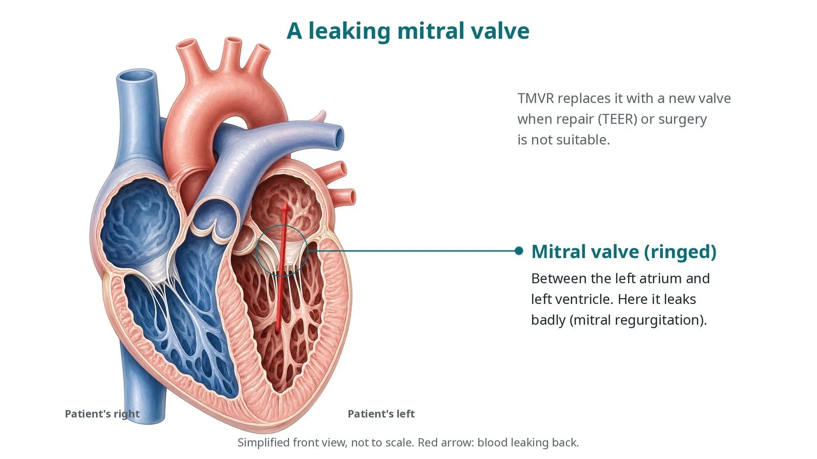
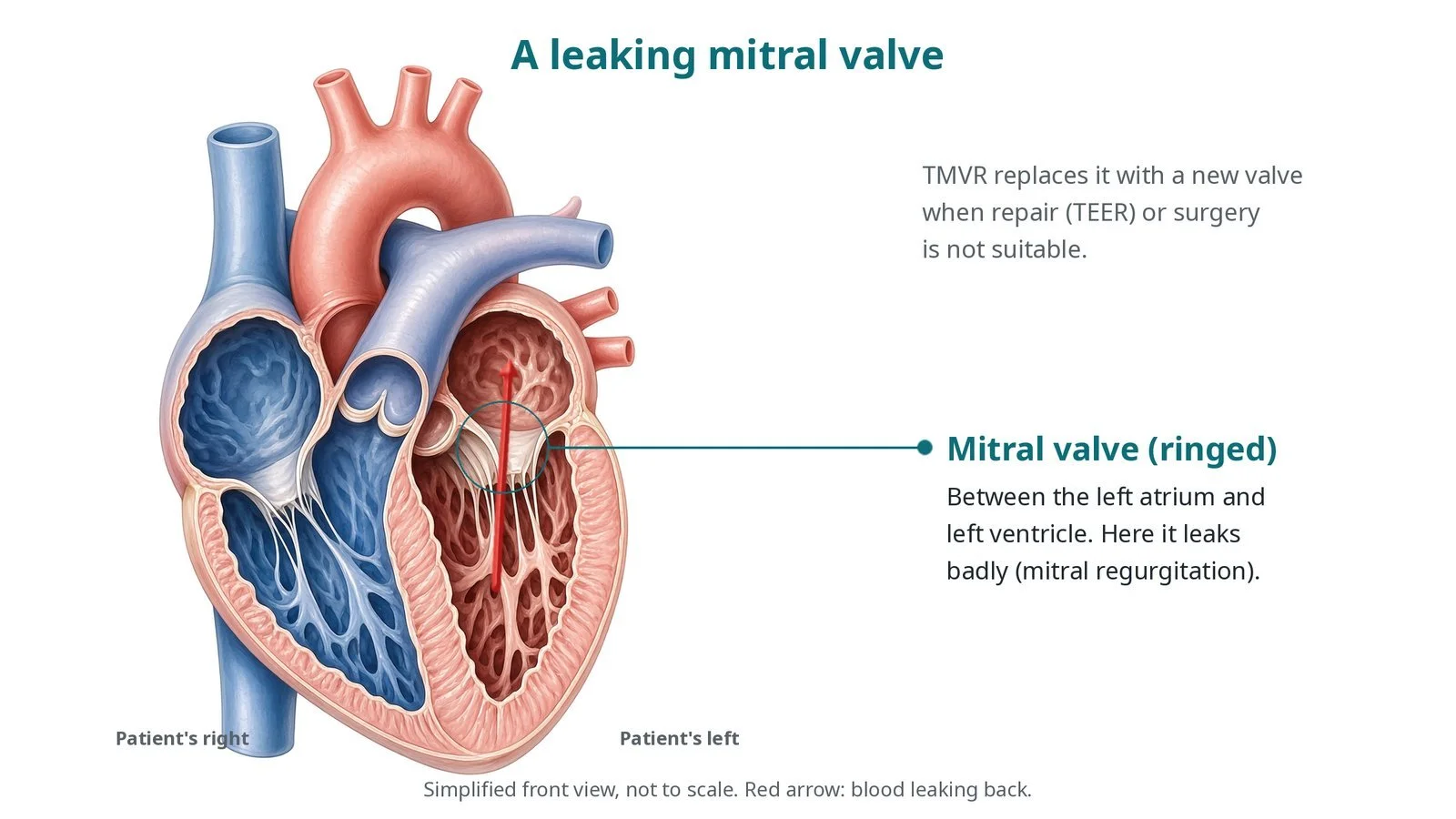
The mitral valve (ringed) lies between the left atrium and left ventricle. Here it leaks badly. TMVR replaces it with a new valve when repair (TEER) or surgery is not suitable.
What is native mitral valve replacement?
The mitral valve lets blood flow from the left atrium into the left ventricle. It has two leaflets attached to a ring (the annulus) and held by cords (chordae) to muscles in the ventricle. The front (anterior) leaflet sits next to the outflow tract (LVOT), the path blood takes out to the aorta.
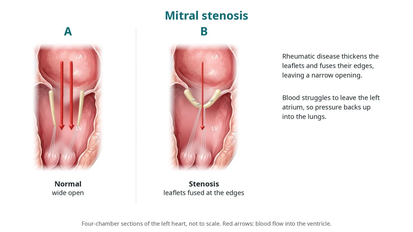
In mitral regurgitation the valve leaks back into the left atrium. Repair with a clip (mitral TEER) suits many people, but not those with very calcified, short or complex leaflets, or where a clip would leave the valve too narrow.
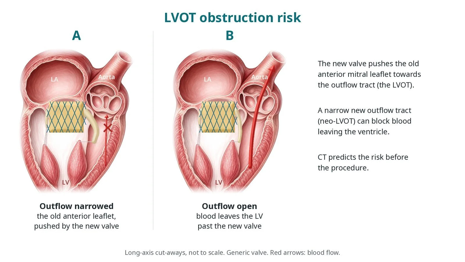
A TMVR valve sits in the annulus and pushes the native leaflets aside. The anterior leaflet is pushed towards the septum, creating a new, narrower outflow called the neo-LVOT. If it is too small, blood cannot leave the ventricle (LVOT obstruction). CT predicts the neo-LVOT before the case. Some valves are anchored by their frame; transapical designs may also use a tether to a pad on the outside of the apex.
New to the acronyms? See the Cath Lab Abbreviations A–Z.
Who is it for?
Adults with severe, symptomatic mitral regurgitation at high risk from surgery whose anatomy is unsuitable for TEER. The heart team checks:
the annulus size and calcium on CT
the predicted neo-LVOT
the access route (transseptal or transapical)
left ventricular function and other illnesses
Who it may not suit: People whose predicted neo-LVOT is too small, whose annulus is outside the device sizes, or who are suitable for surgery or TEER.
The decision is made by your heart team, who will talk through the reasons, the alternatives and the risks with you.
When is it done?
A planned procedure, mostly within trials or at specialist centres, after heart team review.
gives you a chance to ask questions and give consent
arranges blood tests, including kidney function and a blood count, and an ECG
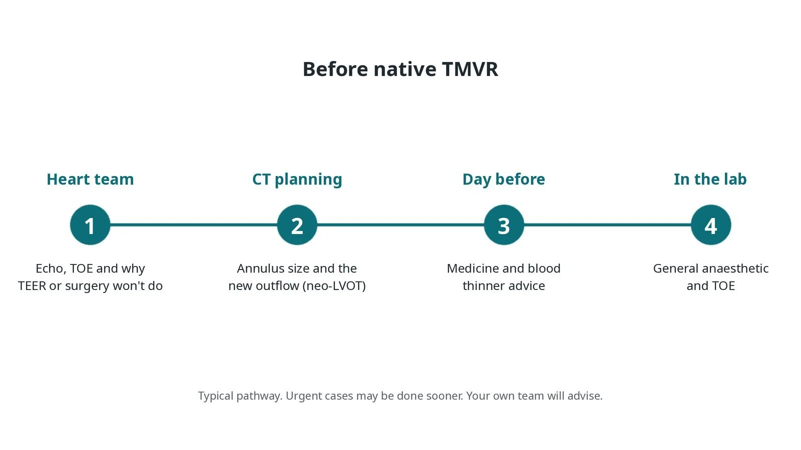
Before native TMVR: heart team review with echo and TOE; CT plans the annulus size and the new outflow tract (neo-LVOT); medicine advice; general anaesthetic and TOE in the lab.
Step by step
1. Getting ready
A general anaesthetic is given and a TOE probe is placed. Monitoring includes ECG, invasive blood pressure and oxygen levels.
2. Access
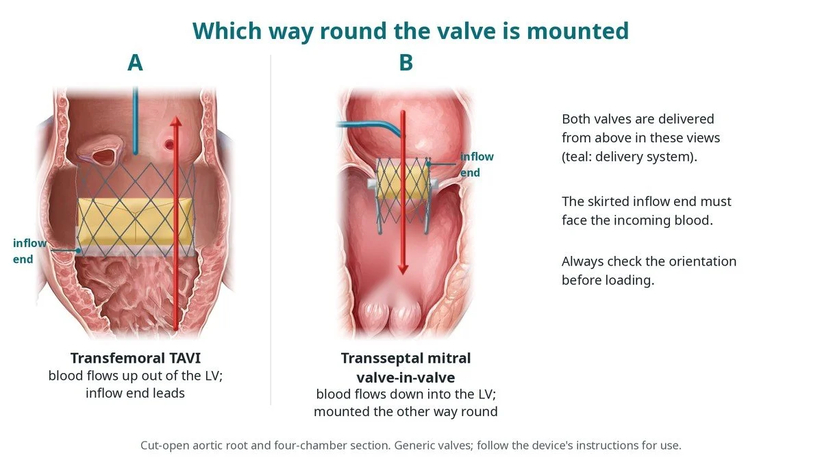
Transseptal: a large sheath in the femoral vein, a transseptal puncture into the left atrium, and the septum may be stretched with a balloon. Transapical: a small cut on the left side of the chest and a sheath into the apex of the heart, with a purse-string stitch. A pacing wire is placed and heparin is given as per centre protocol.
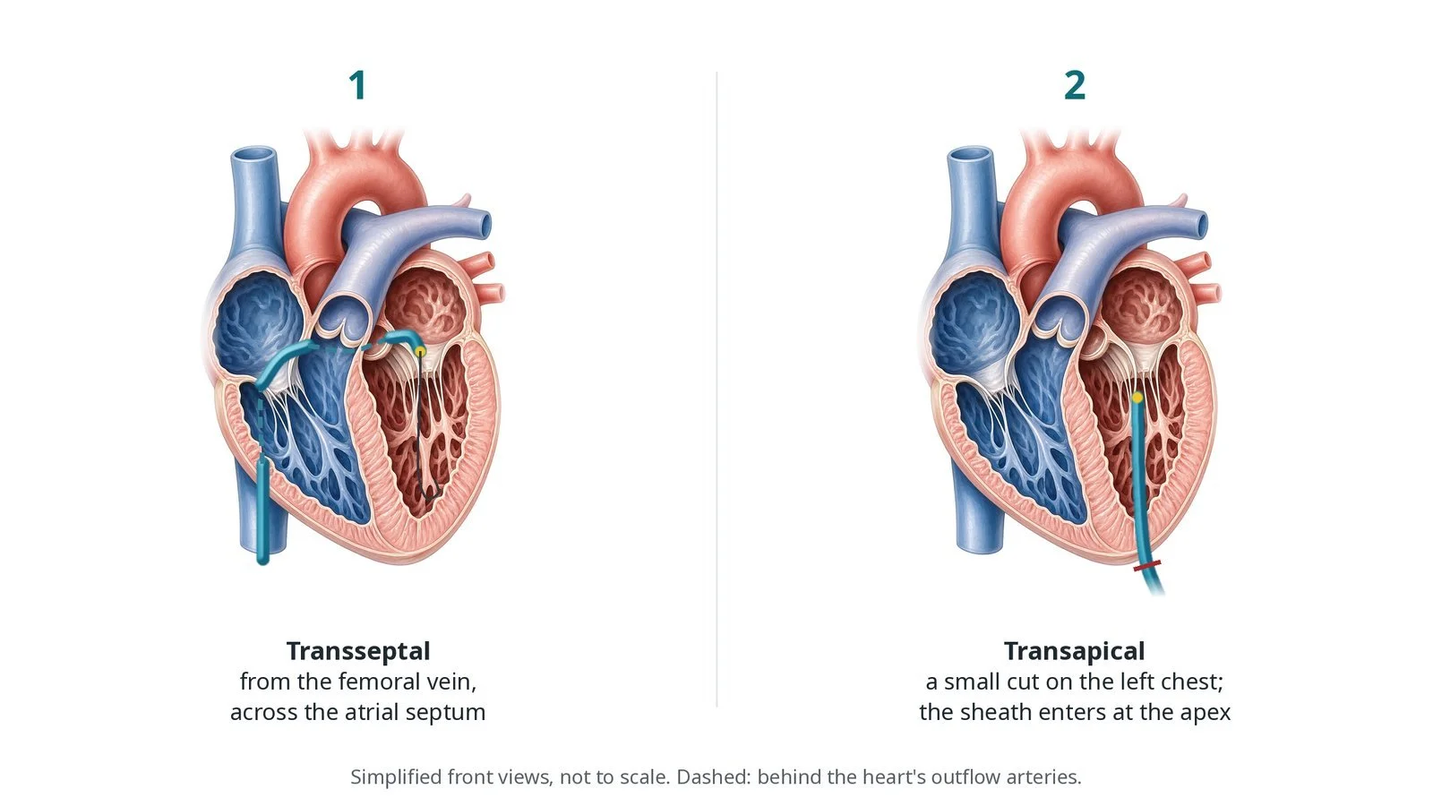
Routes: (1) transseptal, from the femoral vein across the atrial septum; (2) transapical, through a small cut on the left chest with the sheath entering at the apex.
3. Placing the valve
A. Rail. A wire is passed across the mitral valve into the left ventricle to make a rail for the delivery system.
B. Position. The crimped valve is advanced across the mitral annulus, checked on TOE and X-ray.
C. Deploy. The valve is released so the frame grips the annulus and leaflets. Rapid pacing is used as per device; transapical devices may be tethered to a pad at the apex.
D. Check. TOE checks for leaks, valve position and flow through the outflow tract, and pressures are measured.
Watch: Falling blood pressure with a high gradient across the outflow on TOE: possible LVOT obstruction. Call out at once; the team will act per the bail-out plan.
4. Finishing
Transseptal: the large vein sheath is removed and the vein closed; the septal hole is usually left to heal. Transapical: the apex is closed with the purse-string and the chest wound closed, with a drain if needed.
5. Recovery
Most people wake in recovery or intensive care. Echo checks the valve again before going home.
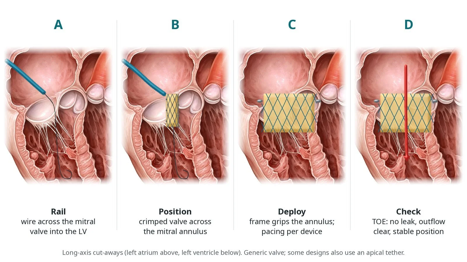
A: wire rail across the mitral valve into the left ventricle. B: crimped valve positioned across the annulus. C: deploy; the frame grips the annulus and leaflets. D: TOE checks for leaks and a clear outflow.
Recovery and aftercare
First hours: recovery or intensive care with monitoring, as per centre protocol.
Going home: often after several days; longer after transapical access.
Medicines: blood thinners, usually an anticoagulant for a period, as your team advises.
Activity: walk early; after transapical access avoid heavy lifting for several weeks.
Driving: do not drive until your team says it is safe, usually several weeks; check DVLA rules.
Follow-up: echo, heart failure clinic and any trial visits.
Call 999 if you have chest pain, sudden breathlessness, fainting, signs of a stroke, or heavy bleeding.
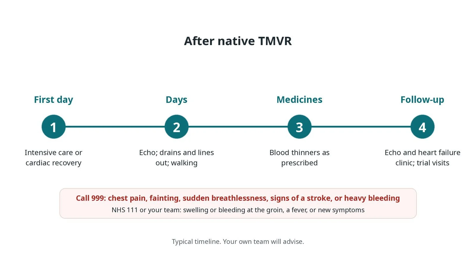
After native TMVR: (1) intensive care or cardiac recovery; (2) echo, lines out and walking over the next days; (3) blood thinners as prescribed; (4) echo, heart failure clinic and trial visits.
Risks and complications
Your team will explain the risks that apply to you. Possible problems include:
Blocked outflow from the left ventricle (LVOT obstruction) (uncommon)
Bleeding, including from the apex in transapical cases
Leak around the valve
Valve moving out of position (rare)
Stroke (uncommon)
Abnormal heart rhythms
Clot on the new valve: Blood thinners help prevent it.
Cath lab kit list (printable)
Barts example kit list
An example from one UK centre, written generically. Every lab has its own protocol, and sizes and doses vary.
large femoral venous sheath or transapical sheath, per device
transseptal needle and sheath, and septal balloon, per operator
stiff guidewires for the rail, per operator
transcatheter mitral valve and delivery system, size per CT plan
apical pad and tether, for devices that use them
temporary pacing wire and pacing box
TOE probe and echo machine
surgical set for transapical access and closure
contrast; heparin and anaesthetic medicines, as per centre protocol
bail-out kit as per centre protocol
Procedural steps (Barts example)
Getting ready
Access
Placing the valve
Finishing
Recovery
First time in the cath lab?
Know the route. Transseptal and transapical cases look very different; set up the room for the planned one.
TOE leads. The imager guides almost every step; keep the screen in the operators' view.
Watch the outflow. LVOT obstruction is the danger after deployment; know the plan if it happens.
It is a team case. Surgeons, anaesthetists and imagers are usually all involved.
Check the CT plan for the neo-LVOT, the device size and the planned access route.
FAQ
Why not a clip?
Some valves are too calcified or complex for a clip, or a clip would leave the valve too narrow.
Is it a new treatment?
Yes. It is mostly offered in trials and specialist centres, and long-term results are still being studied.
What is the neo-LVOT?
The new outflow path past the valve. CT checks it will be wide enough before the procedure.
Will I have a scar?
Only with the transapical route, which needs a small cut on the left side of the chest.
Related Cath Lab 101 guides
Transcatheter mitral valve implantation (TMVI, valve-in-valve)
Transcatheter mitral annuloplasty via the coronary sinus (coming soon)
Sources
All checked on 12 November 2026. Each page's own date is shown where it gives one.
Praz F, et al. 2025 ESC/EACTS Guidelines for the management of valvular heart disease. Eur Heart J 2025
StatPearls (NCBI Bookshelf), search: mitral regurgitation. ncbi.nlm.nih.gov/books
Disclaimer
Education only. This page is for general education for healthcare students, cath lab staff and curious readers. It isn't medical advice and doesn't replace advice from your own doctor, heart team or local unit protocols. Equipment and medicines are described generically. No product or manufacturer is recommended. If you think someone is having a heart emergency, call 999.
Disclosure: I work for a company that makes a device used in this procedure. This page is independent education, isn't sponsored or reviewed by any company, and doesn't recommend any product.
All diagrams on this page are original illustrations created for rodnieoro.com.