Caval valve implantation (CAVI): a Cath Lab 101 guide
Reviewed by Rodnie Oro, cath lab charge nurse · Last reviewed: November 2026 · Education only, not medical advice
Caval valve implantation in one minute
What it is: In severe tricuspid regurgitation, blood is pushed back from the right ventricle into the veins and liver. Caval valve implantation (CAVI) places valves in the superior and inferior vena cava, usually through a vein in the groin, to stop this backflow.
Who it is for: Adults with severe tricuspid regurgitation and symptoms such as swelling and fluid build-up, who cannot have surgery or a valve repair or replacement by catheter, as decided by a heart team. A dedicated system is used, per device and operator.
Main risks: Bleeding at the groin, valve moving, clot on the valve, blocked liver veins and abnormal heart rhythms. Your team will explain your own risk.
Usually: Under sedation or general anaesthetic, about 1 to 2 hours, a few days in hospital.
On this page: What is caval valve implantation? · Who is it for? · When is it done? · Step by step · Recovery · Risks · Kit list · First time in the lab? · FAQ
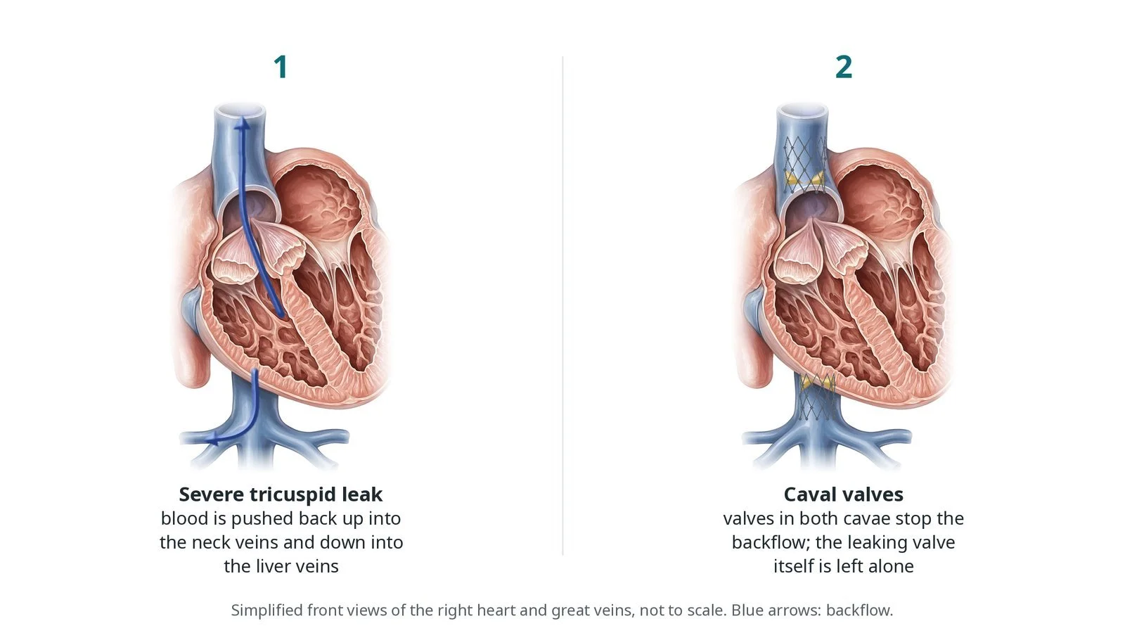
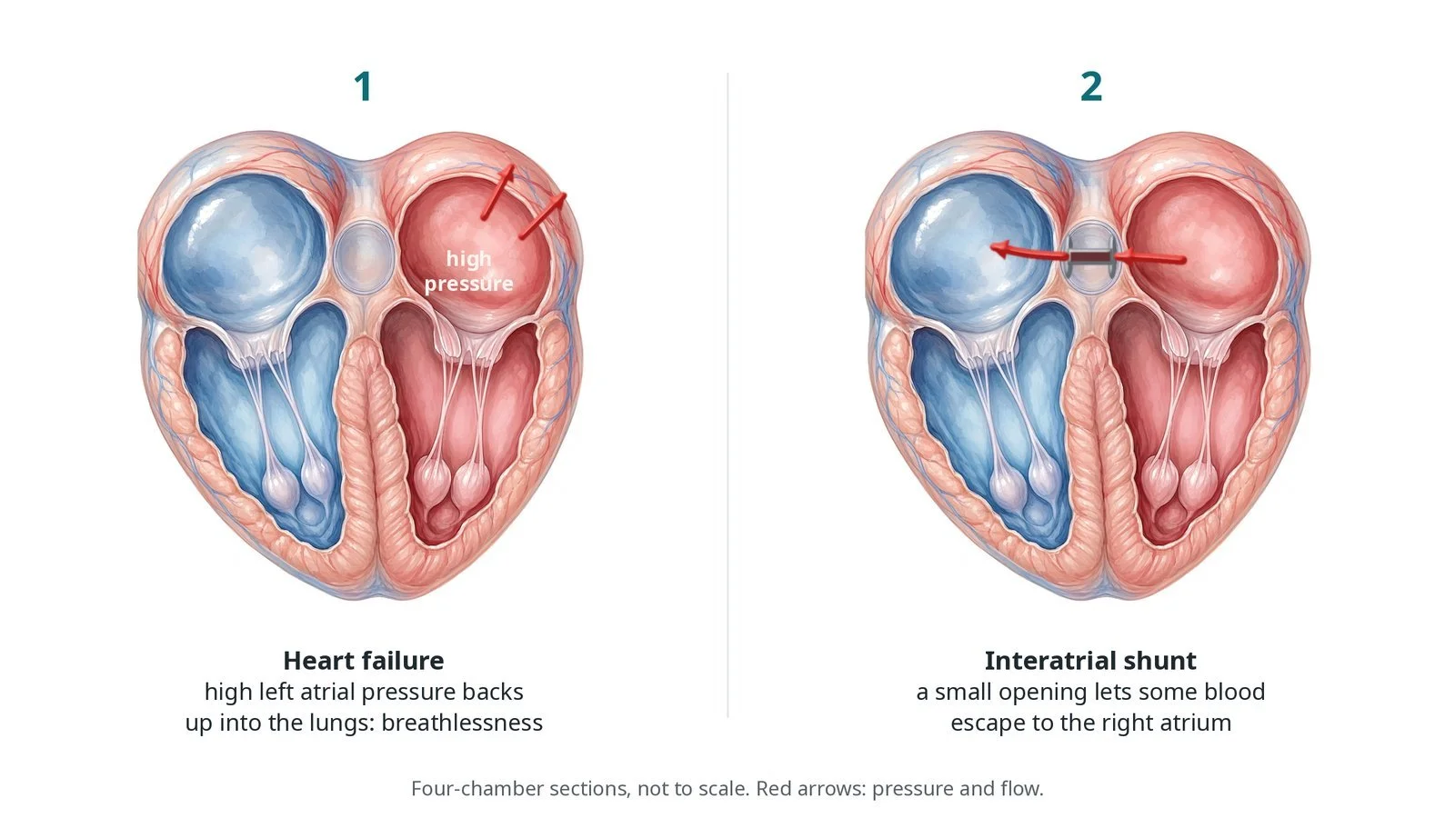
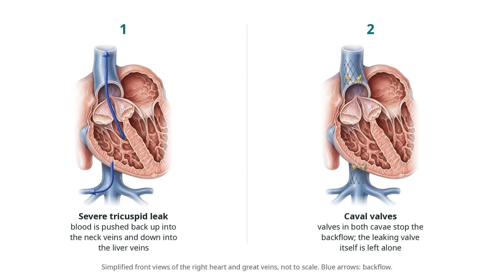
(1) A severe tricuspid leak pushes blood back up into the neck veins and down into the liver veins. (2) Valves in both venae cavae stop the backflow; the leaking valve itself is left alone.
What is caval valve implantation?
The tricuspid valve sits between the right atrium and right ventricle. Blood returns to the right atrium through two large veins: the superior vena cava (SVC) from the head and arms, and the inferior vena cava (IVC) from the body. The liver veins (hepatic veins) drain into the IVC just below the heart.
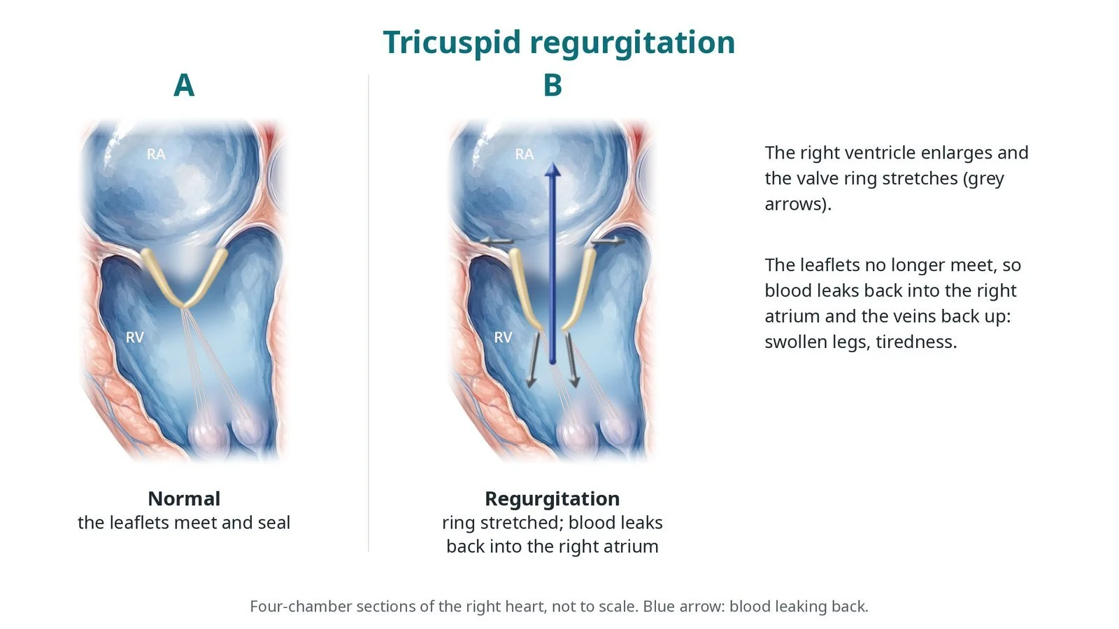
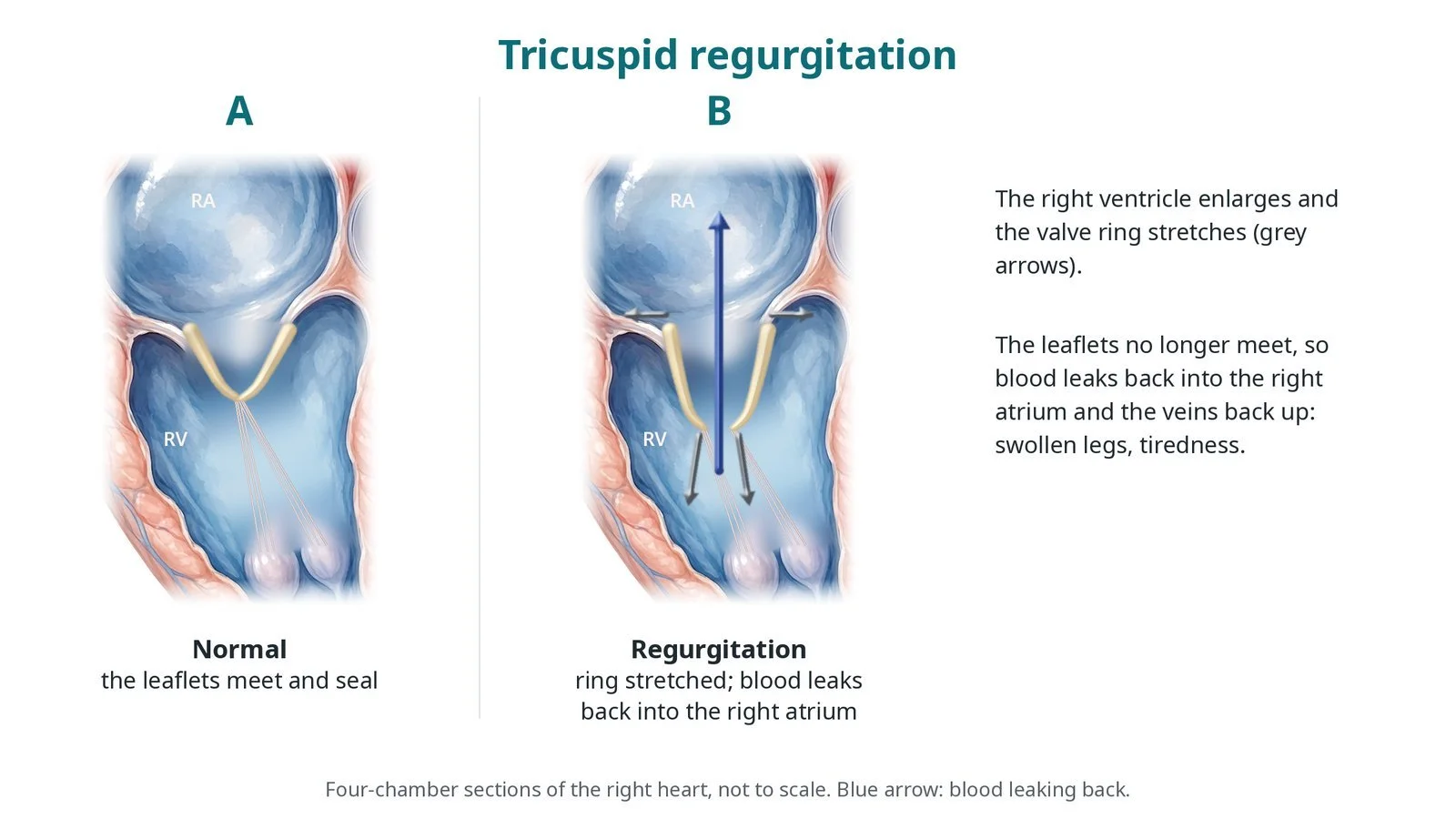
When the tricuspid valve leaks badly, each heartbeat pushes blood back into the right atrium and onwards into the cavae. This raises pressure in the neck veins, liver and legs, causing swelling, fluid in the abdomen, tiredness and poor appetite.
CAVI does not fix the leaking valve. Instead, valves in the SVC and IVC stop the backflow reaching the veins and liver. The IVC valve must sit at the junction with the right atrium without covering the hepatic veins. CT measures both cavae and shows where the hepatic veins join.
New to the acronyms? See the Cath Lab Abbreviations A–Z.
Who is it for?
Adults with severe, symptomatic tricuspid regurgitation who are not suitable for surgery, TEER or transcatheter tricuspid valve replacement. The heart team checks:
the size of the SVC and IVC on CT
the position of the hepatic veins
right ventricular function
kidney and liver function
Who it may not suit: People suitable for treatment at the tricuspid valve itself, those with veins outside the device sizes, or with pacing leads or clots that make the cavae unsuitable.
The decision is made by your heart team, who will talk through the reasons, the alternatives and the risks with you.
When is it done?
A planned procedure in specialist centres after heart team review.
gives you a chance to ask questions and give consent
arranges blood tests, including kidney function and a blood count, and an ECG
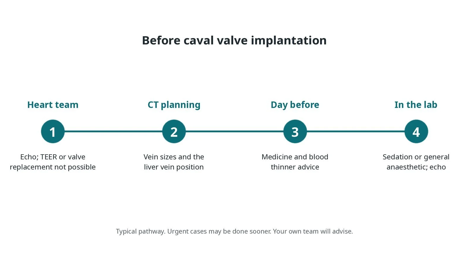
Before caval valve implantation: heart team review; CT measures the veins and the position of the liver veins; medicine advice; sedation or general anaesthetic with echo in the lab.
Step by step
1. Getting ready
You lie on the X-ray table. Sedation or a general anaesthetic is given, as per centre protocol. Echo is used during the case.
2. Access
A large sheath is placed in the femoral vein under ultrasound. Sizes are per device and operator. Heparin is given as per centre protocol. A stiff wire is placed up through the right atrium into the SVC.
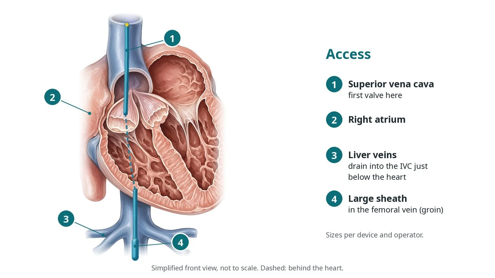
Access: (1) superior vena cava, where the first valve goes; (2) right atrium; (3) liver veins, which drain into the IVC just below the heart; (4) large sheath in the femoral vein.
3. Placing the valves
A. Measure. Venograms and pressure measurements, sometimes with a sizing balloon, confirm the size of each vena cava and where the hepatic veins join.
B. SVC valve. The first valve is released in the SVC just above the right atrium.
C. IVC valve. The second valve is released at the junction of the IVC and right atrium, clear of the hepatic veins.
D. Check. Venograms and echo confirm the valves are in place and backflow into the veins has stopped.
Watch: Contrast not draining from a hepatic vein, or the valve sitting low on the venogram: possible hepatic vein blockage. Tell the operator before the valve is fully released if possible.
4. Finishing
The sheath is removed and the vein closed with a stitch, a closure device or pressure, per operator.
5. After the valves
Echo checks the valves the next day. Fluid tablets and blood thinners are reviewed as pressures in the veins fall.
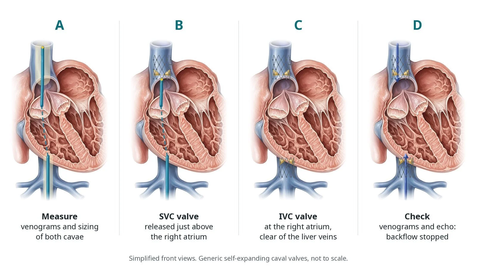
A: venograms and sizing of both cavae. B: SVC valve released just above the right atrium. C: IVC valve at the right atrium, clear of the liver veins. D: venograms and echo confirm the backflow has stopped.
Recovery and aftercare
First hours: rest with checks of the groin, heart rhythm and blood pressure, as per centre protocol.
Going home: after a few days, once fluid tablets are settled.
Medicines: blood thinners as your team advises; fluid tablets are often adjusted.
Activity: walk early; avoid heavy lifting for about a week, or as advised.
Driving: ask your team; usually not for at least a week; check DVLA rules.
Follow-up: echo, blood tests including liver function, and heart failure clinic.
Call 999 if you have chest pain, sudden breathlessness, fainting, or heavy bleeding from the groin.
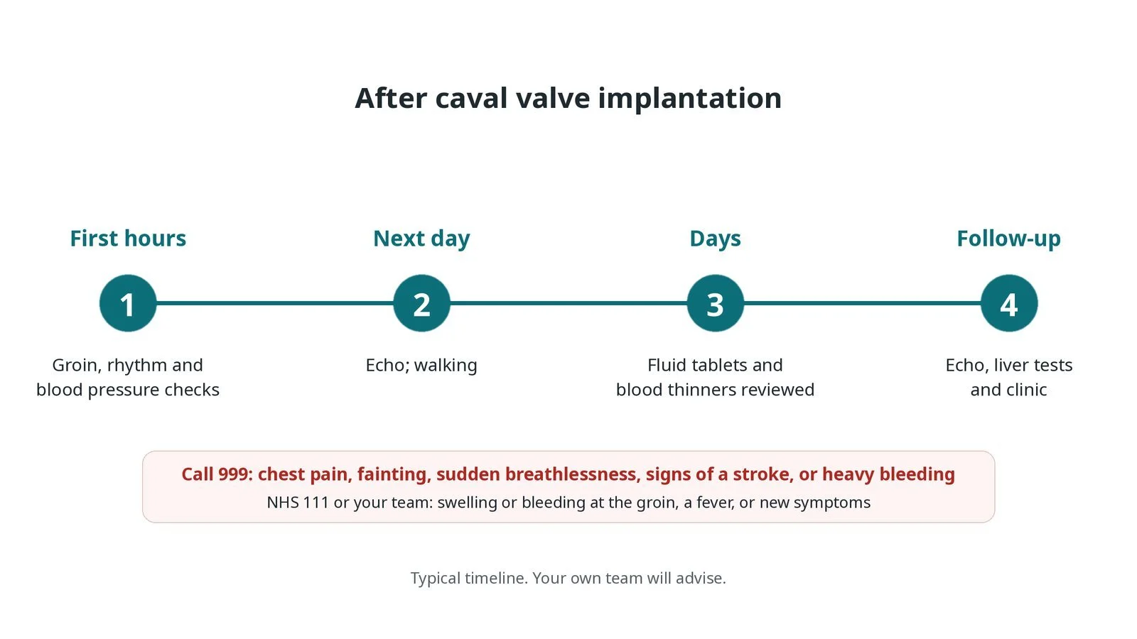
After caval valve implantation: (1) groin, rhythm and blood pressure checks; (2) echo and walking the next day; (3) fluid tablets and blood thinners reviewed; (4) echo, liver tests and clinic.
Risks and complications
Your team will explain the risks that apply to you. Possible problems include:
Bleeding or bruising at the groin
Valve moving out of position (uncommon)
Clot on the valves: Blood thinners are usually needed.
Blocked hepatic veins (rare)
Abnormal heart rhythms
Kidney strain from contrast
Cath lab kit list (printable)
Barts example kit list
An example from one UK centre, written generically. Every lab has its own protocol, and sizes and doses vary.
large femoral venous sheath, size per device
stiff guidewires, per operator
pigtail or marker catheter for venograms
sizing balloon, per operator
caval valve system for the SVC and IVC, per device
pressure transducer
contrast and syringes
echo: TOE or transthoracic
heparin, as per centre protocol
venous closure: stitch, device or pressure, per operator
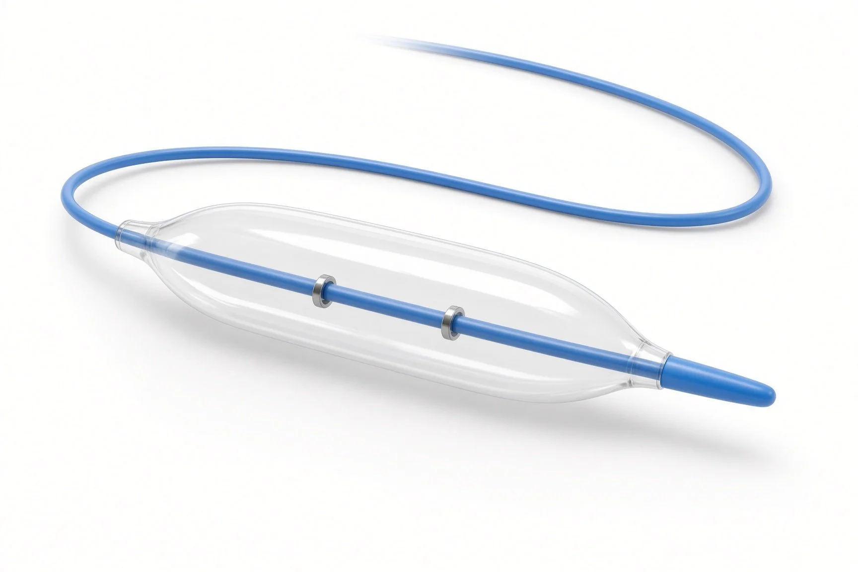
Sizing balloon.
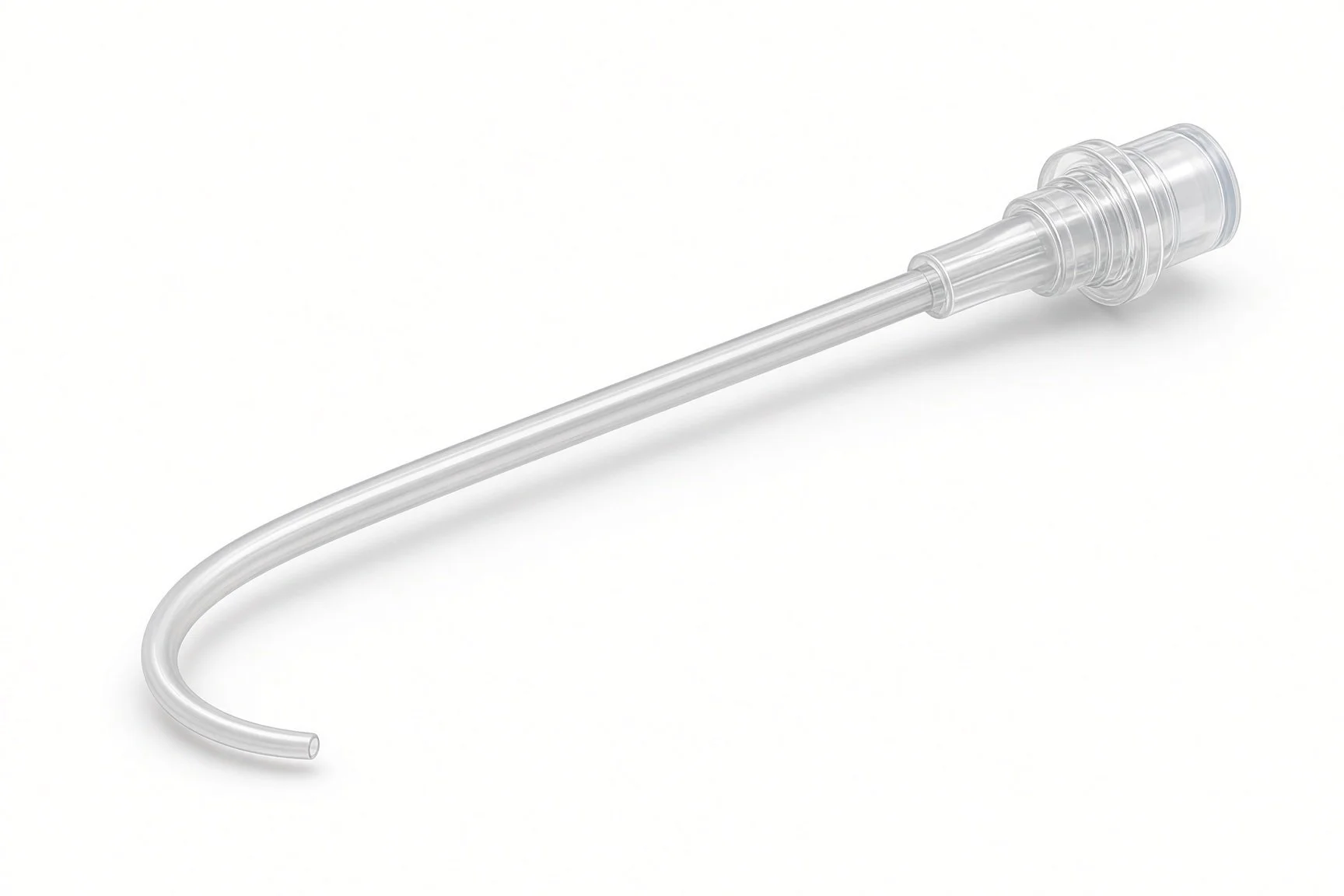
Pigtail or sizing catheter.
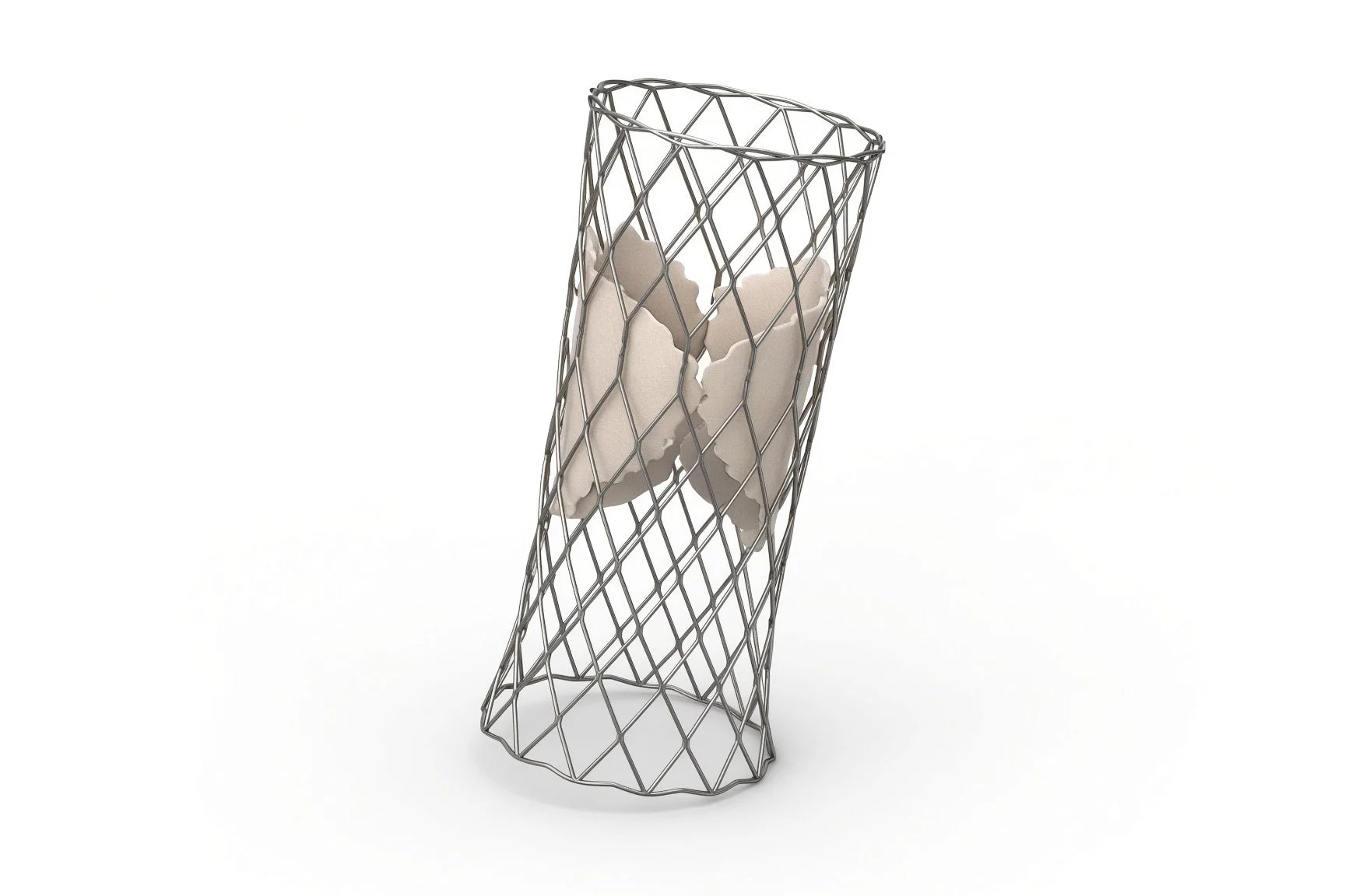
Caval valves on delivery systems.
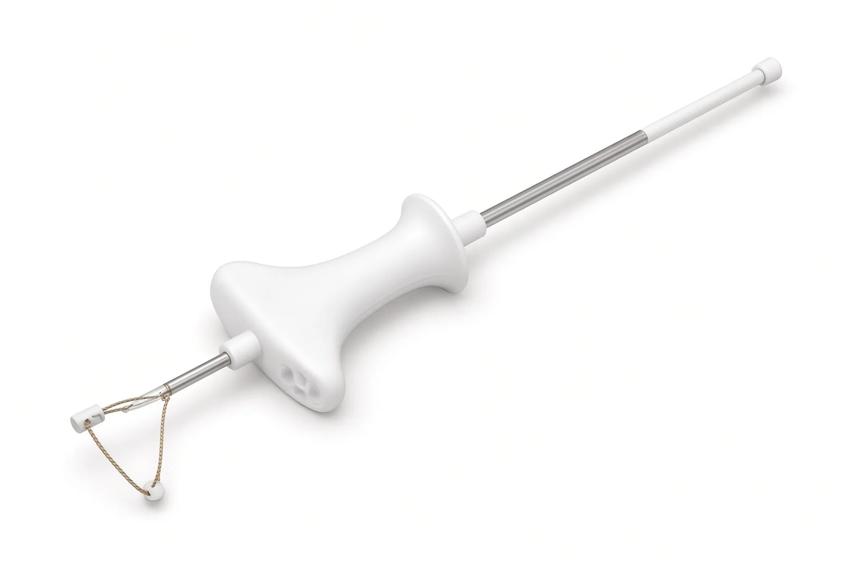
Closure or suture for the vein.
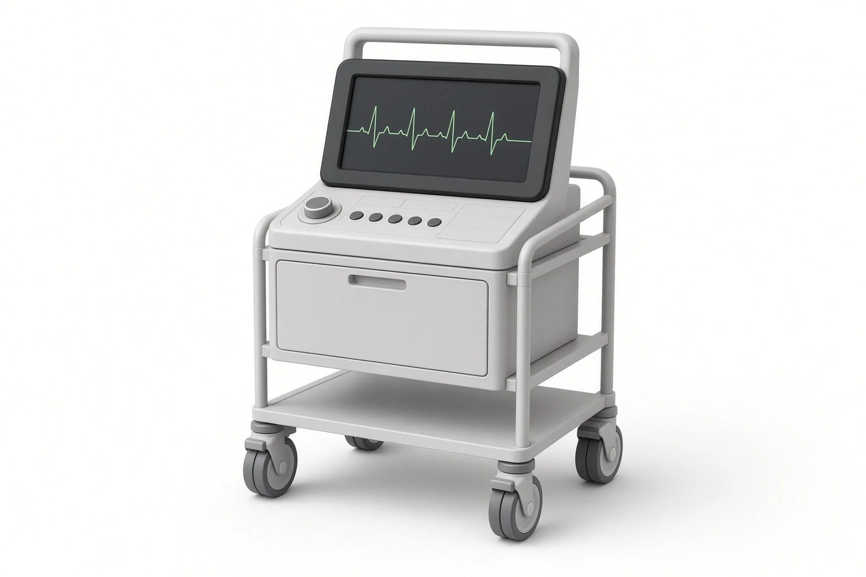
Pressure transducer.
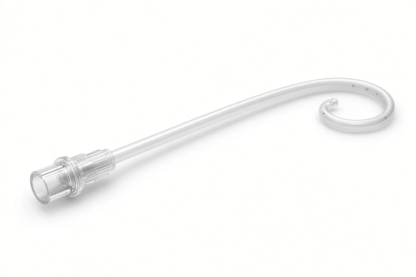
Pigtail or sizing catheter.
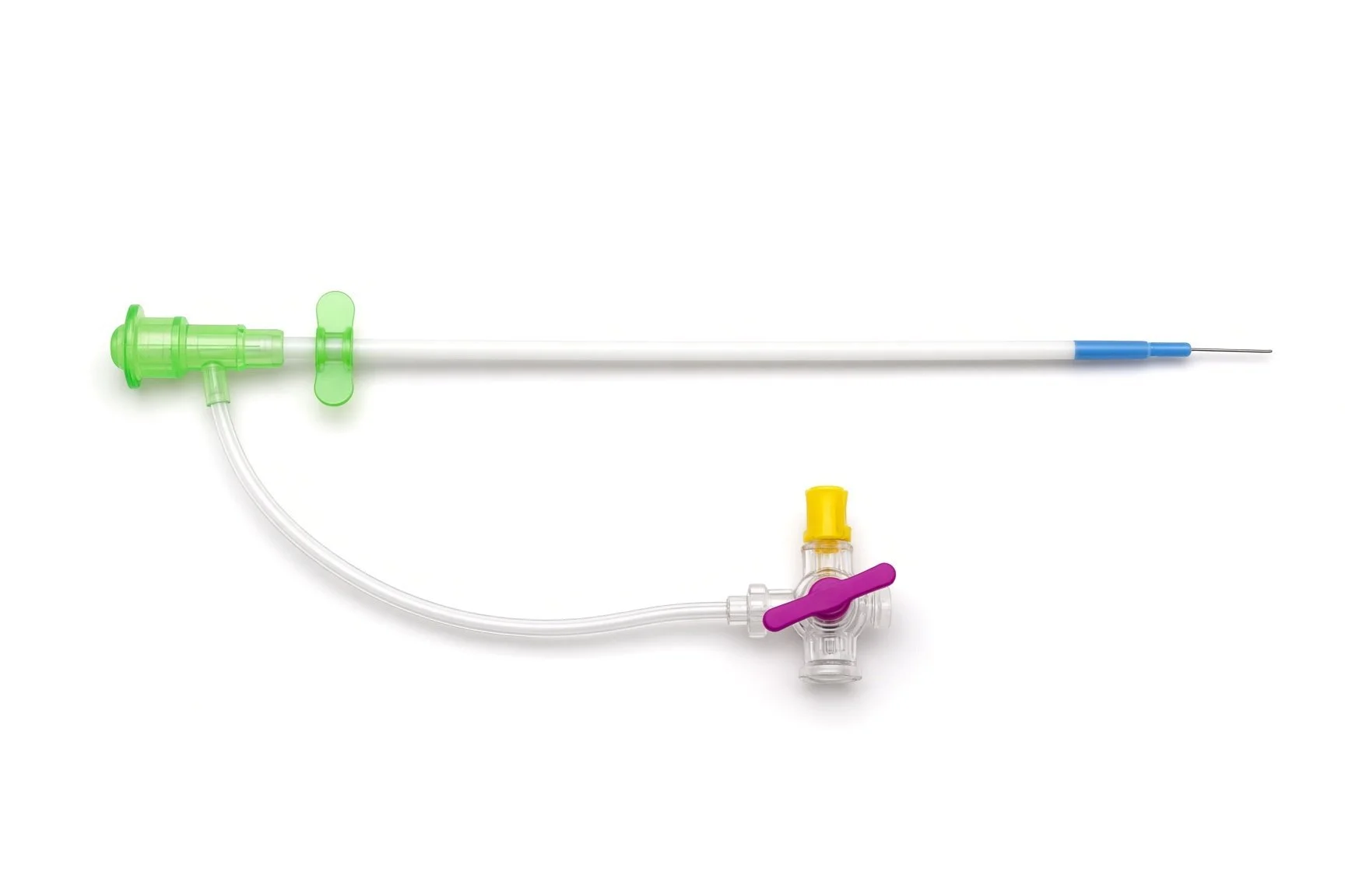
Large femoral venous sheath.
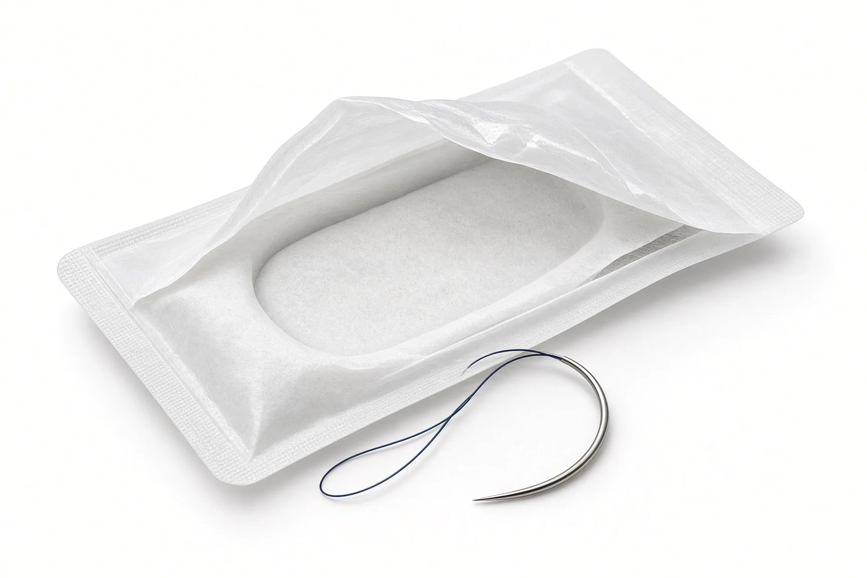
Closure or suture for the vein.
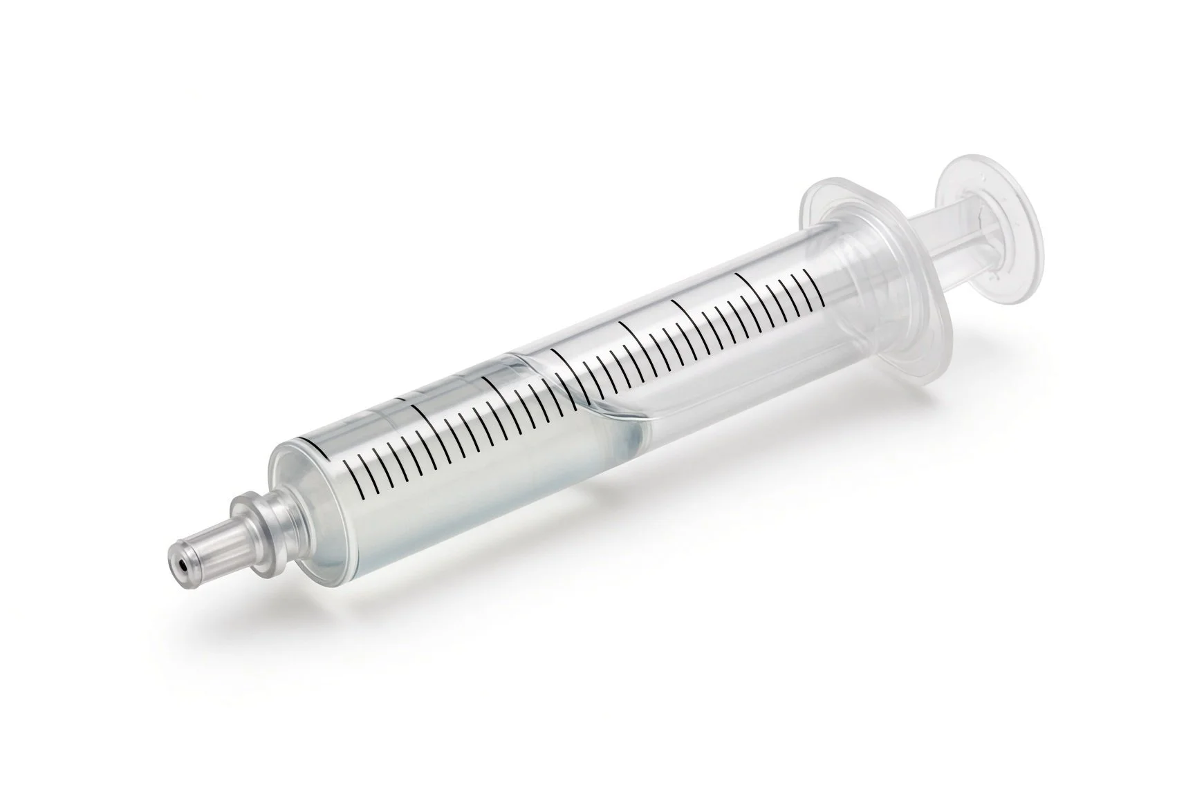
Contrast syringe.
Contrast.
Stiff guidewire.
Procedural steps (Barts example)
Getting ready
Access
Placing the valves
Finishing
After the valves
First time in the cath lab?
It treats the veins, not the valve. The tricuspid leak is still there on echo; success is measured in the veins.
Find the hepatic veins. Their level decides where the IVC valve goes.
Measure twice. Both cavae can be large; sizing is critical.
Expect fluid shifts. Diuretics are often changed in the days afterwards.
Check the CT measurements of both cavae and the hepatic vein level are available on the day.
FAQ
Does it fix my leaking valve?
No. It stops the leak being pushed back into your veins and liver, which can ease swelling and other symptoms.
Why two valves?
Blood returns through two big veins, so each needs a valve to stop backflow.
Will my swelling go down?
Many people find symptoms improve over weeks, but results vary.
Is it widely available?
It is offered in a small number of specialist centres.
Related Cath Lab 101 guides
Sources
All checked on 12 November 2026. Each page's own date is shown where it gives one.
Praz F, et al. 2025 ESC/EACTS Guidelines for the management of valvular heart disease. Eur Heart J 2025
StatPearls (NCBI Bookshelf), search: tricuspid regurgitation. ncbi.nlm.nih.gov/books
Disclaimer
Education only. This page is for general education for healthcare students, cath lab staff and curious readers. It isn't medical advice and doesn't replace advice from your own doctor, heart team or local unit protocols. Equipment and medicines are described generically. No product or manufacturer is recommended. If you think someone is having a heart emergency, call 999.
Disclosure: I work in the medical device industry. This page is independent education and isn't sponsored or reviewed by any company.
All diagrams on this page are original illustrations created for rodnieoro.com.