Interatrial shunt devices for heart failure: a Cath Lab 101 guide
Reviewed by Rodnie Oro, cath lab charge nurse · Last reviewed: November 2026 · Education only, not medical advice
Interatrial shunt devices for heart failure in one minute
What it is: In some people with heart failure, pressure in the left atrium is high and backs up into the lungs. An interatrial shunt creates a small permanent hole in the wall between the atria, so a little blood flows from left to right and left atrial pressure falls.
Who it is for: Selected adults with heart failure who are still breathless despite treatment, mostly within clinical trials. Results so far are mixed: RELIEVE-HF was neutral overall, and RESPONDER-HF is testing the people who may benefit most.
Main risks: Bleeding at the groin, damage from the transseptal puncture, device problems, and strain on the right side of the heart over time. Your team will explain your own risk.
Usually: Under sedation or general anaesthetic, about an hour, often home the next day.
On this page: What is interatrial shunt devices for heart failure? · Who is it for? · When is it done? · Step by step · Recovery · Risks · Kit list · First time in the lab? · FAQ
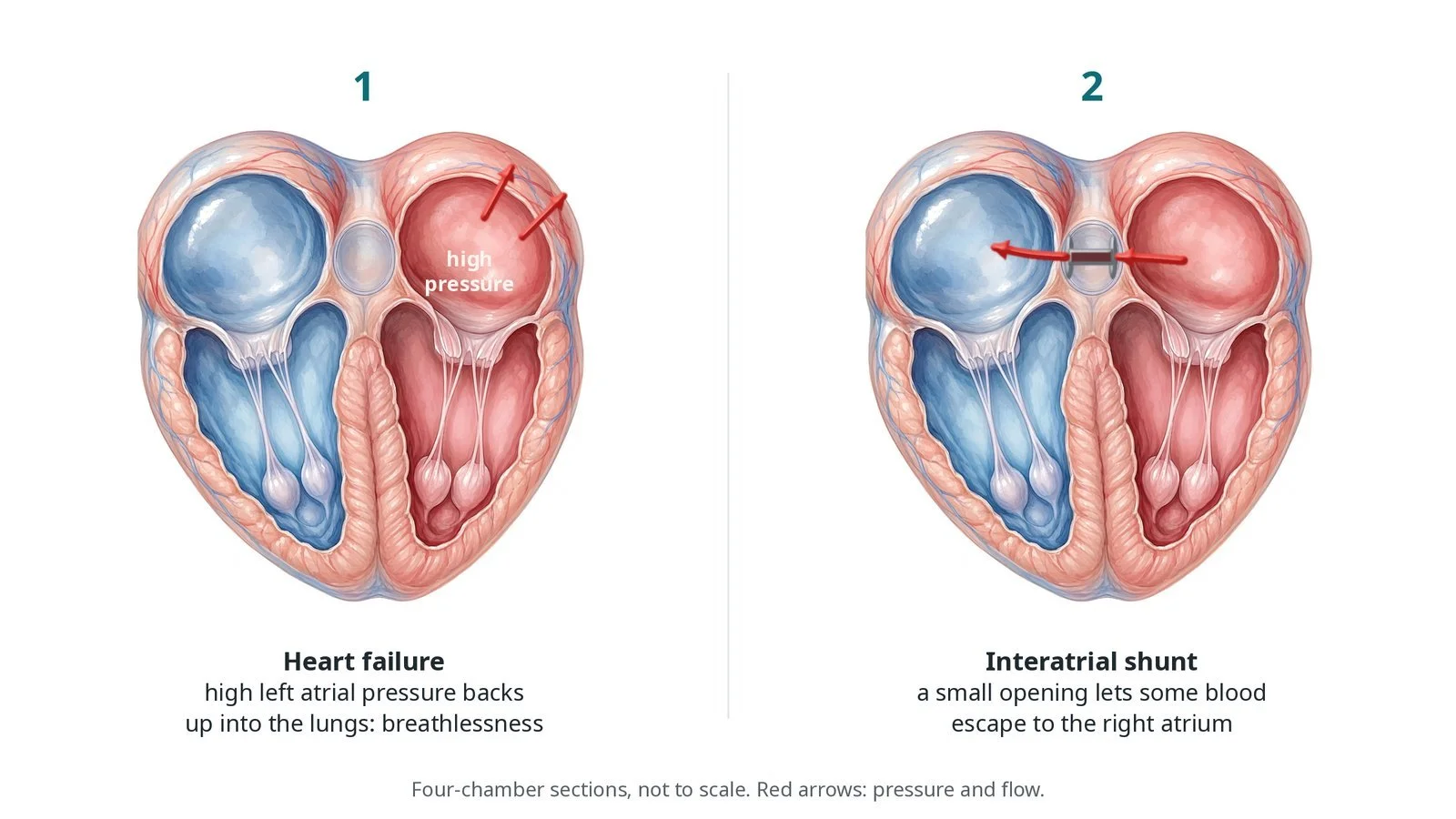
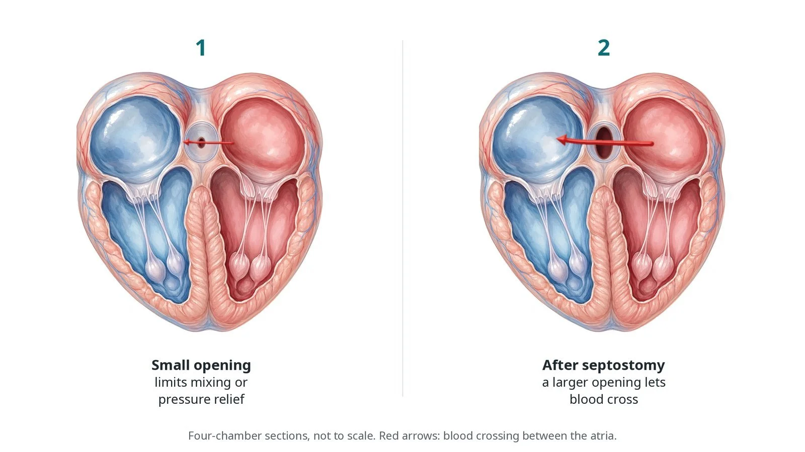
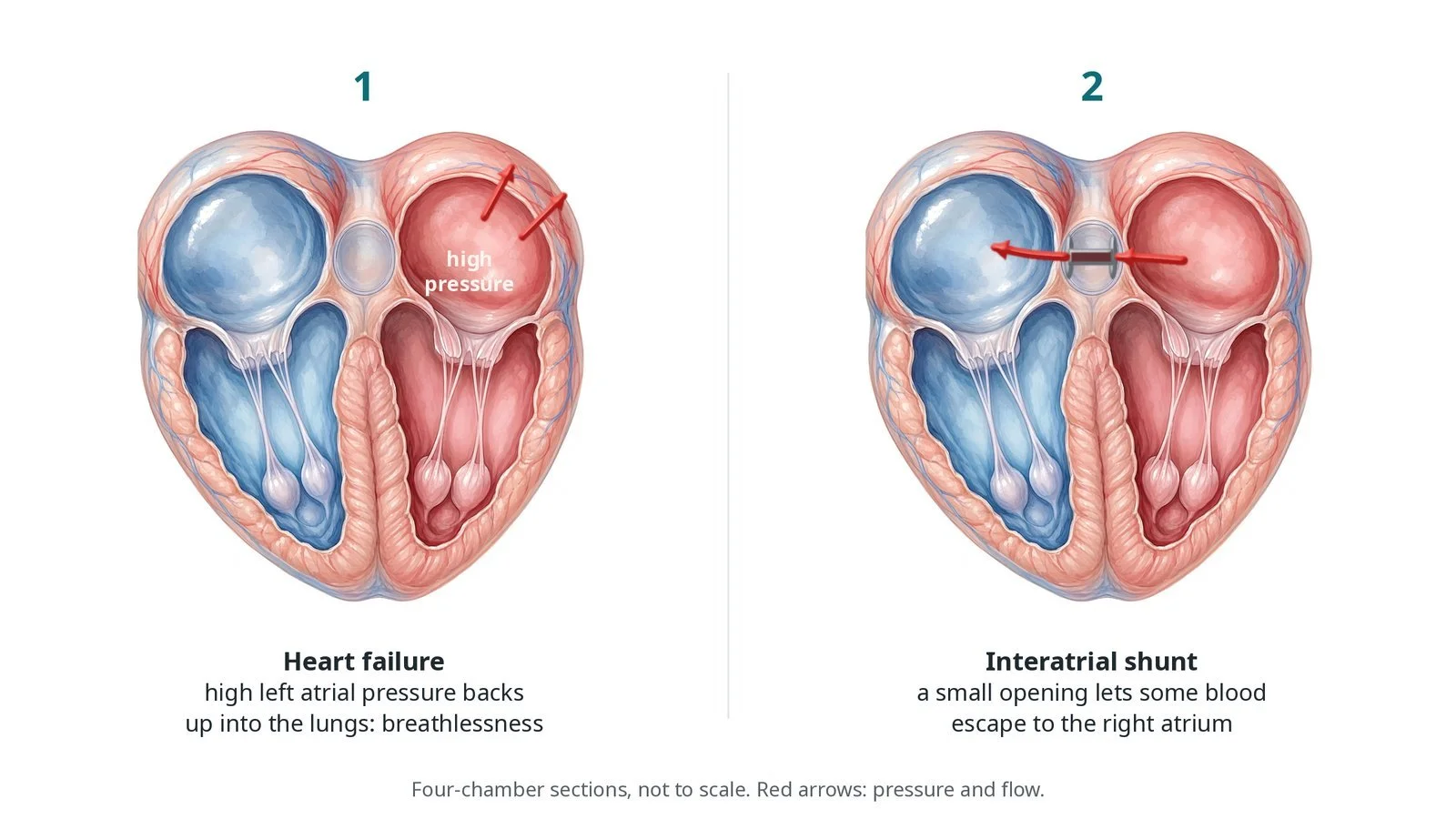
(1) In heart failure, high left atrial pressure backs up into the lungs and causes breathlessness. (2) An interatrial shunt is a small opening that lets some blood escape to the right atrium.
What is interatrial shunt devices for heart failure?
The left and right atria are separated by the interatrial septum. Normally the left atrium has a slightly higher pressure. In heart failure, with reduced or preserved ejection fraction (HFrEF or HFpEF), left atrial pressure can rise sharply, especially on exertion, pushing fluid into the lungs and causing breathlessness.
An interatrial shunt makes a small, controlled left-to-right shunt. Some blood leaves the left atrium for the right atrium, so the left atrial pressure, and the pressure in the lungs, rises less.
Shunts are made with an implant (a small device that holds the hole open) or without one, by removing or ablating a small disc of septum. Too large a shunt can overload the right heart, so patient selection matters. Trial results are mixed and the treatment is used in a research setting.
New to the acronyms? See the Cath Lab Abbreviations A–Z.
Who is it for?
Adults with heart failure and ongoing symptoms despite the best tolerated medicines, usually within a clinical trial. Before it, the team checks:
echocardiography and a right heart catheter, often with exercise
right ventricular function and lung pressures
the anatomy of the septum
Who it may not suit: People with a weak right ventricle, high pulmonary vascular resistance, or a septum unsuitable for puncture, and anyone who might later need the septum crossed for other procedures (discussed case by case).
The decision is made by your heart team, who will talk through the reasons, the alternatives and the risks with you.
When is it done?
A planned procedure, mostly within trials at specialist centres.
gives you a chance to ask questions and give consent
arranges blood tests, including kidney function and a blood count, and an ECG
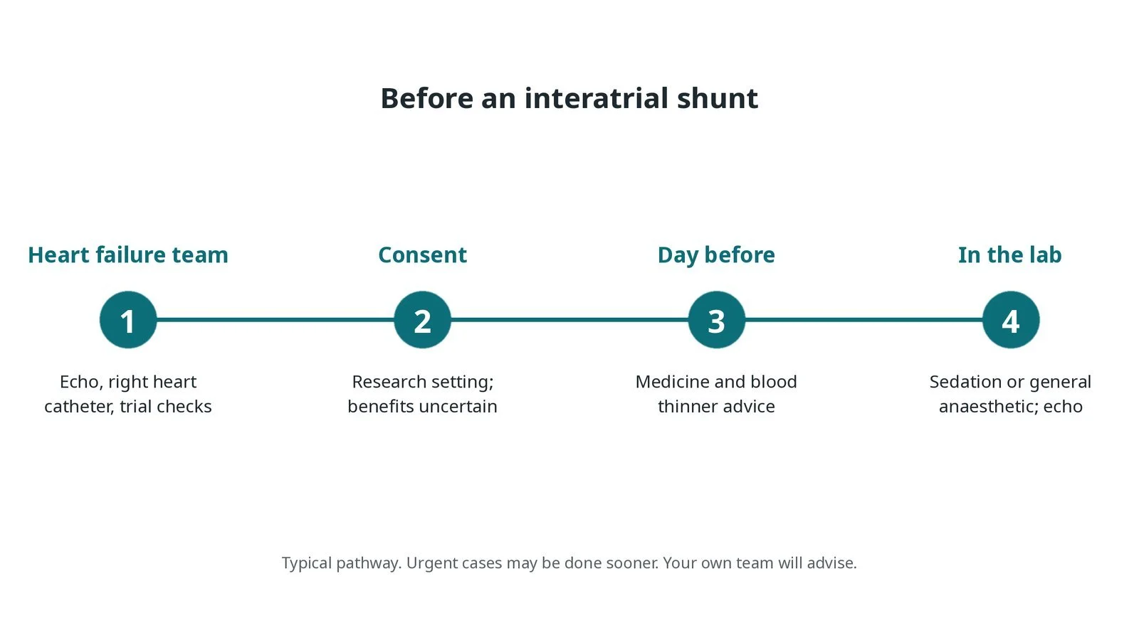
Before an interatrial shunt: heart failure team review with echo and a right heart catheter; consent in a research setting, where benefits are uncertain; medicine advice; sedation or anaesthetic with echo.
Step by step
1. Getting ready
You lie on the X-ray table. Sedation or a general anaesthetic is given, as per centre protocol. Echo (TOE or intracardiac echo) guides the case.
2. Access and pressures
A sheath is placed in the femoral vein under ultrasound. Sizes are per device and operator. Right heart pressures are often measured first. Heparin is given after the transseptal puncture, as per centre protocol.
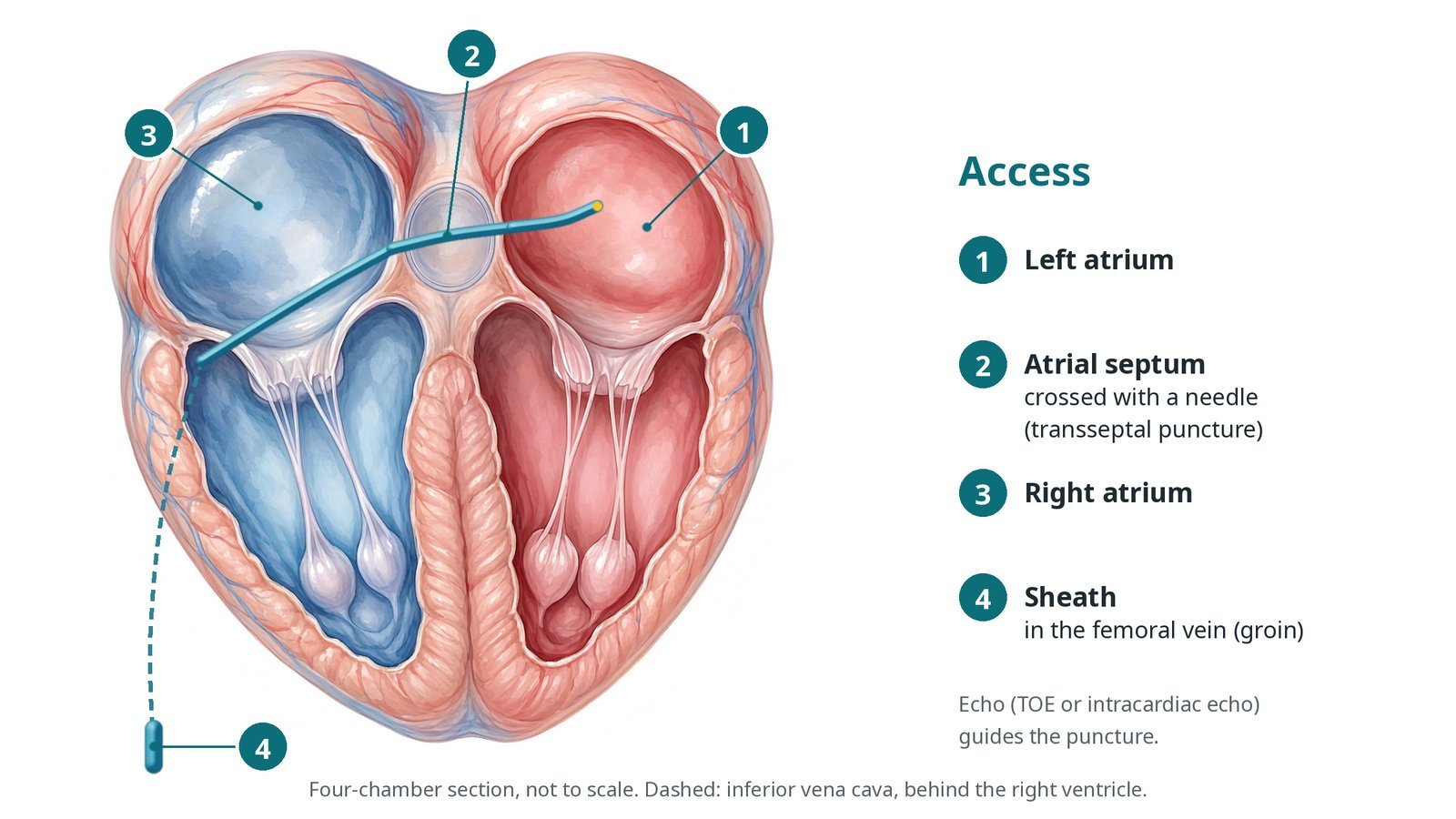
Access: (1) left atrium; (2) atrial septum, crossed with a needle (transseptal puncture); (3) right atrium; (4) sheath in the femoral vein.
3. Making the shunt
A. Puncture. A transseptal needle crosses the septum under echo and X-ray guidance.
B. Prepare. A wire is placed in the left atrium and a balloon may stretch the puncture.
C. Shunt. A small device is placed across the septum, or a disc of septum is removed or ablated (no-implant approach).
D. Check. Echo shows left-to-right flow through the shunt and pressures are measured.
Watch: A falling blood pressure with fluid around the heart on echo: possible perforation and tamponade. Tell the operator at once; the pericardiocentesis kit should be ready.
4. Finishing
The sheath is removed and the vein closed with pressure or a stitch, per operator.
5. Follow-up
Echo checks that the shunt is open. Symptoms, walking distance and heart failure admissions are followed, often as part of the trial.
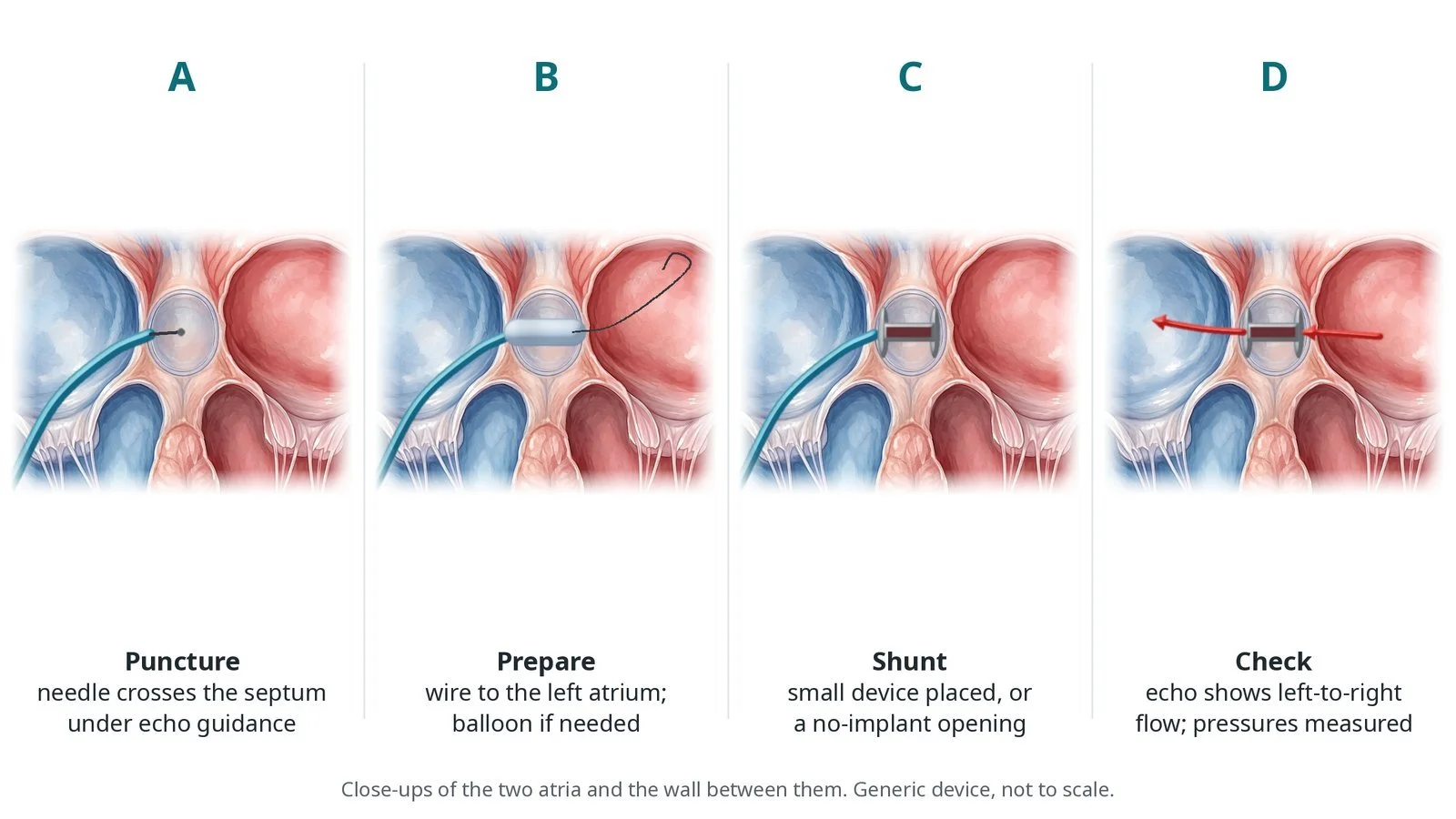
A: needle puncture of the septum under echo guidance. B: wire to the left atrium and balloon if needed. C: small device placed, or a no-implant opening made. D: echo shows left-to-right flow.
Recovery and aftercare
First hours: rest with checks of the groin, heart rhythm and blood pressure, as per centre protocol.
Going home: often the next day.
Medicines: blood thinners for a period, as your team advises; continue heart failure medicines.
Activity: walk as normal; avoid heavy lifting for a few days.
Driving: ask your team; usually after a few days if well; check DVLA rules for your heart condition.
Follow-up: echo and heart failure clinic, often with trial visits.
Call 999 if you have chest pain, sudden breathlessness, fainting, signs of a stroke, or heavy bleeding from the groin.
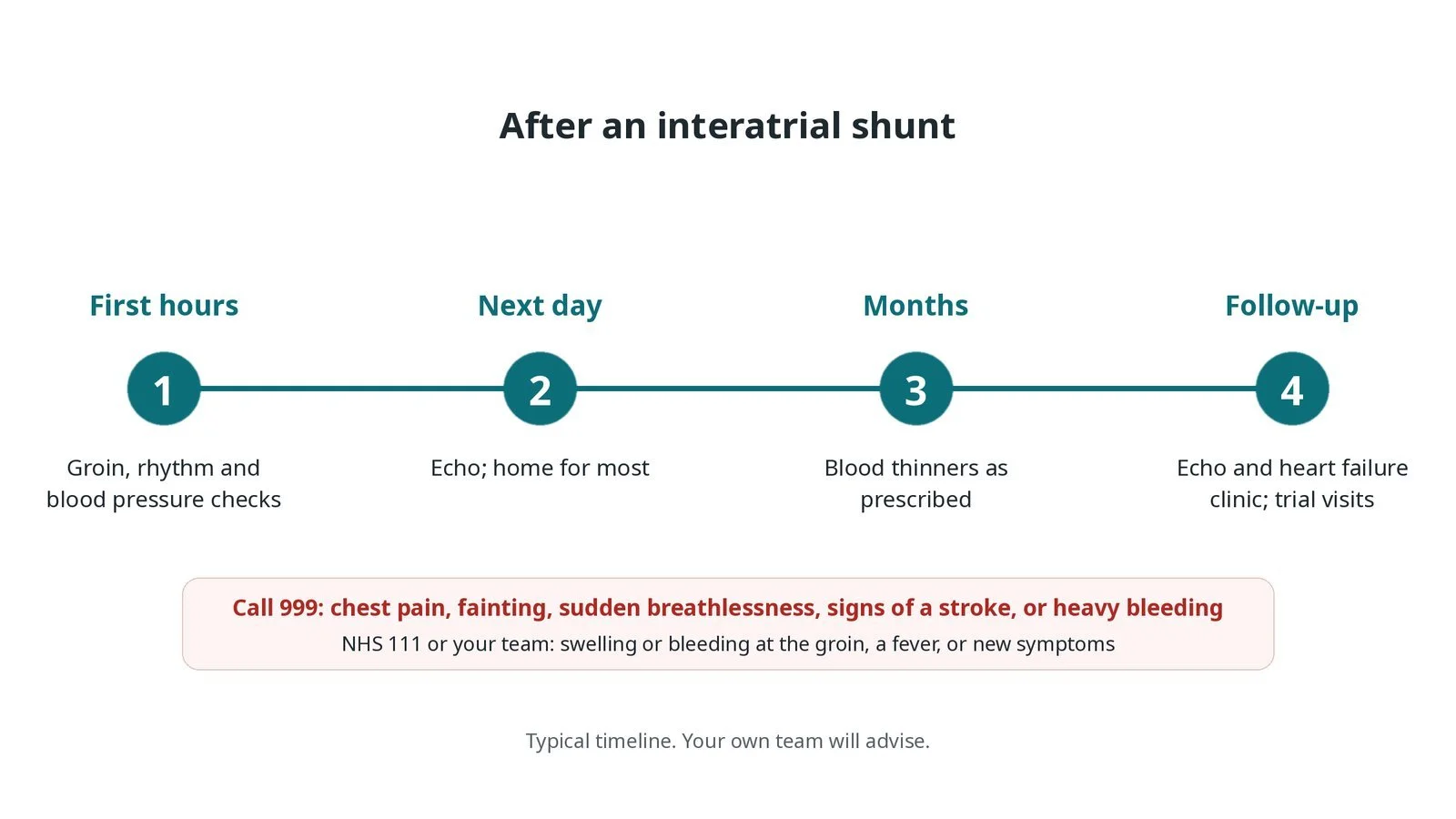
After an interatrial shunt: (1) groin, rhythm and blood pressure checks; (2) echo and home the next day for most; (3) blood thinners as prescribed; (4) echo, heart failure clinic and trial visits.
Risks and complications
Your team will explain the risks that apply to you. Possible problems include:
Bleeding or bruising at the groin
Perforation and fluid around the heart (rare)
Device moving or the shunt closing over time (uncommon)
Strain on the right side of the heart
Stroke or clot passing across the shunt (rare)
Symptoms not improving
Cath lab kit list (printable)
Barts example kit list
An example from one UK centre, written generically. Every lab has its own protocol, and sizes and doses vary.
femoral venous sheath, size per device
transseptal sheath and needle, per operator
guidewires, per operator
balloon catheter, per device
interatrial shunt device and delivery system, or no-implant shunt system, per device
pressure transducer
TOE or intracardiac echo
heparin, as per centre protocol
pericardiocentesis kit available
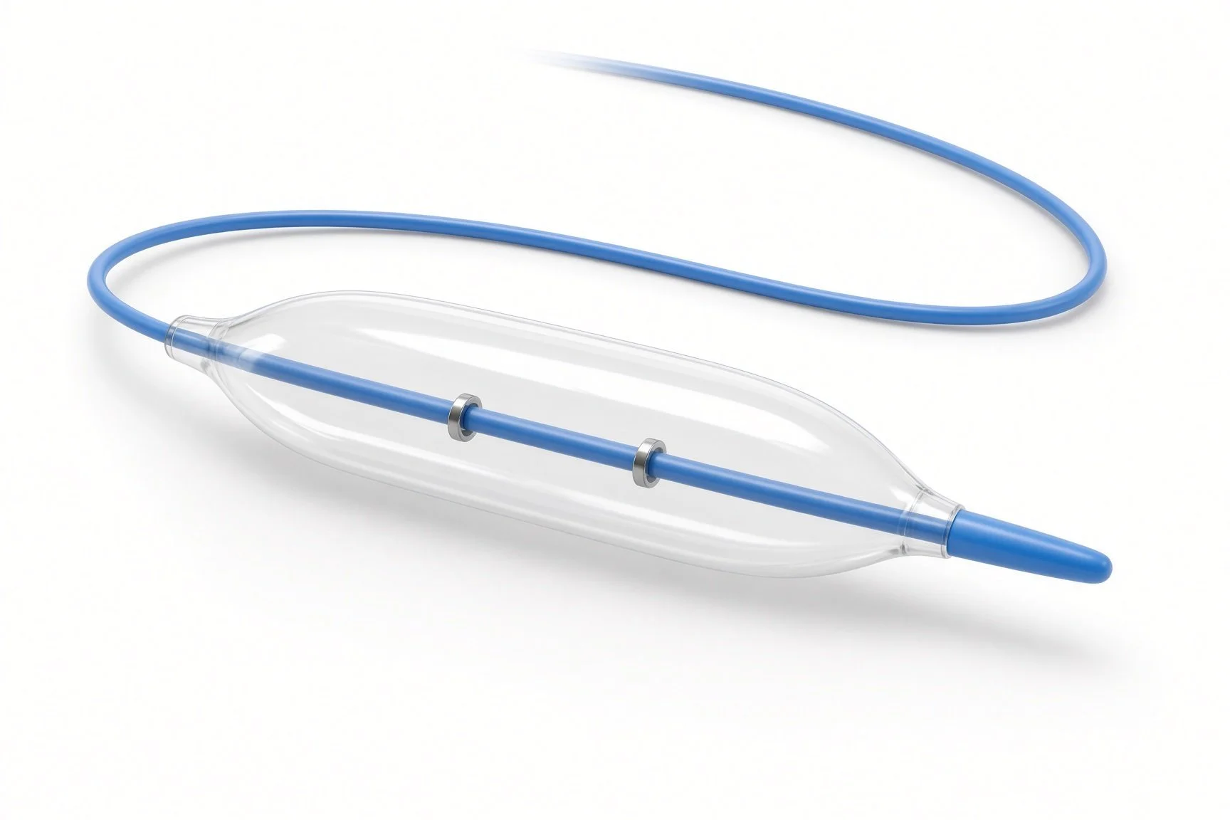
Balloon catheter.
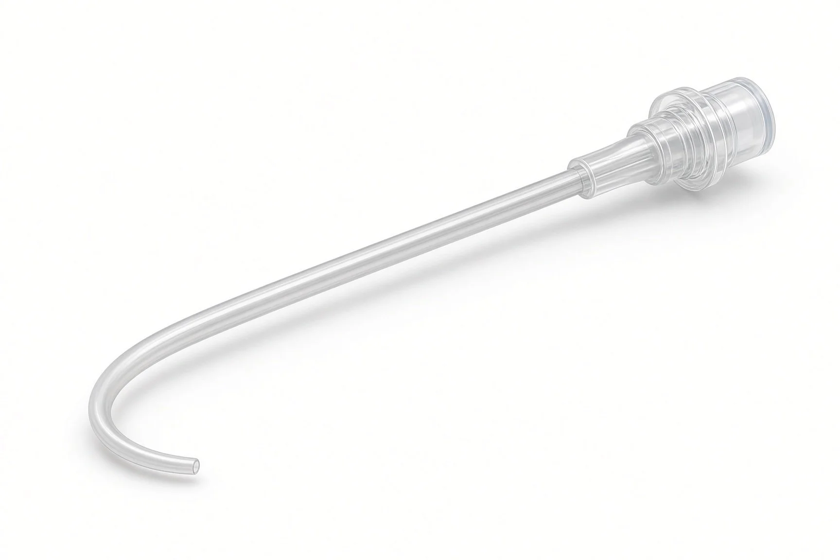
Balloon catheter.
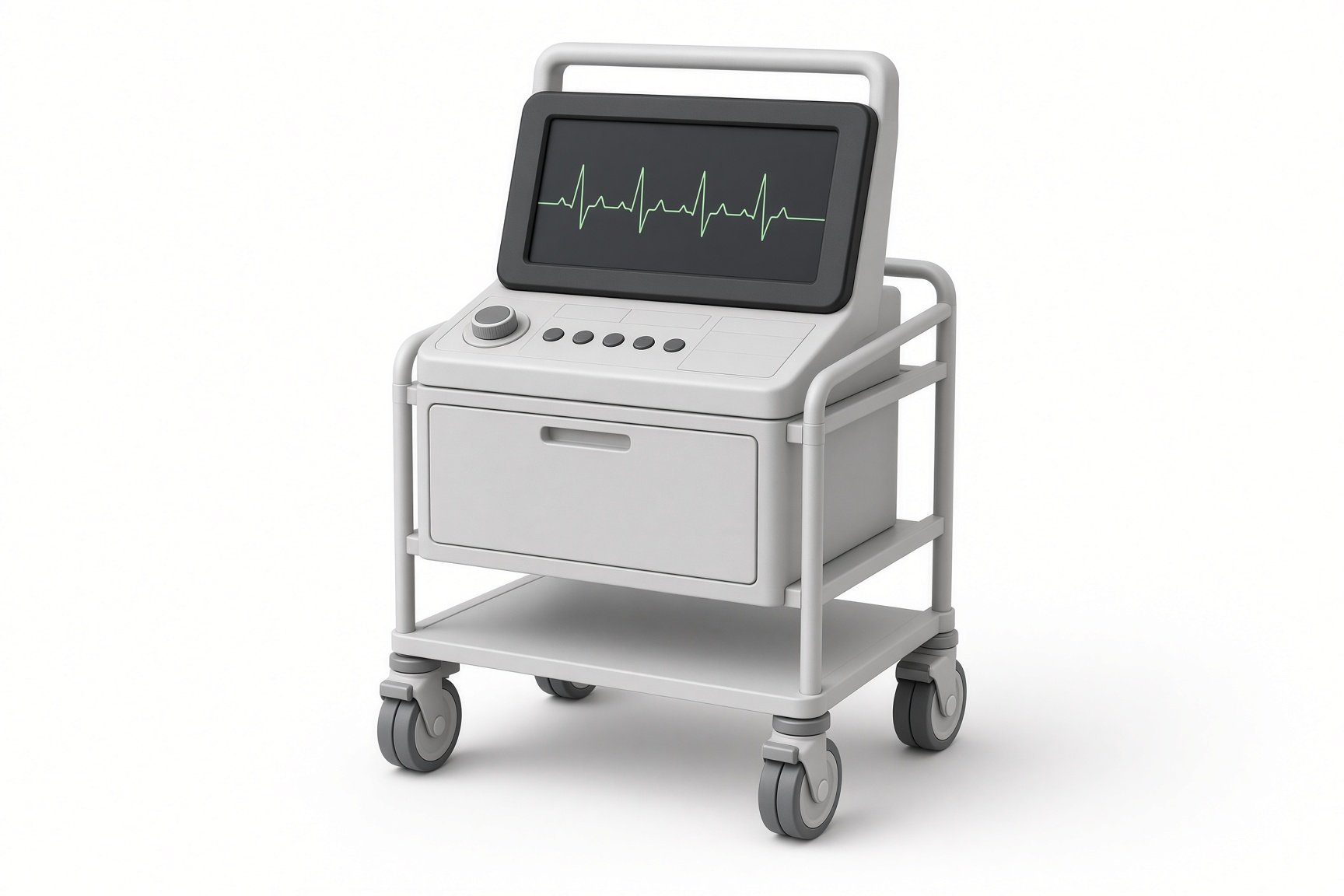
Pressure transducer.
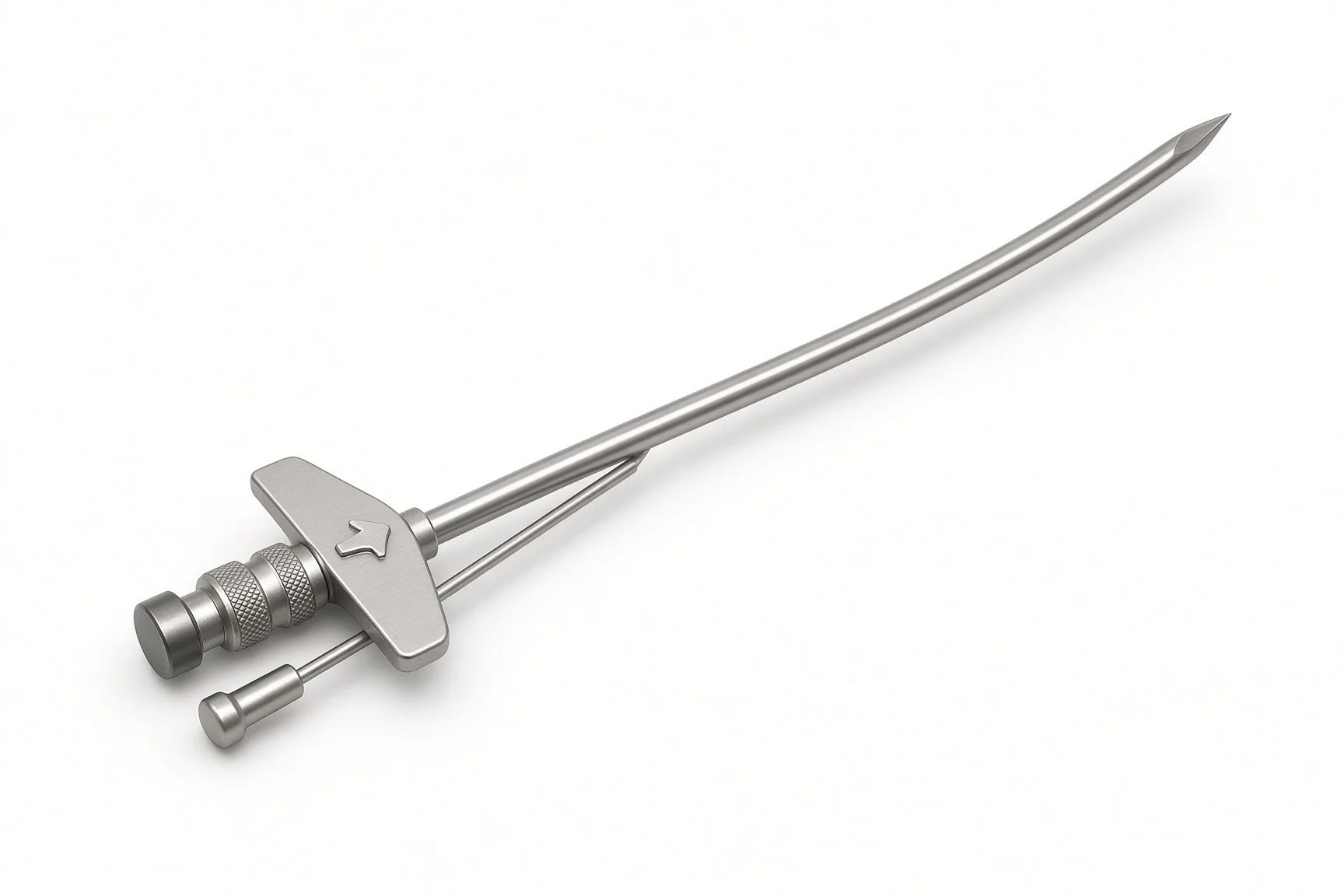
Transseptal sheath and needle.
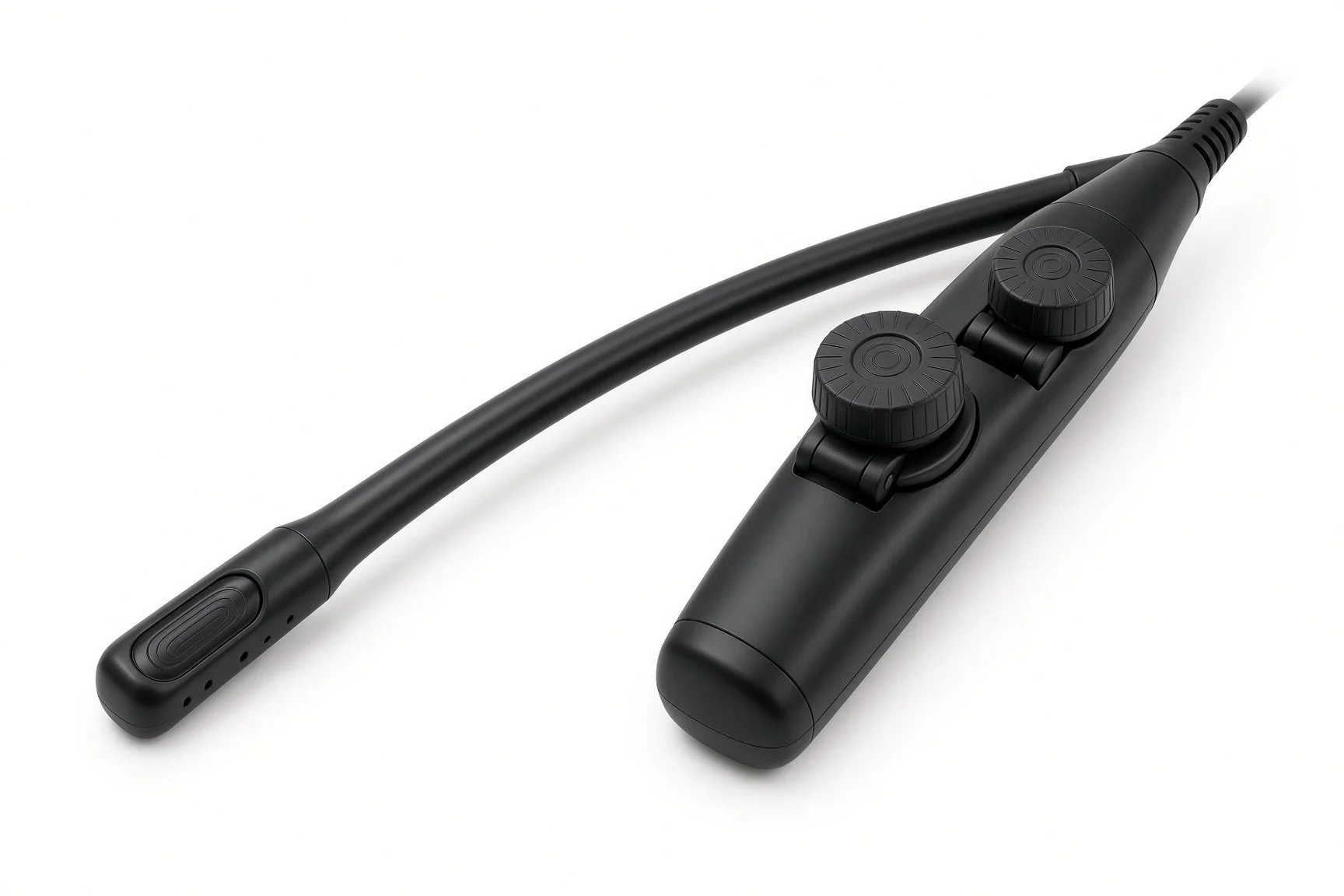
TOE or intracardiac echo.
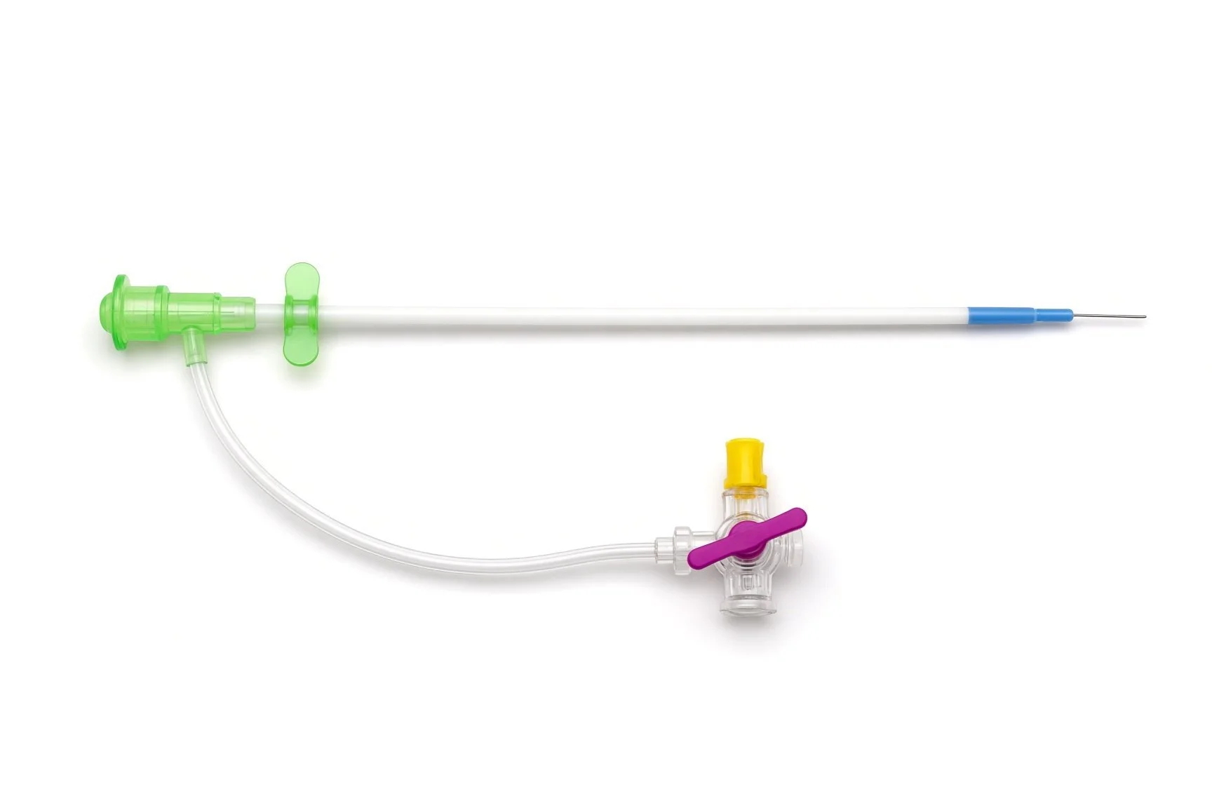
Femoral venous sheath.
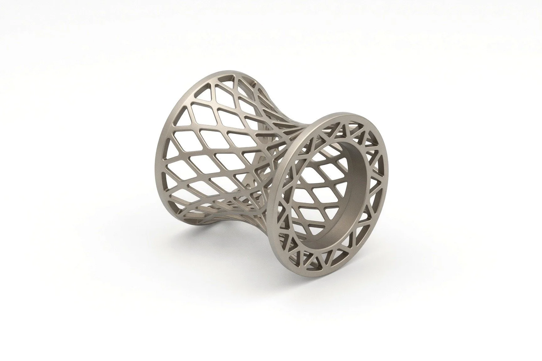
Shunt device on delivery system.

TOE or intracardiac echo.
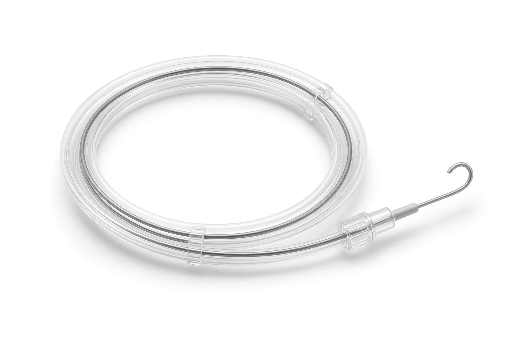
Guidewire.
Procedural steps (Barts example)
Getting ready
Access and pressures
Making the shunt
Finishing
Follow-up
First time in the cath lab?
It is research. Check consent, the trial protocol and any data that must be recorded.
Transseptal skills matter. The puncture is the same as for other left atrial procedures.
Have the pericardiocentesis kit ready. Perforation is uncommon but needs quick action.
Measure before and after. Pressures help show the shunt is working.
Trial protocols may need extra measurements and timings. Have the protocol to hand.
FAQ
Why make a hole in my heart?
A small controlled opening lets some blood escape from the high-pressure left atrium, aiming to ease breathlessness.
Does it work?
Trial results are mixed. Some people may benefit more than others, and research is ongoing.
Is it permanent?
It is designed to stay open, although some shunts narrow over time.
Can I still have other heart procedures?
Usually, but tell any team that you have a shunt; it can affect procedures that cross the septum.
Related Cath Lab 101 guides
CardioMEMS pulmonary artery pressure sensor (coming soon)
Exercise and fluid-challenge right heart catheterisation (coming soon)
Sources
All checked on 12 November 2026. Each page's own date is shown where it gives one.
McDonagh TA, et al. 2021 ESC Guidelines for the diagnosis and treatment of acute and chronic heart failure. Eur Heart J 2021;42:3599-3726
StatPearls (NCBI Bookshelf), search: heart failure with preserved ejection fraction. ncbi.nlm.nih.gov/books
Disclaimer
Education only. This page is for general education for healthcare students, cath lab staff and curious readers. It isn't medical advice and doesn't replace advice from your own doctor, heart team or local unit protocols. Equipment and medicines are described generically. No product or manufacturer is recommended. If you think someone is having a heart emergency, call 999.
Disclosure: I work in the medical device industry. This page is independent education and isn't sponsored or reviewed by any company.
All diagrams on this page are original illustrations created for rodnieoro.com.