Post-infarct ventricular septal rupture and LV pseudoaneurysm closure: a Cath Lab 101 guide
Reviewed by Rodnie Oro, cath lab charge nurse · Last reviewed: November 2026 · Education only, not medical advice
Post-infarct ventricular septal rupture and LV pseudoaneurysm closure in one minute
What it is: After a large heart attack, dead muscle can tear. A ventricular septal rupture (VSR) is a tear between the ventricles; an LV pseudoaneurysm is a tear in the outer wall held in by scar and the pericardium. A device can close the tear by catheter, either as a bridge to surgery or as the final treatment.
Who it is for: Very unwell patients after a heart attack, often already on a balloon pump, a heart pump or ECMO, as decided by the heart team with cardiac surgeons. Honest conversations with the patient and family, including palliative care, are part of the plan.
Main risks: Death, bleeding, the device moving or not sealing fully, damage to heart valves, and stroke. The risk is high whichever treatment is chosen.
Usually: Under general anaesthetic with TOE, often as an emergency, followed by intensive care.
On this page: What is post-infarct ventricular septal rupture and LV pseudoaneurysm closure? · Who is it for? · When is it done? · Step by step · Recovery · Risks · Kit list · First time in the lab? · FAQ
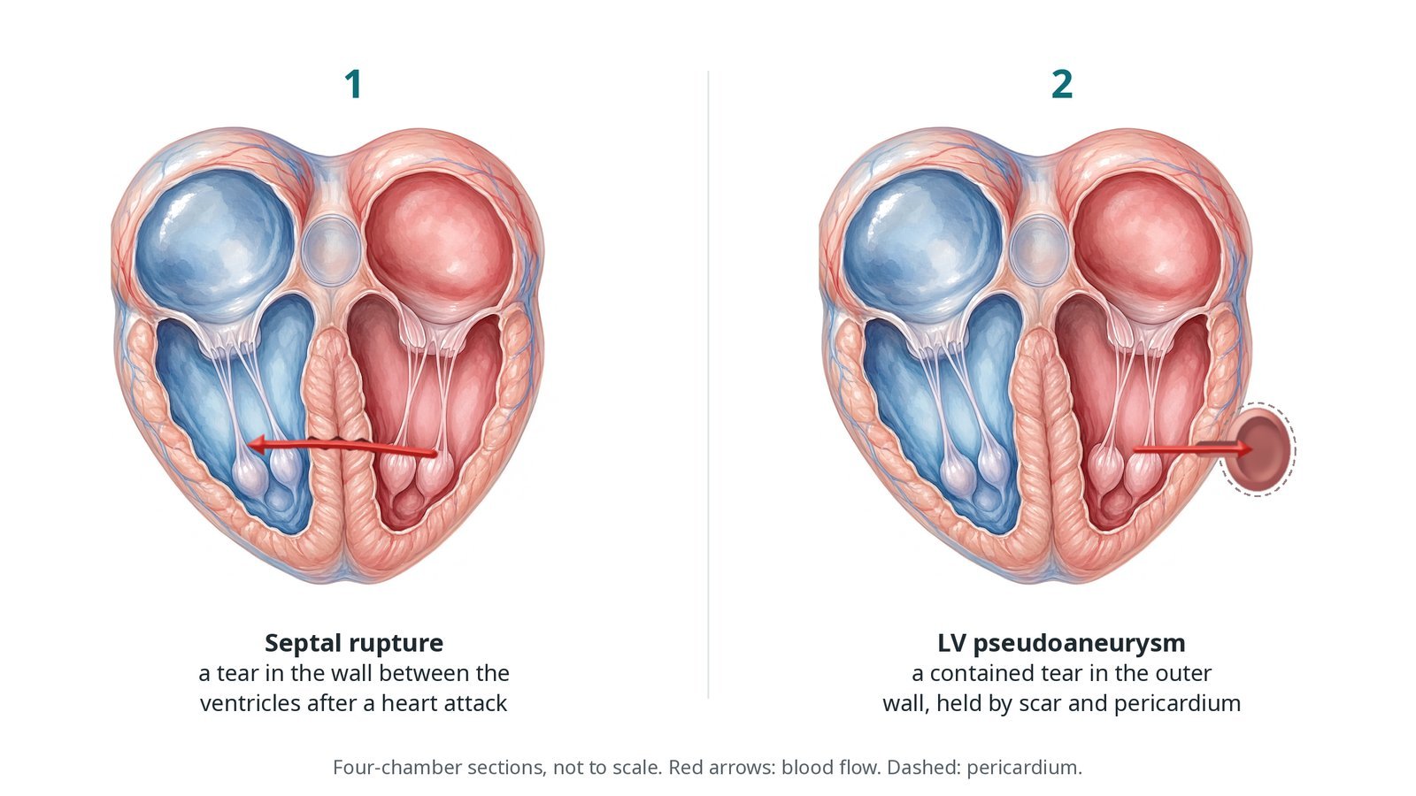
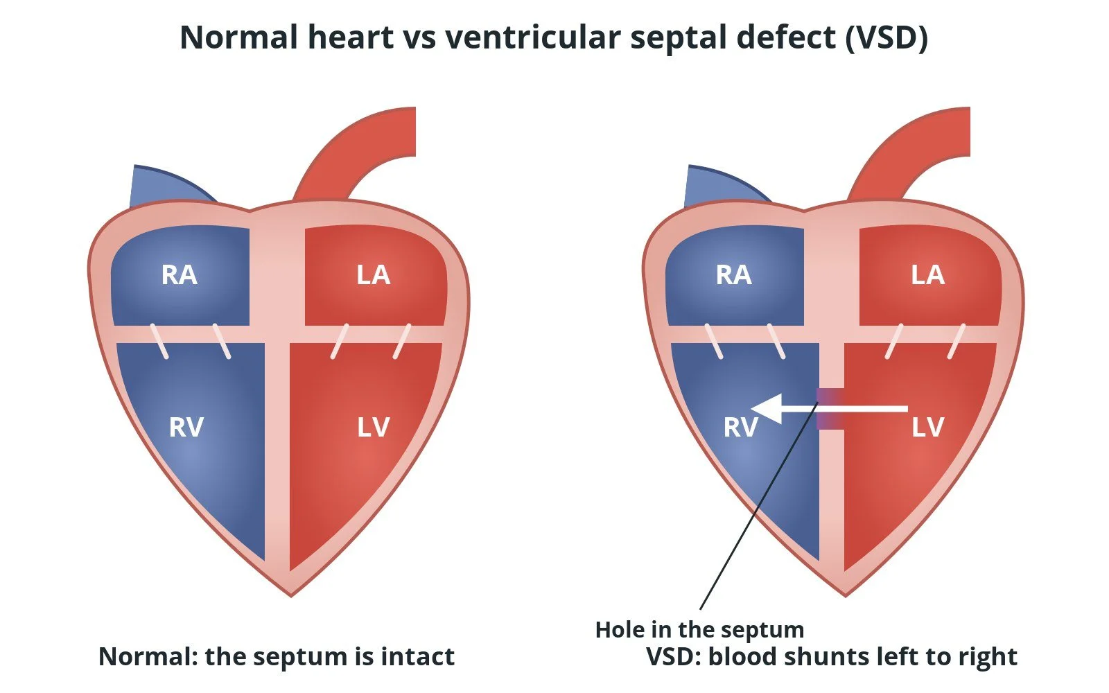
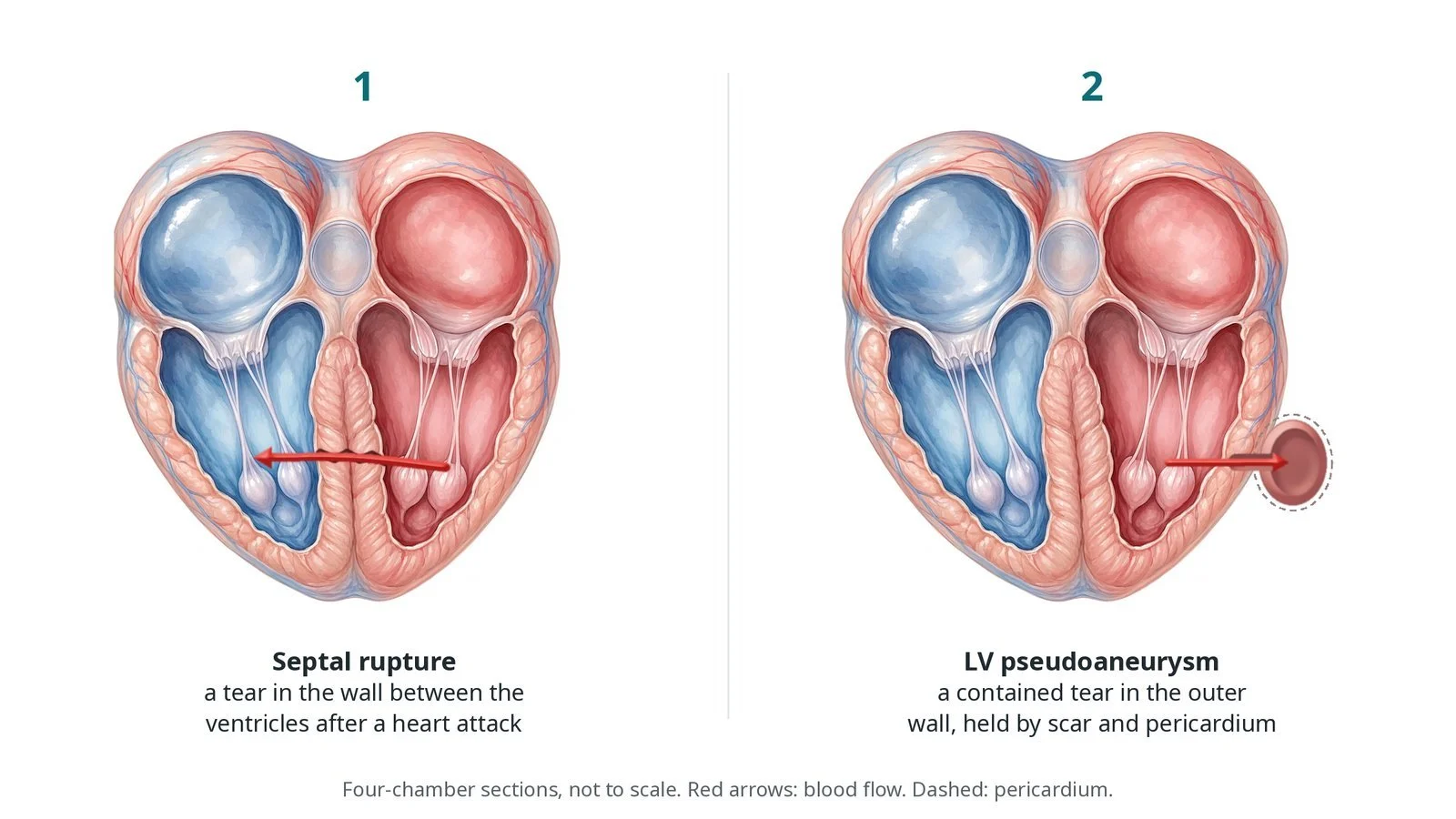
(1) Septal rupture: a tear in the wall between the ventricles after a heart attack. (2) LV pseudoaneurysm: a contained tear in the outer wall, held only by scar and pericardium.
What is post-infarct ventricular septal rupture and LV pseudoaneurysm closure?
The ventricular septum is the muscular wall between the right and left ventricles. After a heart attack, the damaged muscle softens over the following days and can tear. A VSR lets blood rush from the high-pressure left ventricle into the right ventricle, flooding the lungs and causing shock.
A tear in the outer (free) wall usually causes rapid death from bleeding into the pericardium. Sometimes it is contained by clot, scar and pericardium, forming a pseudoaneurysm with a narrow neck. It can still rupture at any time.
Catheter closure uses an occluder device, like those for congenital holes, to plug the tear. The edges of a post-infarct tear are soft and irregular, so the device may not seal completely. For a VSR, a wire is passed from the left ventricle through the tear and snared from the venous side to make an arteriovenous rail. A pseudoaneurysm can be closed through the apex (transapical) or from the aorta across the aortic valve (retrograde).
New to the acronyms? See the Cath Lab Abbreviations A–Z.
Who is it for?
People with a post-infarct VSR or LV pseudoaneurysm where the heart team, including surgeons, decides a catheter approach is best, for example:
as a bridge to stabilise before later surgery
as the final treatment when surgery is too high risk
for a leftover leak after surgical repair
Who it may not suit: Very large or complex tears, tears too close to valves, and patients for whom treatment would not be in their best interests after discussion with them and their family.
The decision is made by your heart team, who will talk through the reasons, the alternatives and the risks with you.
When is it done?
Often urgently, once the patient has been stabilised with mechanical support, ventilation and medicines. Timing is a team decision, balancing a sick patient against tissue that becomes firmer with time.
gives you a chance to ask questions and give consent
arranges blood tests, including kidney function and a blood count, and an ECG
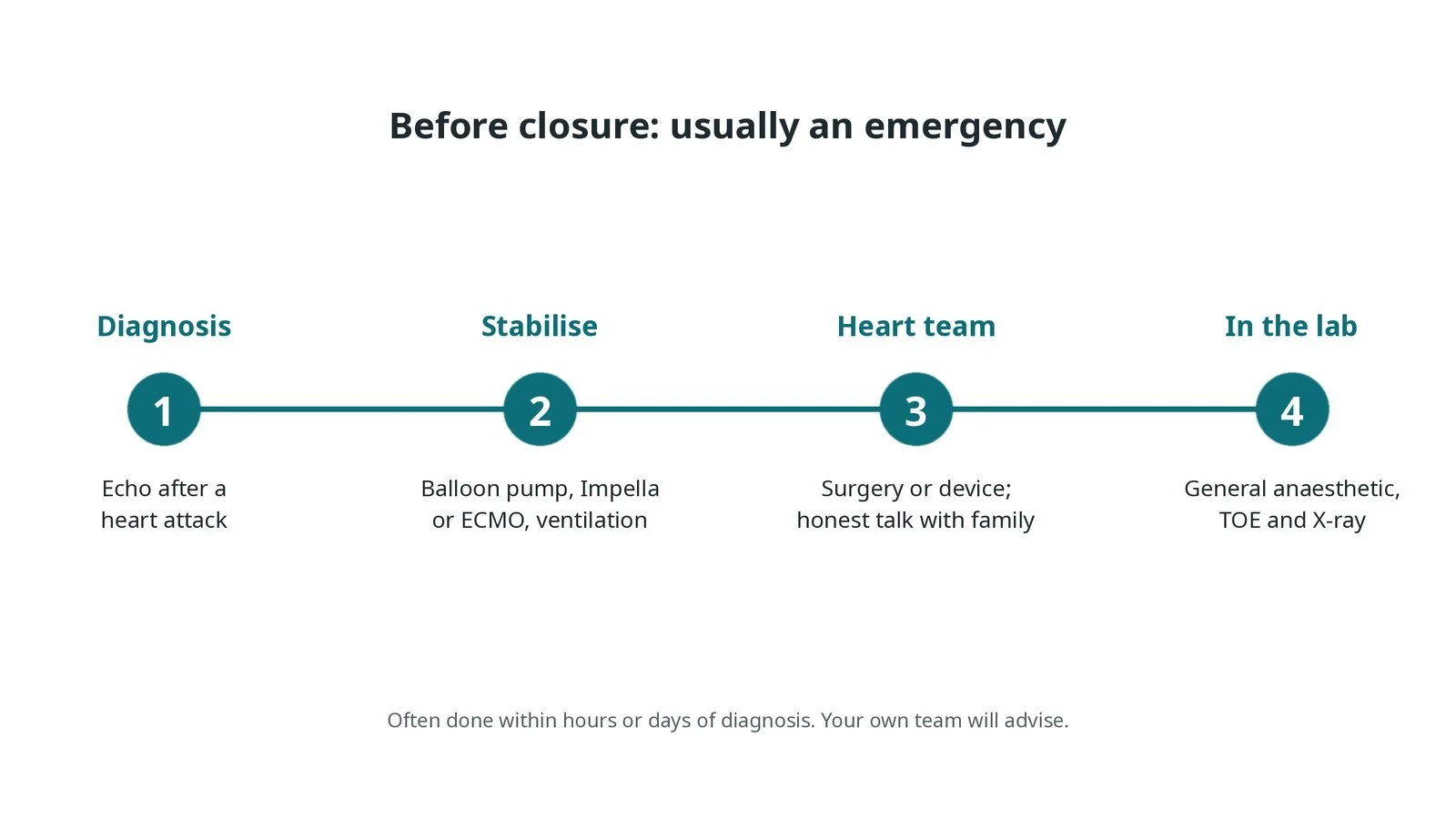
Before closure, usually an emergency: echo diagnosis after a heart attack; stabilisation with a balloon pump, Impella or ECMO; heart team decision and an honest talk with the family; general anaesthetic and TOE.
Step by step
1. Getting ready
The patient usually comes from intensive care, ventilated and on mechanical support. A general anaesthetic and TOE are used. Blood products and the surgical team should be available.
2. Access
Femoral arterial and venous sheaths are placed under ultrasound, sometimes a jugular venous sheath. Sizes are per device and operator. Heparin is given as per centre protocol, balanced against the bleeding risk.
3. Closing a VSR
A. Cross. A catheter from the artery goes across the aortic valve into the left ventricle, and a wire is passed through the tear into the right ventricle.
B. Rail. The wire is snared from the vein and brought out, making an arteriovenous rail; the delivery sheath is advanced from the vein across the tear.
C. LV disc. The occluder's left ventricular disc is opened and pulled back against the septum.
D. Release. The right ventricular disc is opened, TOE checks position and leak, then the device is released.
Watch: A sudden drop in blood pressure or a new valve leak on TOE while the rail is in place: the wire can pull on valve tissue. Tell the operator, who may relax the rail.
4. Pseudoaneurysm closure
The neck of the pseudoaneurysm is crossed either through the apex (transapical) or from the aorta across the aortic valve (retrograde), and an occluder or plug is placed in the neck, guided by TOE and angiography.
5. Finishing
Sheaths are usually left in or removed per operator, and the patient returns to intensive care. Support devices are weaned as the patient improves.
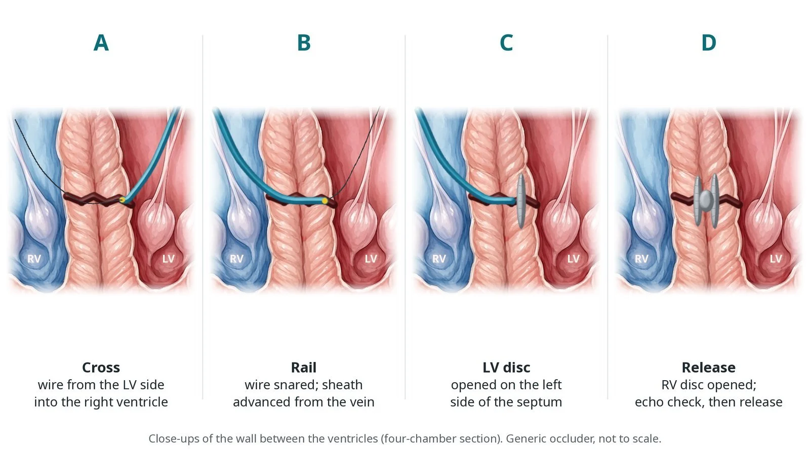
A: wire crosses from the left ventricle. B: wire snared and a sheath advanced from the vein. C: left disc opened. D: right disc opened, echo check, then release.
Recovery and aftercare
First hours: intensive care with ventilation and support devices, as per centre protocol.
Next days: echo checks for any remaining leak; support devices are weaned as possible.
Medicines: heart failure medicines and blood thinners, as the team advises.
Next steps: some patients go on to surgery; others recover on the ward and in rehabilitation.
Driving: no driving for a long period after a heart attack and this procedure; your team will advise in line with DVLA rules.
Follow-up: echo, heart failure and cardiology clinic.
Call 999 after discharge if you have chest pain, sudden breathlessness, fainting, or signs of a stroke.
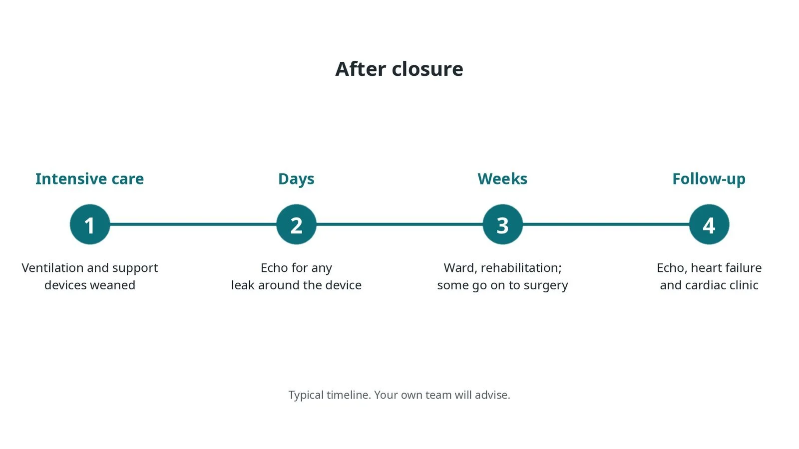
After closure: (1) intensive care, ventilation and support devices weaned; (2) echo for any leak around the device; (3) ward and rehabilitation, some go on to surgery; (4) echo and clinic follow-up.
Risks and complications
Your team will explain the risks that apply to you. Possible problems include:
Death, which is common with this condition whatever the treatment
Bleeding
Device moving or not sealing completely
Damage to heart valves or the rhythm system
Stroke (uncommon)
Kidney problems
Cath lab kit list (printable)
Barts example kit list
An example from one UK centre, written generically. Every lab has its own protocol, and sizes and doses vary.
femoral arterial and large venous sheaths, per device and operator
catheters to cross the defect, per operator
exchange-length guidewire for the rail
snare, per operator
septal or muscular VSD occluder or vascular plug, size per TOE and angiography
TOE probe and echo machine
mechanical support device in place (IABP, a heart pump or ECMO)
blood products and cell salvage, as per centre protocol
heparin and anaesthetic medicines, as per centre protocol
pericardiocentesis kit and surgical back-up
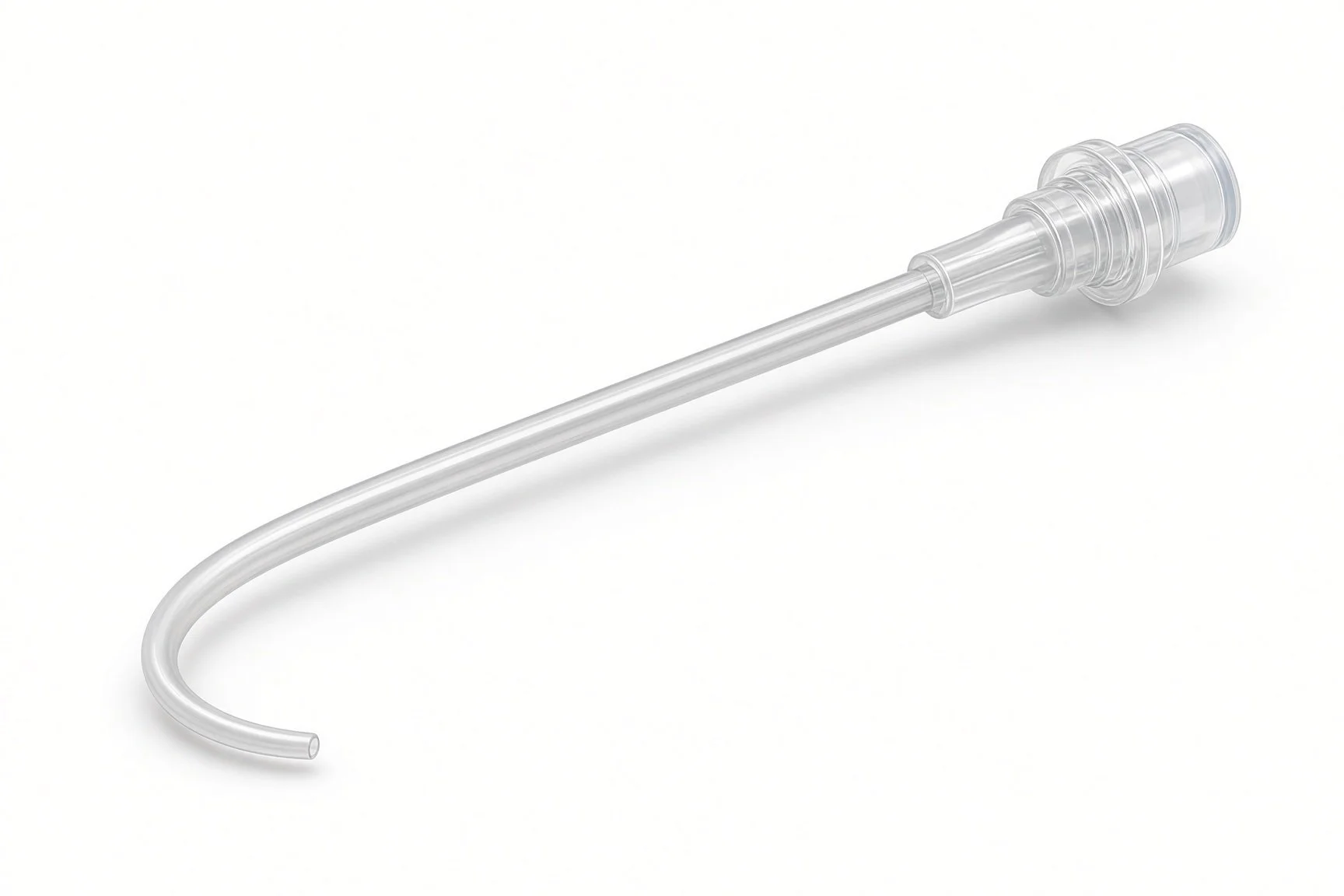
Catheters.
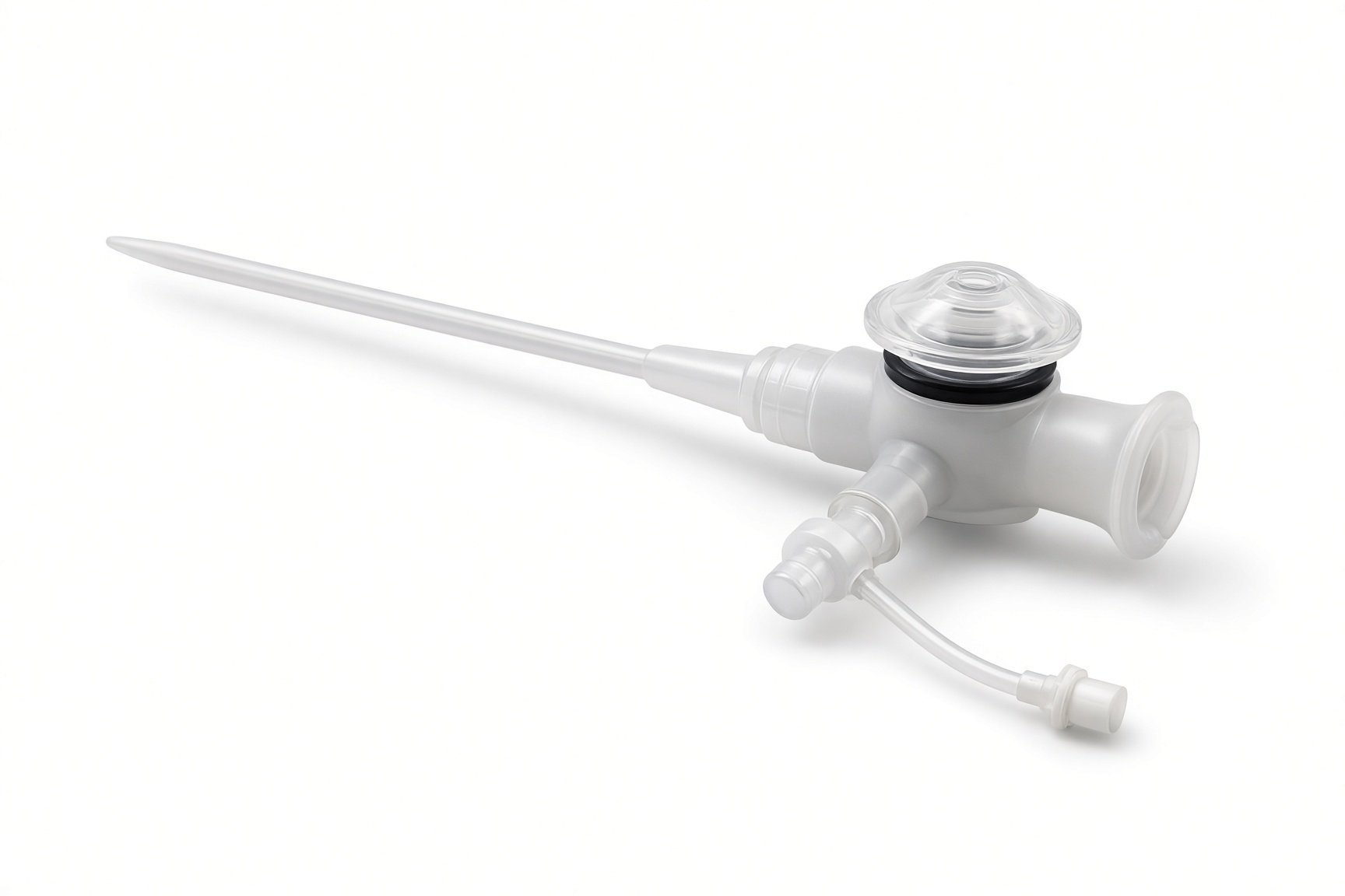
Arterial and large venous sheaths.
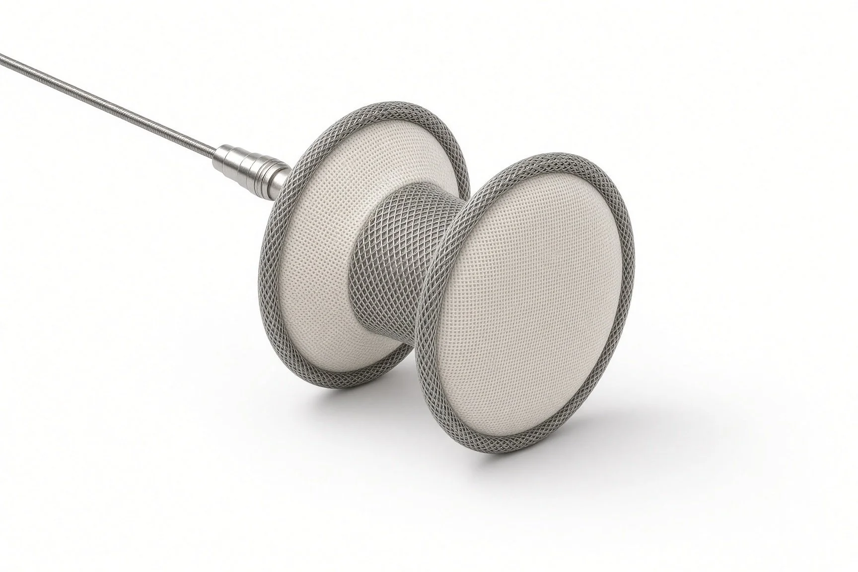
Septal occluder on delivery cable.
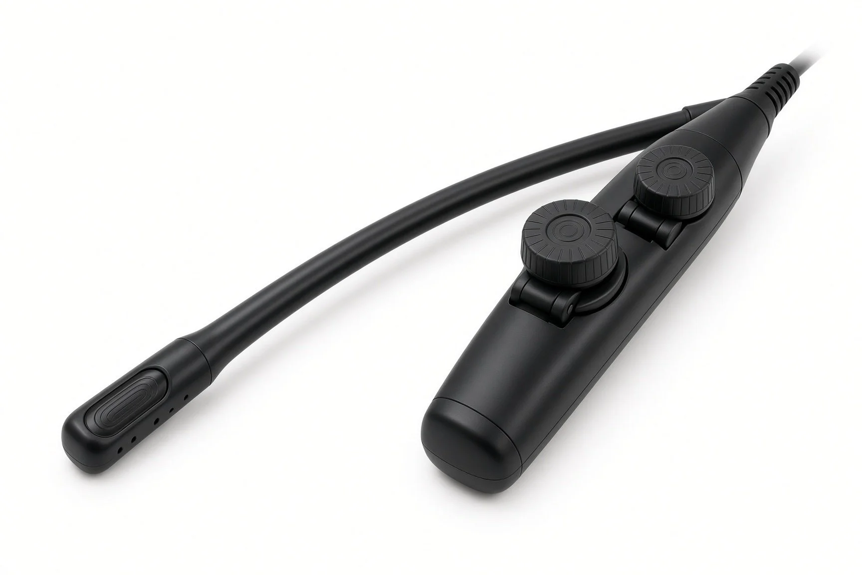
TOE probe.
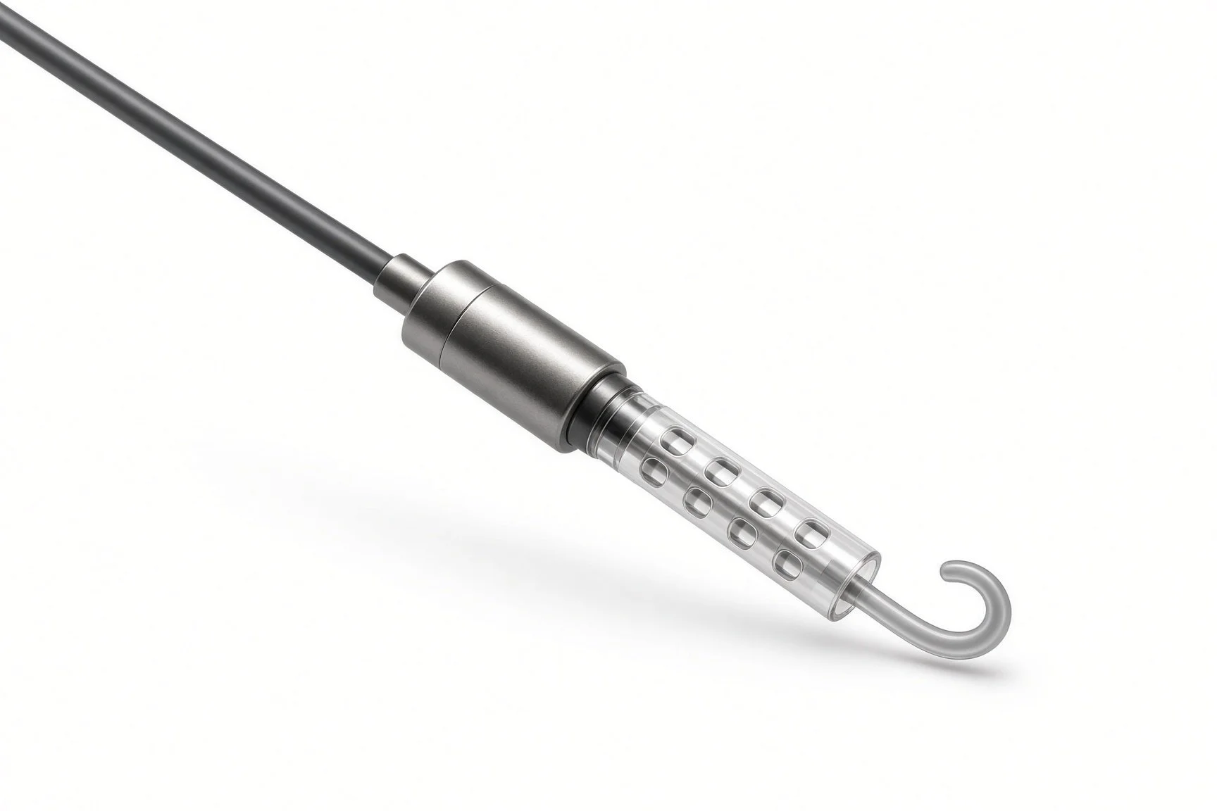
Support device already running.
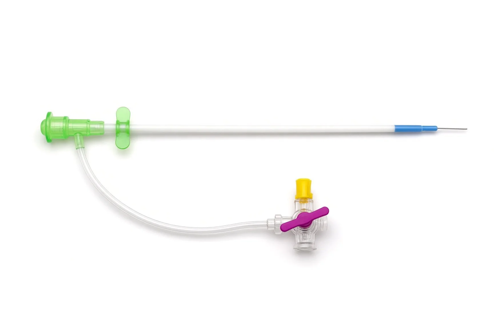
Arterial and large venous sheaths.
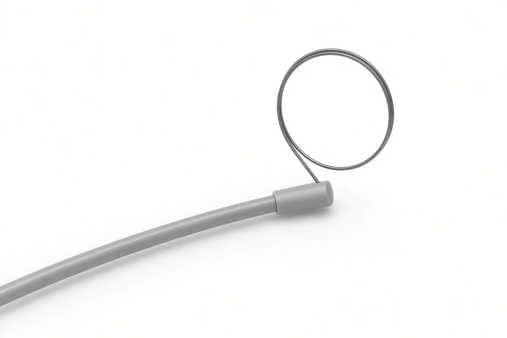
Snare.
Support device already running.
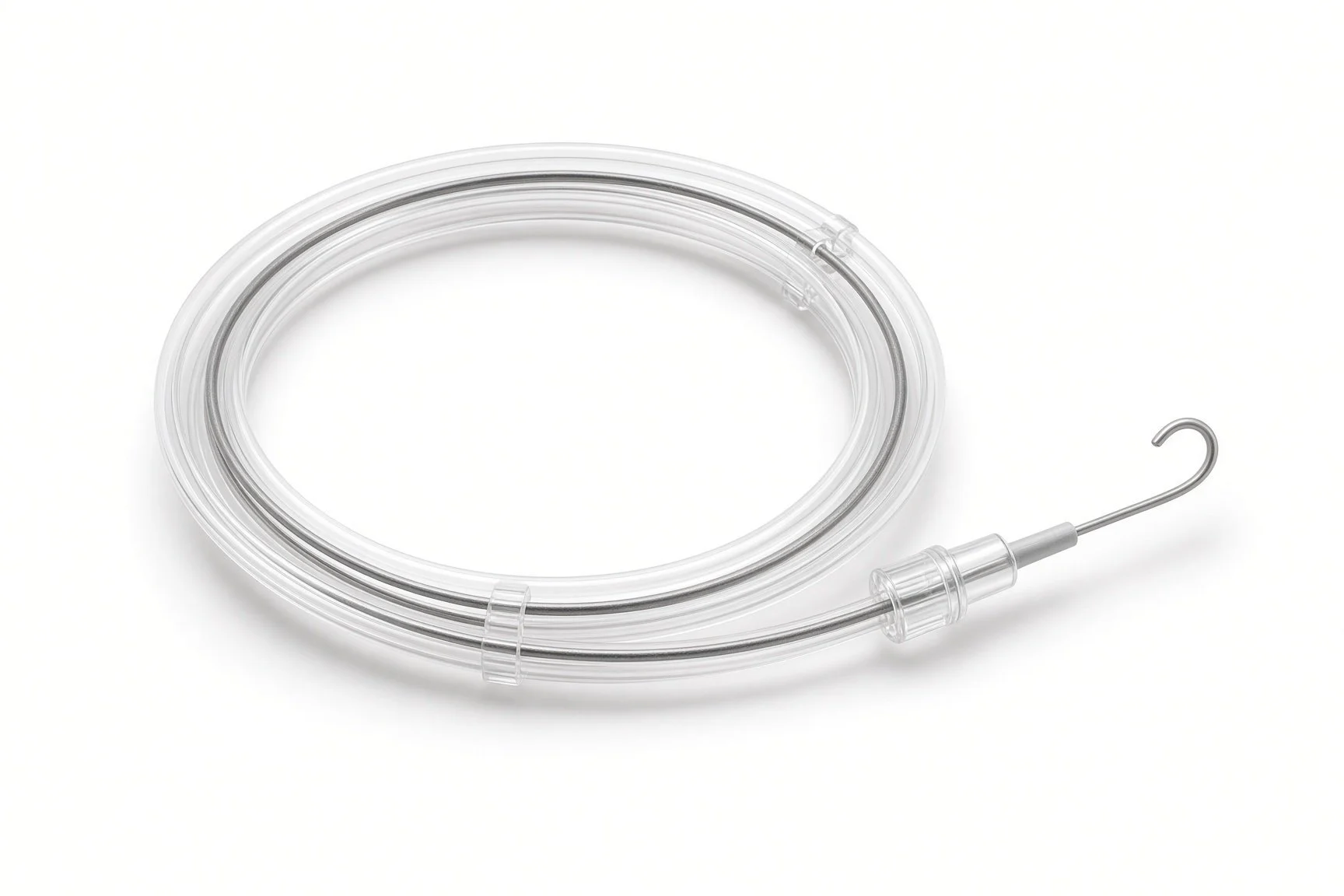
Exchange-length wire.
Procedural steps (Barts example)
Getting ready
Access
Closing a VSR
Pseudoaneurysm closure
Finishing
First time in the cath lab?
These patients are critically ill. Expect drug infusions, a ventilator and a support device; plan space and power.
Know the rail. The arteriovenous rail is key to VSR closure; watch the wire on both ends.
Be ready to bleed. Blood products and the surgical team should be ready before the case starts.
Remember the family. The team will have had honest conversations; be kind and clear with relatives.
Plan the transfer: support devices, drug infusions and the ventilator all move with the patient. Assign one person to lines and cables.
FAQ
Why did the heart tear?
A heart attack damages muscle, which softens over days and can tear.
Is surgery better?
It depends on the person and the tear. The heart team, including surgeons, decides which approach gives the best chance.
How risky is it?
This condition is very serious and the risk is high with any treatment. The team will be honest with you and your family.
What does palliative care mean here?
Care focused on comfort and what matters most to the patient. It can sit alongside active treatment or replace it if treatment is not in the patient's interests.
Related Cath Lab 101 guides
Sources
All checked on 12 November 2026. Each page's own date is shown where it gives one.
Byrne RA, et al. 2023 ESC Guidelines for the management of acute coronary syndromes. Eur Heart J 2023;44:3720-3826
StatPearls (NCBI Bookshelf), search: ventricular septal rupture. ncbi.nlm.nih.gov/books
Disclaimer
Education only. This page is for general education for healthcare students, cath lab staff and curious readers. It isn't medical advice and doesn't replace advice from your own doctor, heart team or local unit protocols. Equipment and medicines are described generically. No product or manufacturer is recommended. If you think someone is having a heart emergency, call 999.
Disclosure: I work for a company that makes a device used in this procedure. This page is independent education, isn't sponsored or reviewed by any company, and doesn't recommend any product.
All diagrams on this page are original illustrations created for rodnieoro.com.