ECMO (extracorporeal membrane oxygenation): a Cath Lab 101 guide
Reviewed by Rodnie Oro, cath lab charge nurse · Last reviewed: October 2026 · Education only, not medical advice
ECMO in one minute
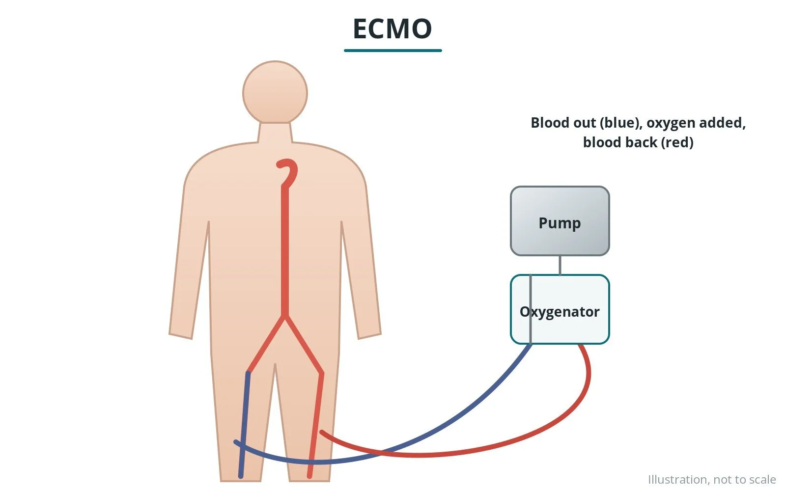
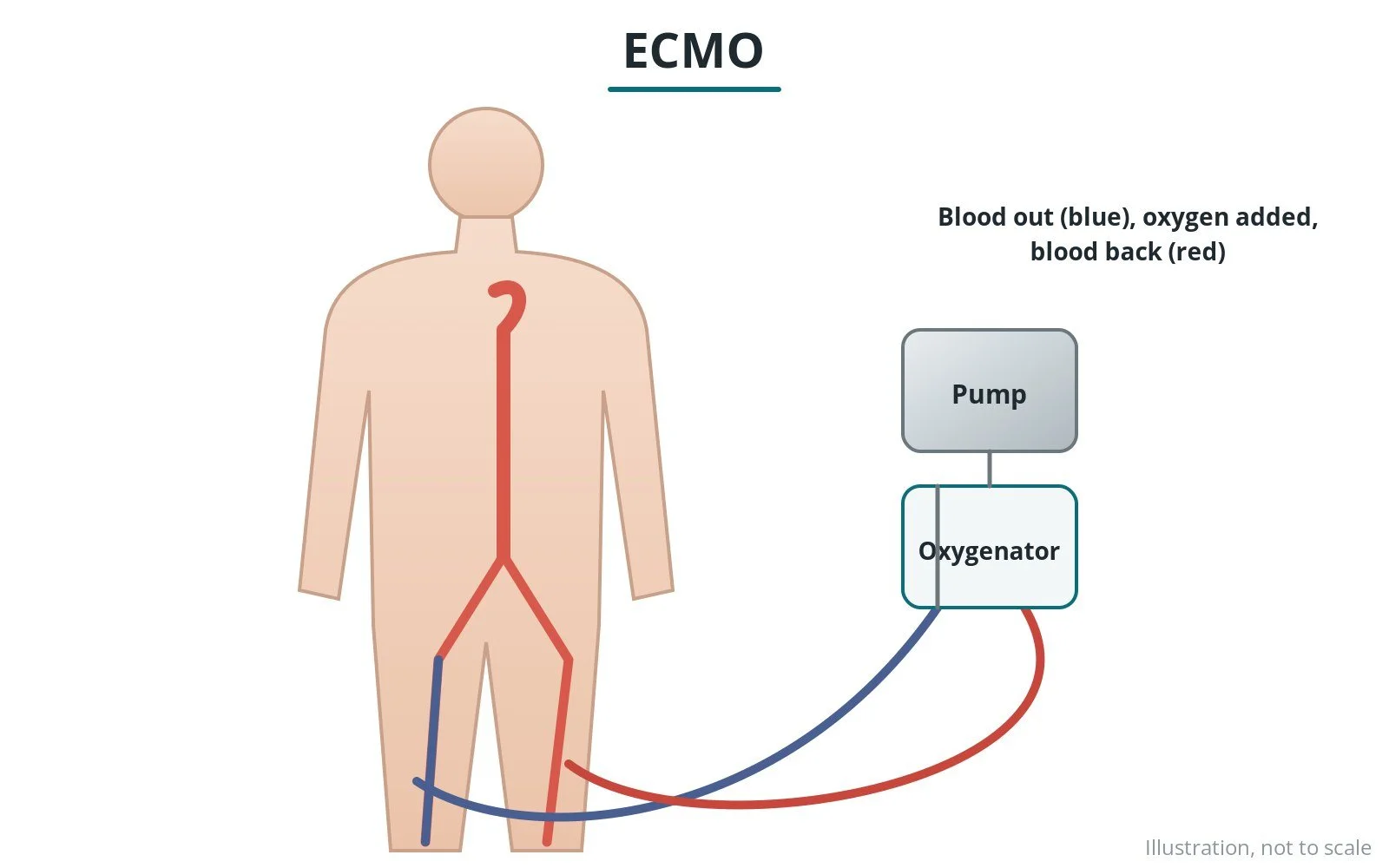
What it is: a machine that takes blood out, adds oxygen, removes carbon dioxide and pumps it back, taking over some or all of the work of the heart and lungs for a time.
Who it is for: people with severe cardiogenic shock or cardiac arrest (VA ECMO), or severe lung failure (VV ECMO), in specialist centres.
Main risks: bleeding, reduced blood flow to the leg, clots and stroke, infection, and damage to blood vessels.
Usually: started in the cath lab, emergency department or intensive care; it stays in for days to weeks.
On this page: What is ECMO? · Who is it for? · When is it done? · Step by step · Recovery · Risks · Kit list · First time in the lab? · FAQ
What is ECMO?
ECMO stands for extracorporeal membrane oxygenation.
- VA ECMO (veno-arterial) supports the heart and lungs: blood is taken from a large vein and returned to an artery, usually in the groin
- VV ECMO (veno-venous) supports the lungs only
In the cath lab, VA ECMO may be started for cardiogenic shock or during cardiac arrest, often while the cause, such as a blocked artery, is treated. It is run by a specialist team, including perfusionists.
New to the acronyms? See the Cath Lab Abbreviations A–Z.
Who is it for?
It may be considered for people with:
- cardiogenic shock not responding to other treatments
- cardiac arrest in selected cases (ECPR)
- support during very high-risk procedures
- bridge to recovery, a heart pump or transplant
The decision is made by your heart team, who will talk through the reasons, the alternatives and the risks with you.
When is it done?
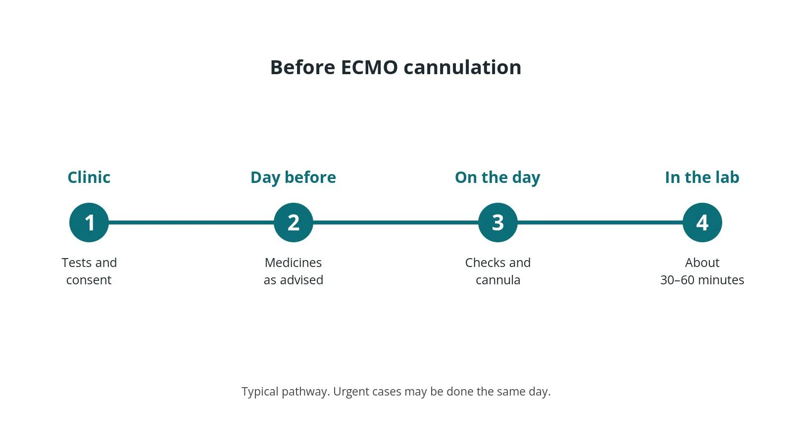
It is usually an emergency. When possible, the team will:
- assess suitability quickly as a team
- inform the family
- prepare the circuit with the perfusion team
Step by step
1. Getting ready
You are usually sedated or anaesthetised and on a ventilator. Monitoring includes arterial pressure and oxygen levels.
2. Access
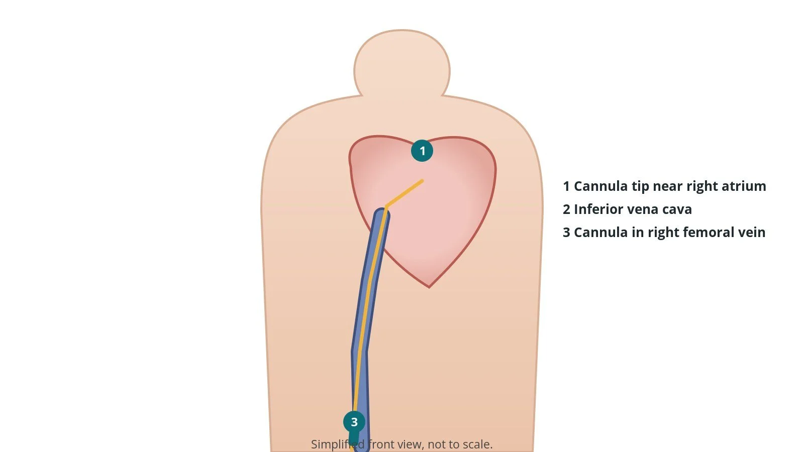
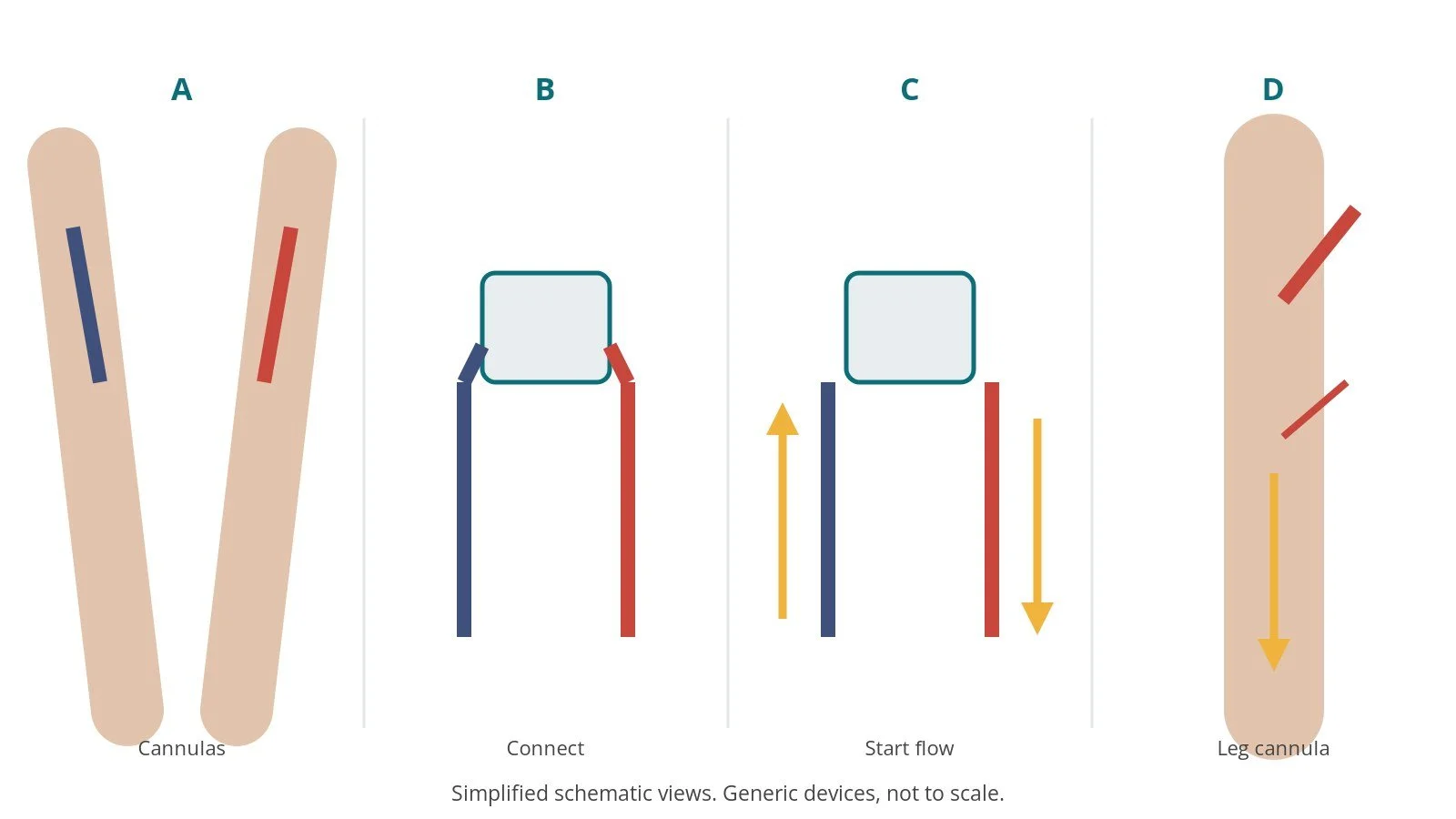
Under ultrasound, a large drainage cannula is placed in the femoral vein and a return cannula in the femoral artery. A smaller cannula often supplies blood to the leg. Sizes per device and operator.
3. Starting ECMO
- A. Cannulas. Venous and arterial cannulas are placed.
- B. Connect. They are connected to the primed circuit without air.
- C. Start flow. Flow is started slowly and increased.
- D. Leg cannula. A small cannula supplies blood to the leg.
4. Finishing
The cannulas are secured. Care continues on intensive care with the ECMO team.
Recovery and aftercare
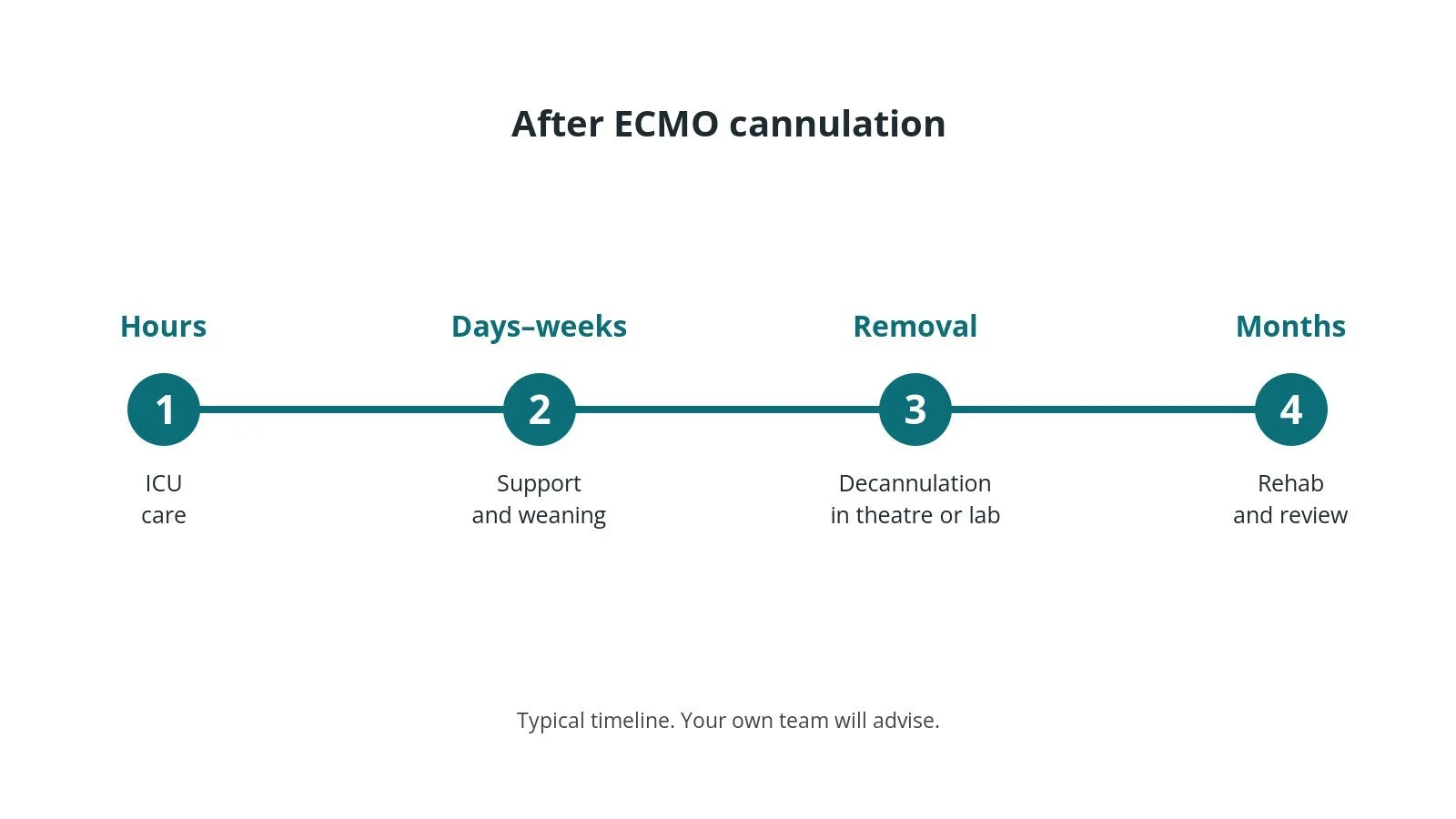
- Intensive care: continuous monitoring by the ECMO team.
- Blood thinners: given and monitored closely, as per centre protocol.
- Leg checks: frequent checks of the leg.
- Weaning: flow is reduced as the heart or lungs recover; removal may need surgery or closure devices.
- Rehabilitation: recovery can be long; support is offered.
Call 999 if you have chest pain, breathlessness, fainting, signs of a stroke, or heavy bleeding or swelling at the access site.
Risks and complications
Your team will explain the risks that apply to you. Possible problems include:
- bleeding
- reduced blood flow to the leg
- clots in the circuit or body, including stroke
- infection
- damage to blood vessels
- kidney problems
Cath lab kit list (printable)
Barts example kit list
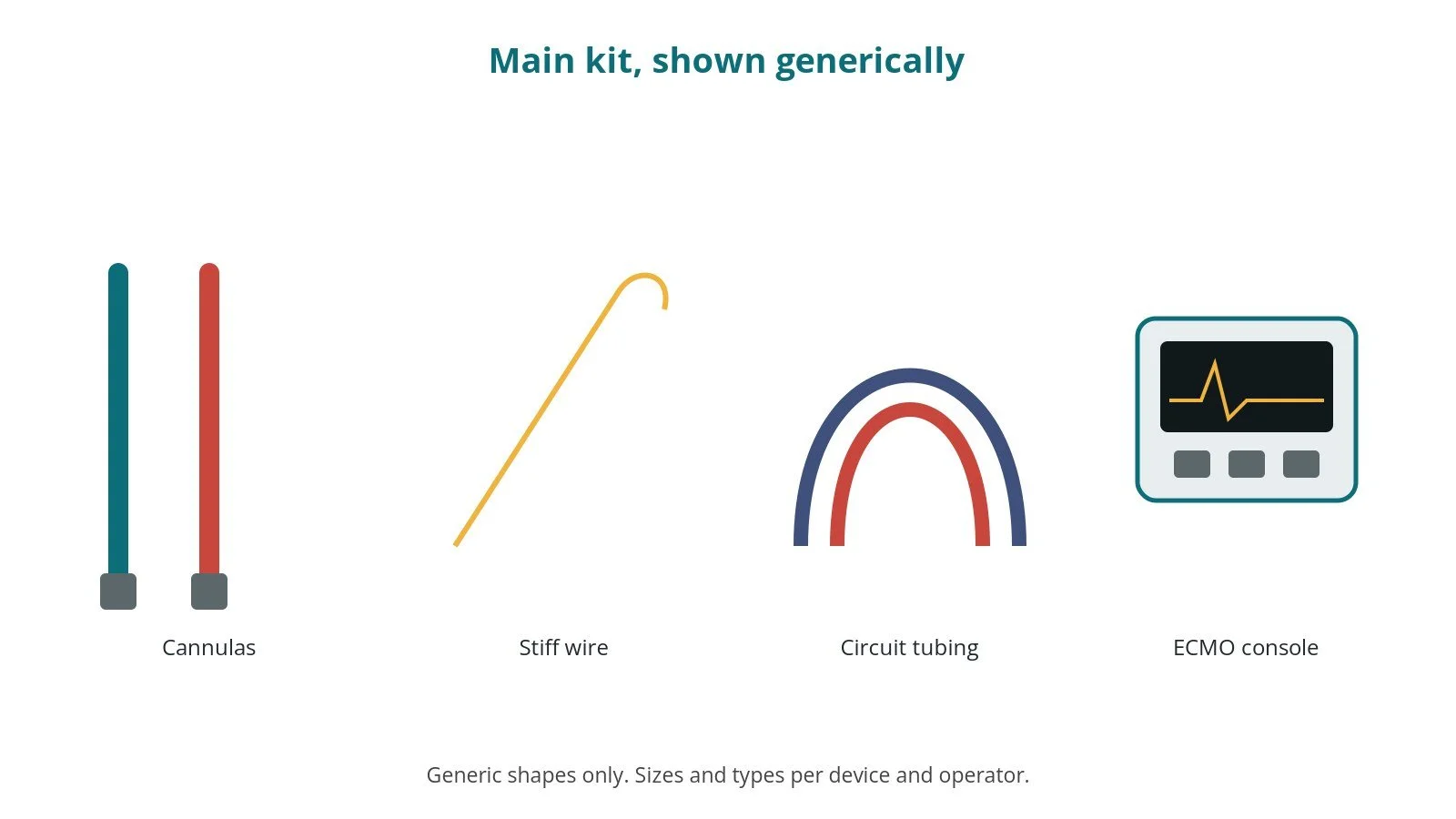
An example from one UK centre, written generically. Every lab has its own protocol, and sizes and doses vary.
- ECMO console and primed circuit with oxygenator
- venous drainage and arterial return cannulas, per device and operator
- distal leg perfusion cannula
- ultrasound
- stiff guidewires and dilators
- heparin, as per centre protocol
- perfusion team
Procedural steps (Barts example)
- Team brief; circuit primed
- Ultrasound-guided venous and arterial access
- Dilate; place cannulas
- Connect without air; start flow
- Distal leg perfusion cannula
- Secure; hand over to ICU
First time in the cath lab?
💡 First-time tip
- Know the roles. Each team member has a set job.
- No air. Connections are made wet-to-wet.
- Leg perfusion. Plan the leg cannula early.
- Secure everything. Cannulas must not move.
FAQ
Is ECMO a cure?
No. It buys time for recovery or other treatments.
How long can it run?
Days to weeks.
Can family visit?
Usually yes on intensive care.
Related Cath Lab 101 guides
Sources
All checked on 10 October 2026. Each page's own date is shown where it gives one.
- McDonagh TA, et al. 2021 ESC Guidelines for the diagnosis and treatment of acute and chronic heart failure. Eur Heart J 2021;42(36):3599–3726. PubMed 34447992
- Byrne RA, et al. 2023 ESC Guidelines for the management of acute coronary syndromes. Eur Heart J 2023;44(38):3720–3826. PubMed 37622654
- Extracorporeal Life Support Organization (ELSO). Guidelines. elso.org
- StatPearls (NCBI Bookshelf), search: VA ECMO. ncbi.nlm.nih.gov/books
Disclaimer
Education only. This page is for general education for healthcare students, cath lab staff and curious readers. It isn't medical advice and doesn't replace advice from your own doctor, heart team or local unit protocols. Equipment and medicines are described generically. No product or manufacturer is recommended. If you think someone is having a heart emergency, call 999.
Disclosure: I work in the medical device industry. This page is independent education and isn't sponsored or reviewed by any company.
All diagrams on this page are original illustrations created for rodnieoro.com.