Transcatheter mitral annuloplasty via the coronary sinus: a Cath Lab 101 guide
Reviewed by Rodnie Oro, cath lab charge nurse · Last reviewed: November 2026 · Education only, not medical advice
Transcatheter mitral annuloplasty via the coronary sinus in one minute
What it is: In functional mitral regurgitation the valve leaks because the heart and the valve ring (annulus) have stretched. A device placed through a neck vein into the coronary sinus, which runs close to the back of the annulus, is anchored at both ends and cinched to pull the annulus in (a cinching system, per device).
Who it is for: Selected adults with heart failure and functional mitral regurgitation, as decided by a heart team, usually when other treatments are not suitable.
Main risks: Squashing of the circumflex coronary artery, damage to the coronary sinus, the device moving, and bleeding at the neck. Your team will explain your own risk.
Usually: Under sedation or general anaesthetic, about 1 to 2 hours, often home the next day.
On this page: What is transcatheter mitral annuloplasty via the coronary sinus? · Who is it for? · When is it done? · Step by step · Recovery · Risks · Kit list · First time in the lab? · FAQ
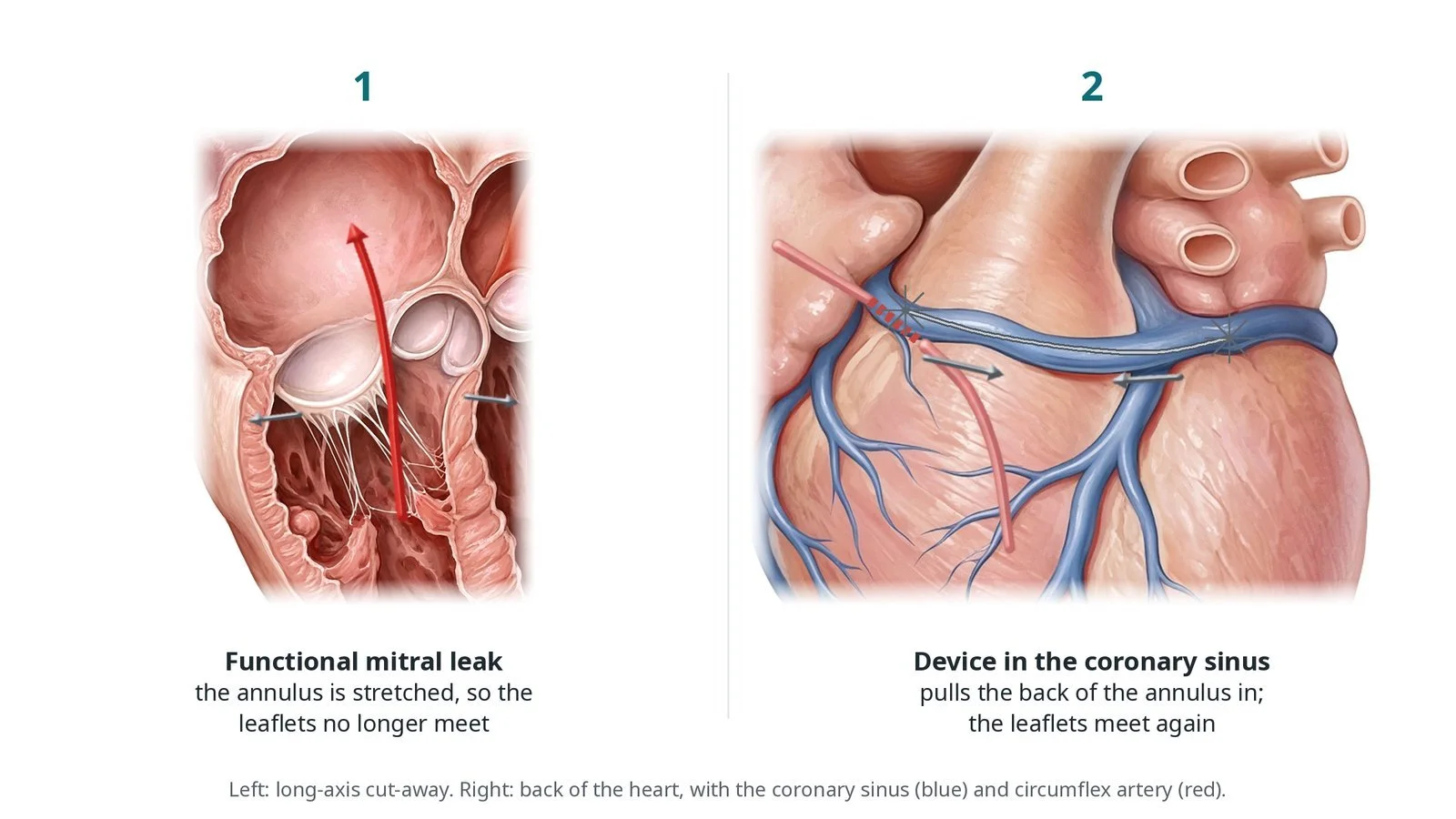
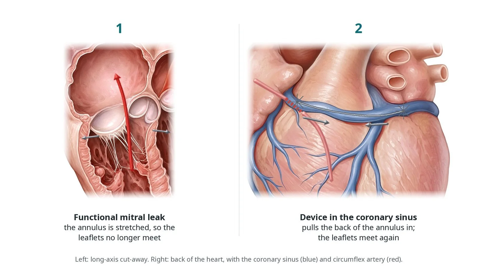
(1) Functional mitral leak: the annulus is stretched, so the leaflets no longer meet. (2) A device in the coronary sinus pulls the back of the annulus in, so the leaflets meet again.
What is transcatheter mitral annuloplasty via the coronary sinus?
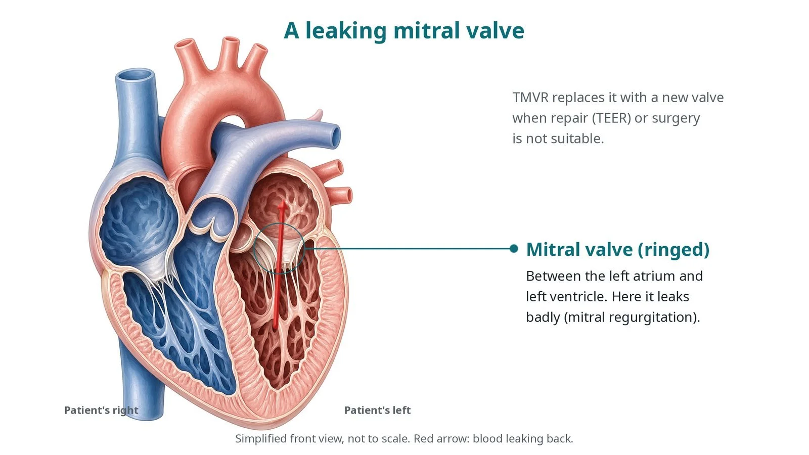
The mitral annulus is the ring the two mitral leaflets hang from. In functional (secondary) mitral regurgitation the leaflets themselves are normal, but the left ventricle and annulus have enlarged, so the leaflets no longer meet.
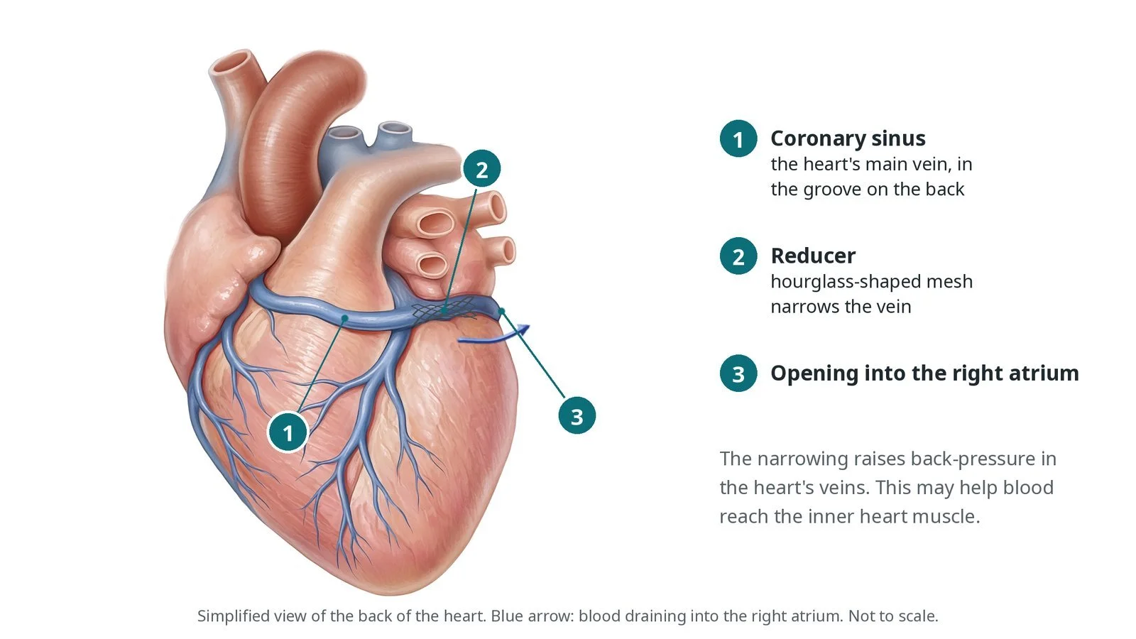
The coronary sinus is the main vein of the heart. It runs in the groove between the left atrium and left ventricle, close to the back (posterior) part of the mitral annulus, and drains into the right atrium. It can be reached from the right internal jugular vein.
A coronary sinus annuloplasty device has an anchor at each end joined by a shaped wire. With the distal anchor fixed deep in the vein, the device is pulled back to cinch the tissue and reduce the size of the annulus, so the leaflets meet better. The circumflex coronary artery often crosses under the coronary sinus, so a coronary angiogram checks it is not squashed before the device is released.
New to the acronyms? See the Cath Lab Abbreviations A–Z.
Who is it for?
Adults with heart failure and functional mitral regurgitation who still have symptoms despite the best tolerated medicines, chosen by a heart team. The team checks:
the mechanism of the leak on echo
the size and course of the coronary sinus
the relationship of the circumflex artery to the coronary sinus
Who it may not suit: People with a primary (leaflet) cause of the leak, pacing leads in the coronary sinus that would be in the way, a coronary sinus outside the device sizes, or a circumflex artery that would be squashed.
The decision is made by your heart team, who will talk through the reasons, the alternatives and the risks with you.
When is it done?
A planned procedure in specialist centres.
gives you a chance to ask questions and give consent
arranges blood tests, including kidney function and a blood count, and an ECG
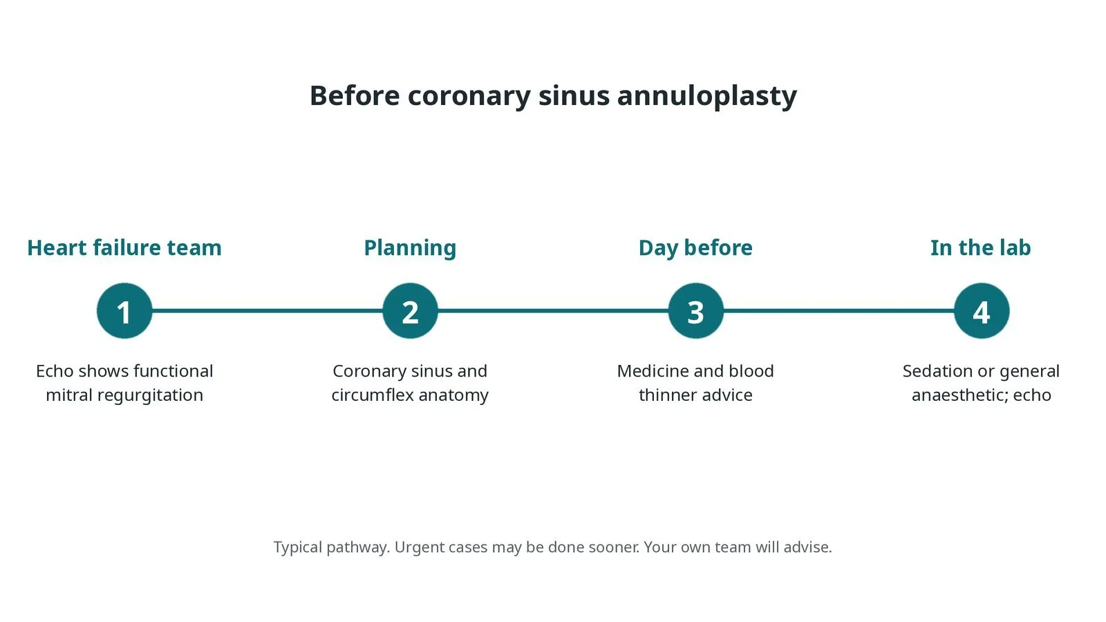
Before coronary sinus annuloplasty: heart failure team review with echo; planning of the coronary sinus and circumflex anatomy; medicine advice; sedation or general anaesthetic with echo.
Step by step
1. Getting ready
You lie on the X-ray table. Sedation or a general anaesthetic is given, as per centre protocol. Echo (transthoracic or TOE) is used to judge the leak.
2. Access
A sheath is placed in the right internal jugular vein under ultrasound, and an arterial sheath for coronary angiography. Sizes are per device and operator. Heparin is given as per centre protocol.
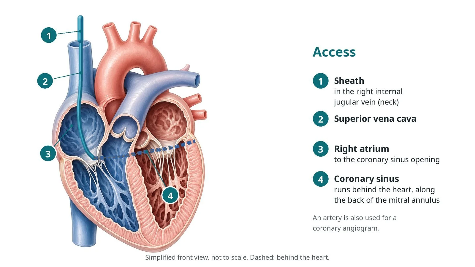
Access: (1) sheath in the right internal jugular vein; (2) superior vena cava; (3) right atrium, to the coronary sinus opening; (4) coronary sinus, behind the heart along the back of the mitral annulus.
3. Placing the device
A. Venogram. A catheter is placed in the coronary sinus and a venogram measures the vein; a coronary angiogram shows the circumflex.
B. Distal anchor. The device is advanced and its distal anchor opened deep in the vein.
C. Cinch. Tension is applied to pull the annulus in while echo checks the leak, and the proximal anchor is opened.
D. Check the artery. A coronary angiogram checks the circumflex is not squashed, then the device is released. If it is, the device can be recaptured.
Watch: Narrowing or slow flow in the circumflex on the angiogram, ST changes or chest pain: possible artery compression. Tell the operator before release; the device can be recaptured.
4. Finishing
The sheaths are removed and pressure applied, per operator.
5. Follow-up
Echo checks the leak and the heart size over the following months.
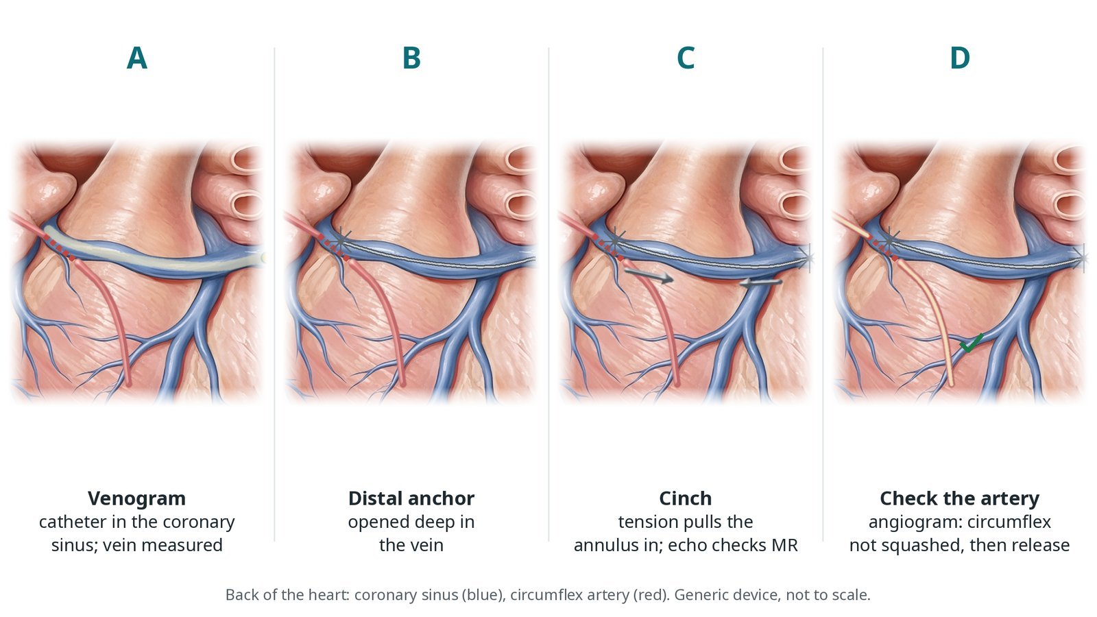
A: venogram and measurements. B: distal anchor opened deep in the vein. C: tension pulls the annulus in while echo checks the leak. D: angiogram confirms the circumflex is not squashed, then release.
Recovery and aftercare
First hours: rest with checks of the neck and arterial sites, heart rhythm and blood pressure, as per centre protocol.
Going home: often the next day.
Medicines: continue heart failure medicines; blood thinners as your team advises.
Activity: walk as normal; avoid heavy lifting for a few days.
Driving: ask your team; usually after a few days if well; check DVLA rules for your heart condition.
Follow-up: echo and heart failure clinic.
Call 999 if you have chest pain, sudden breathlessness, fainting, or heavy bleeding.
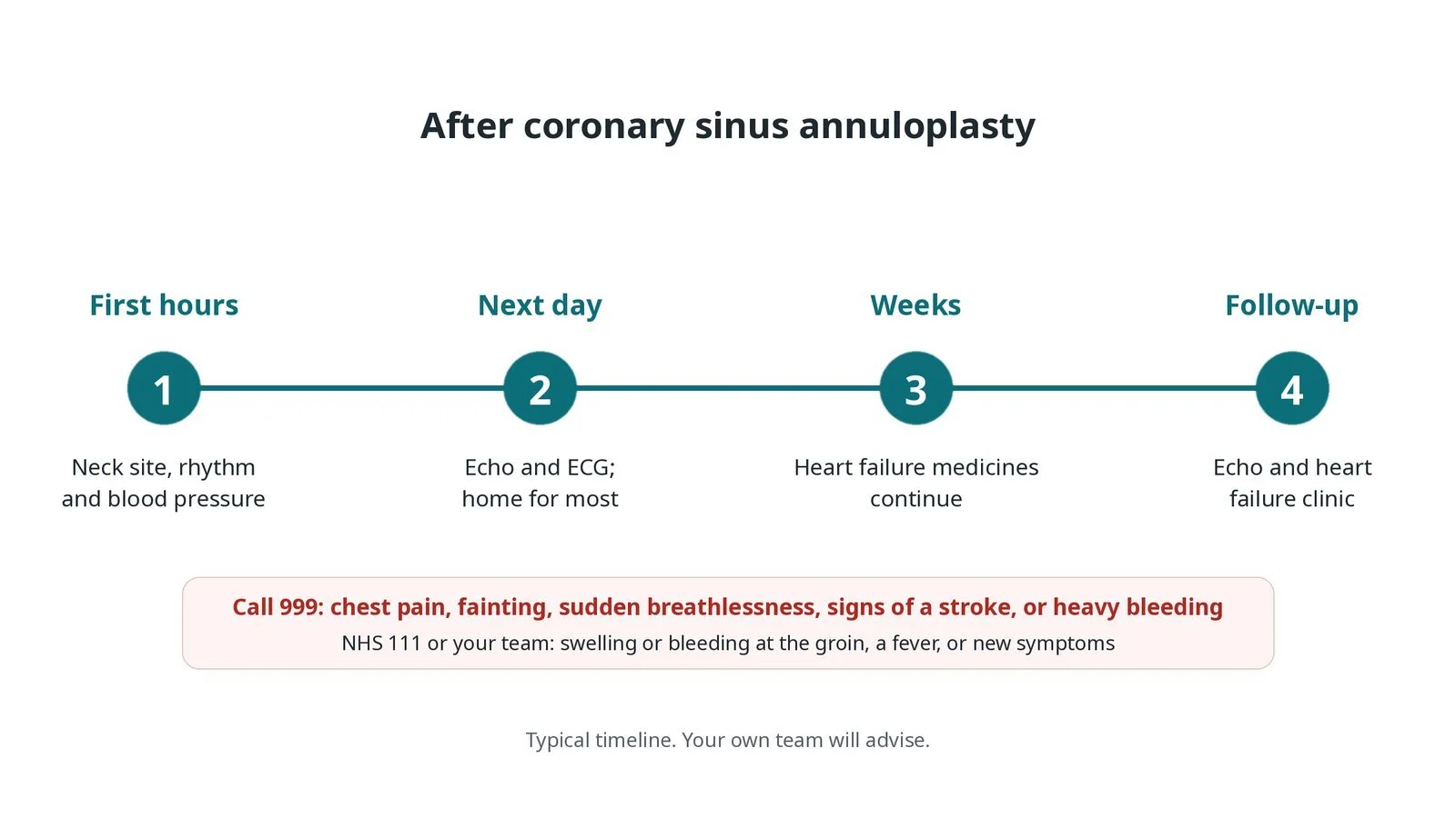
After coronary sinus annuloplasty: (1) neck site, rhythm and blood pressure checks; (2) echo, ECG and home the next day for most; (3) heart failure medicines continue; (4) echo and heart failure clinic.
Risks and complications
Your team will explain the risks that apply to you. Possible problems include:
Squashing of the circumflex coronary artery (uncommon): Checked before release.
Damage to the coronary sinus (rare)
Device moving or breaking over time (rare)
Bleeding or bruising at the neck or access sites
Abnormal heart rhythms
Leak not improving as much as hoped
Cath lab kit list (printable)
Barts example kit list
An example from one UK centre, written generically. Every lab has its own protocol, and sizes and doses vary.
jugular venous sheath, size per device
coronary sinus catheter, per operator
guidewires, per operator
coronary sinus annuloplasty device and delivery system, per device
arterial sheath and coronary catheters for angiography
contrast and syringes
echo: transthoracic or TOE
heparin, as per centre protocol
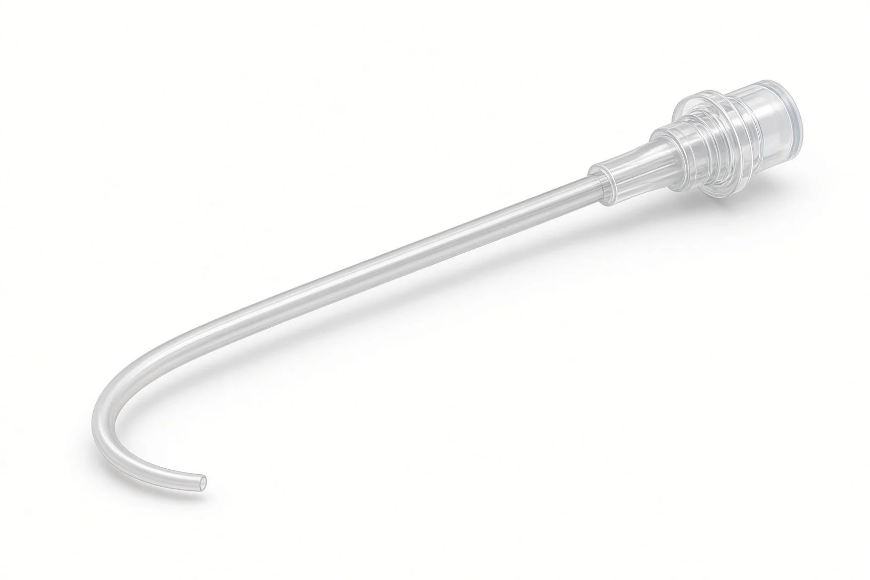
Coronary sinus catheter.
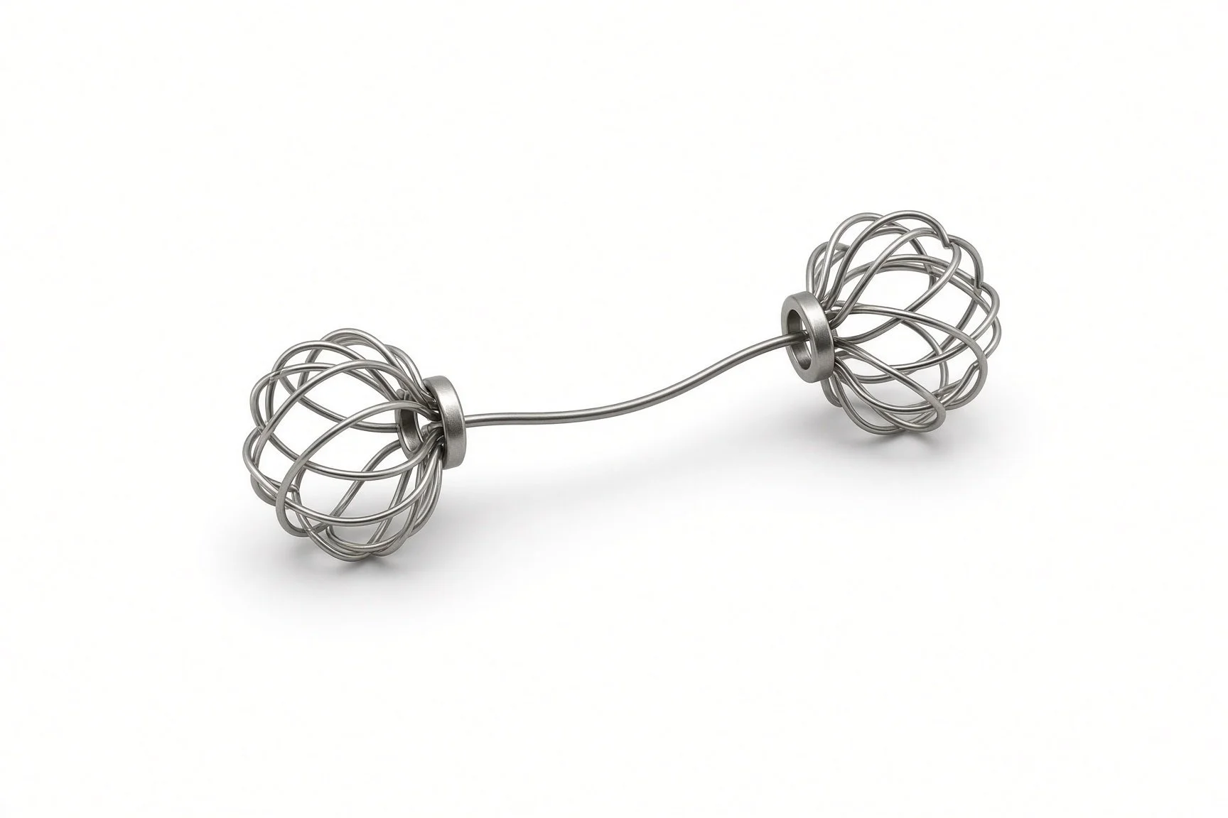
Annuloplasty device.
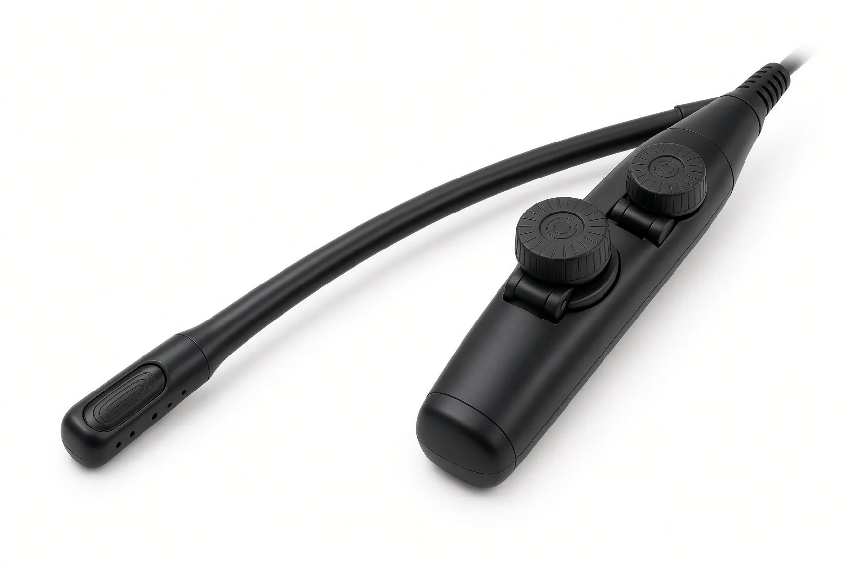
Echo (TOE or transthoracic).
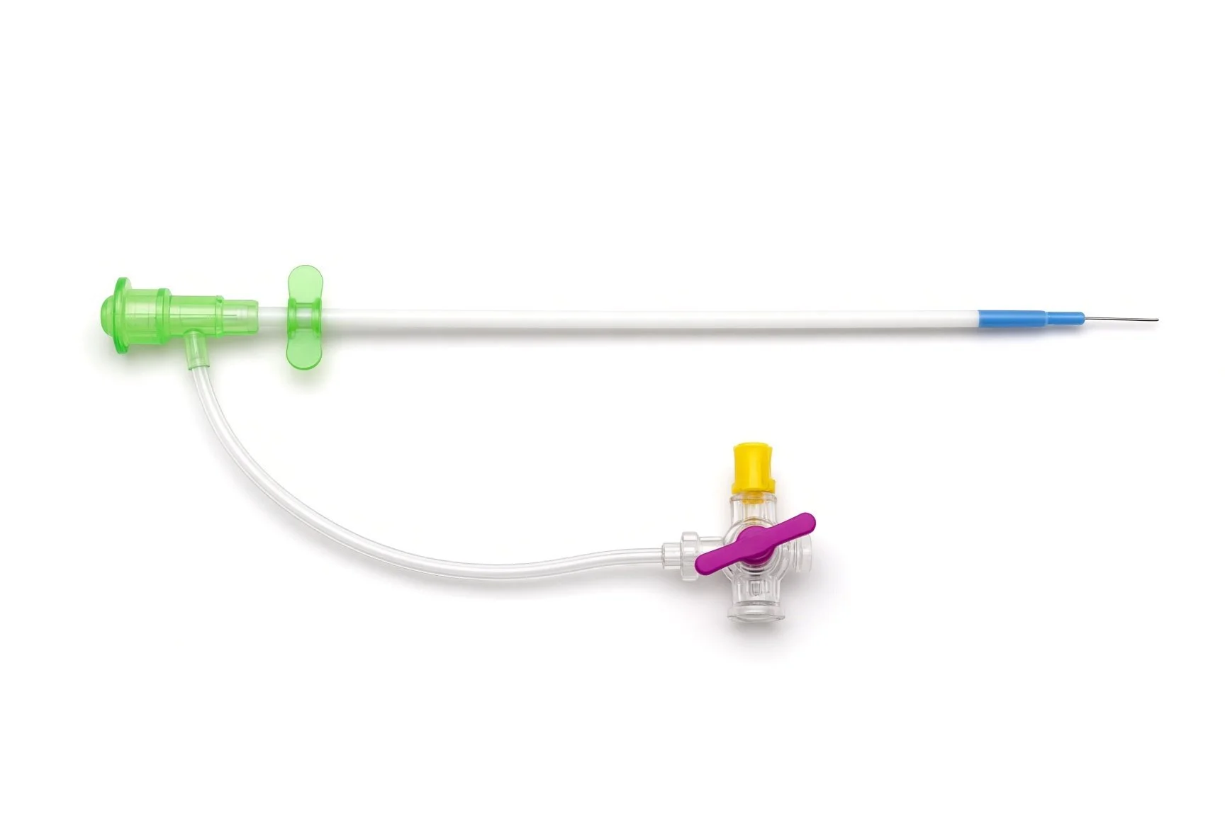
Jugular venous sheath.
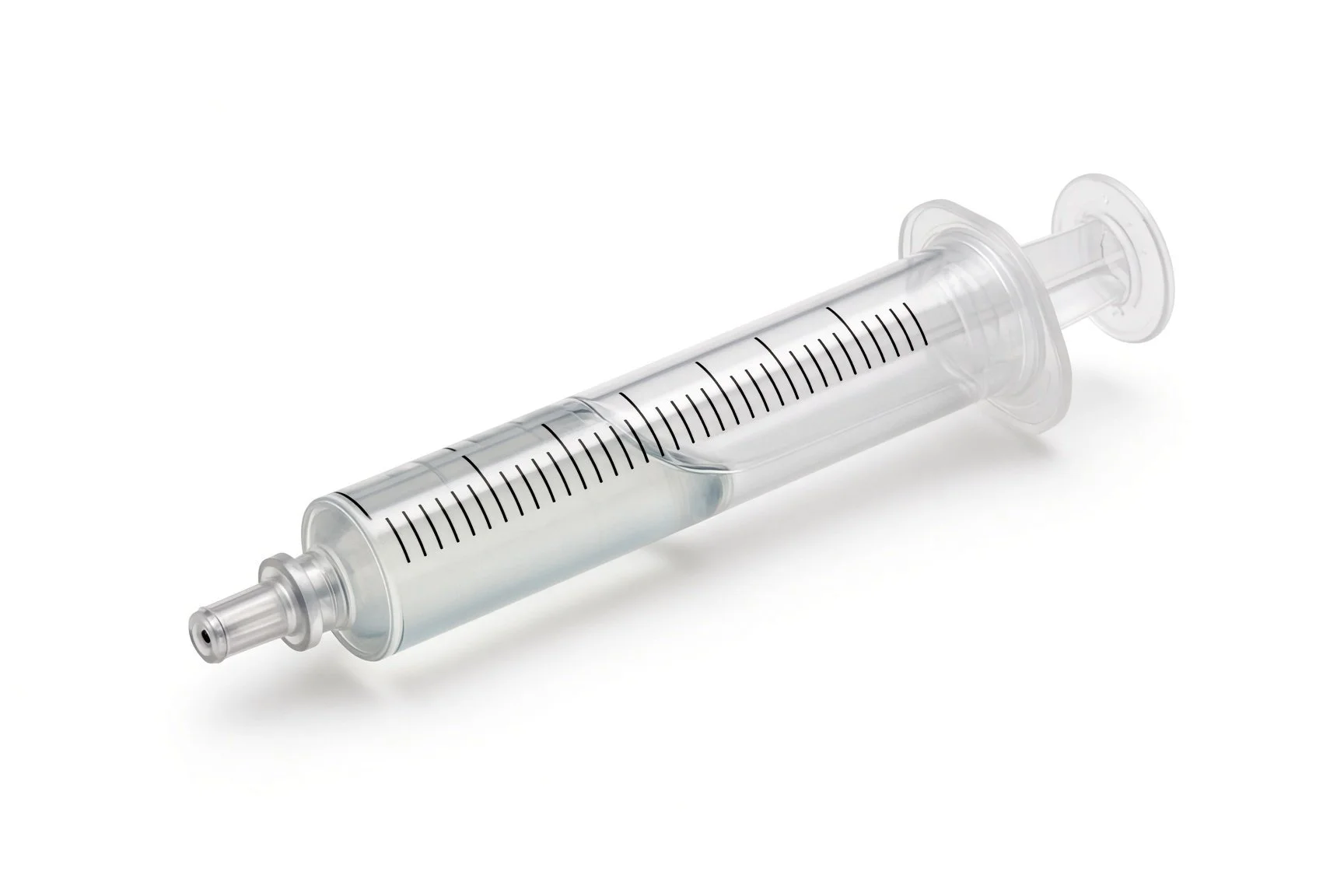
Contrast syringe.

Contrast.
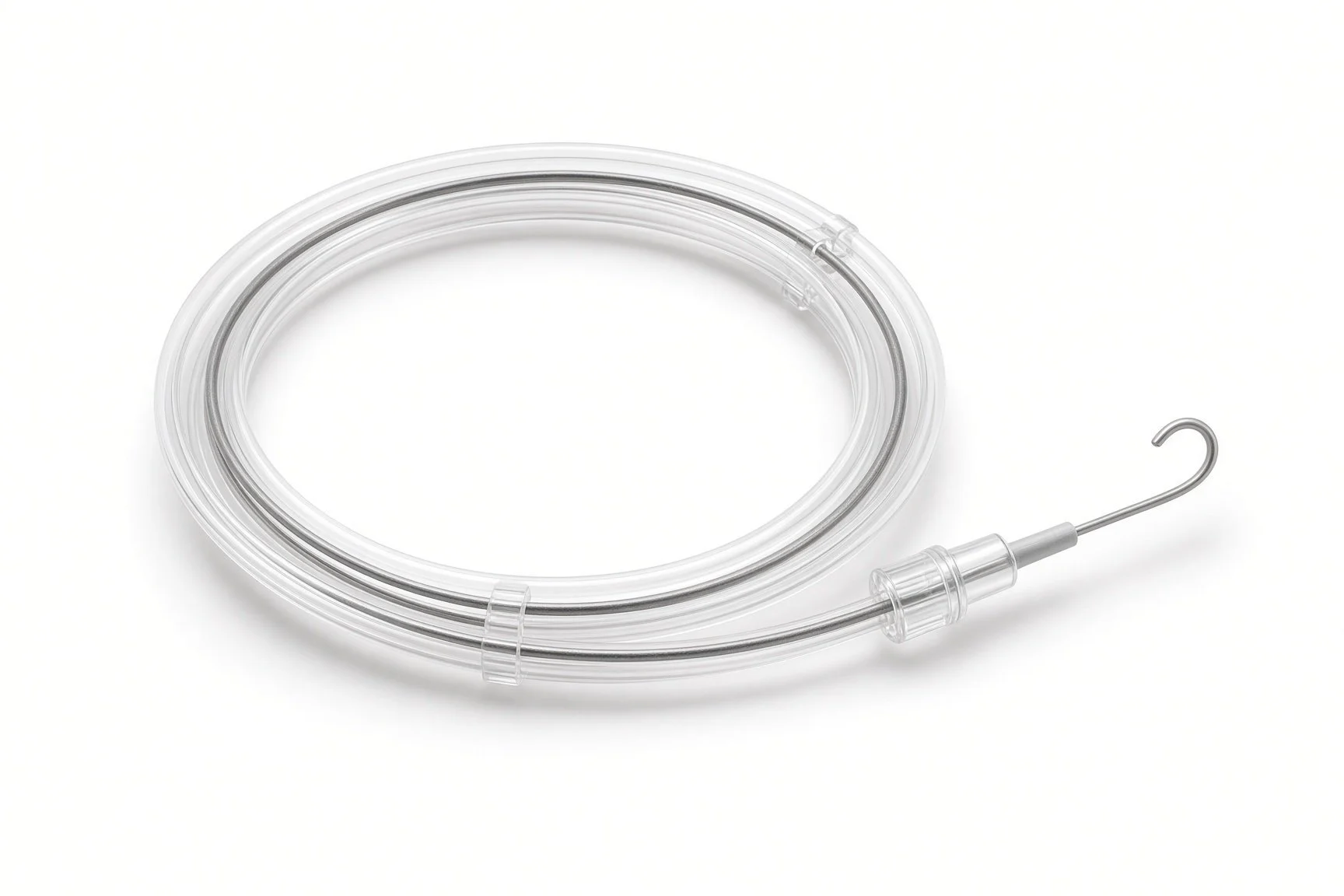
Guidewire.
Procedural steps (Barts example)
Getting ready
Access
Placing the device
Finishing
Follow-up
First time in the cath lab?
Two routes at once. The device goes in from the neck; the angiogram from an artery.
The circumflex check is the key moment. Make sure good angiogram pictures are recorded before release.
The device can be recaptured. If the artery is squashed or the result is poor, it can be taken back.
Echo shows the effect. Watch the leak change as the device is cinched.
Prepare the neck as well as the groin or wrist. The device goes in through the jugular vein and the angiogram through an artery.
FAQ
How does a device in a vein help the valve?
The coronary sinus runs close to the back of the valve ring, so tightening a device there pulls the ring in.
Will my leak go away?
It usually becomes smaller rather than disappearing.
Why is my neck used?
The jugular vein gives the most direct path to the coronary sinus.
Can I still have other mitral treatments later?
Often yes. Your team will discuss future options.
Related Cath Lab 101 guides
Native mitral valve replacement (TMVR) (coming soon)
Cardiac resynchronisation therapy (CRT-P and CRT-D) (coming soon)
Sources
All checked on 12 November 2026. Each page's own date is shown where it gives one.
Praz F, et al. 2025 ESC/EACTS Guidelines for the management of valvular heart disease. Eur Heart J 2025
McDonagh TA, et al. 2021 ESC Guidelines for the diagnosis and treatment of acute and chronic heart failure. Eur Heart J 2021;42:3599-3726
StatPearls (NCBI Bookshelf), search: mitral regurgitation. ncbi.nlm.nih.gov/books
Disclaimer
Education only. This page is for general education for healthcare students, cath lab staff and curious readers. It isn't medical advice and doesn't replace advice from your own doctor, heart team or local unit protocols. Equipment and medicines are described generically. No product or manufacturer is recommended. If you think someone is having a heart emergency, call 999.
Disclosure: I work in the medical device industry. This page is independent education and isn't sponsored or reviewed by any company.
All diagrams on this page are original illustrations created for rodnieoro.com.