TAVI for aortic regurgitation: a Cath Lab 101 guide
Reviewed by Rodnie Oro, cath lab charge nurse · Last reviewed: November 2026 · Education only, not medical advice
TAVI for aortic regurgitation in one minute
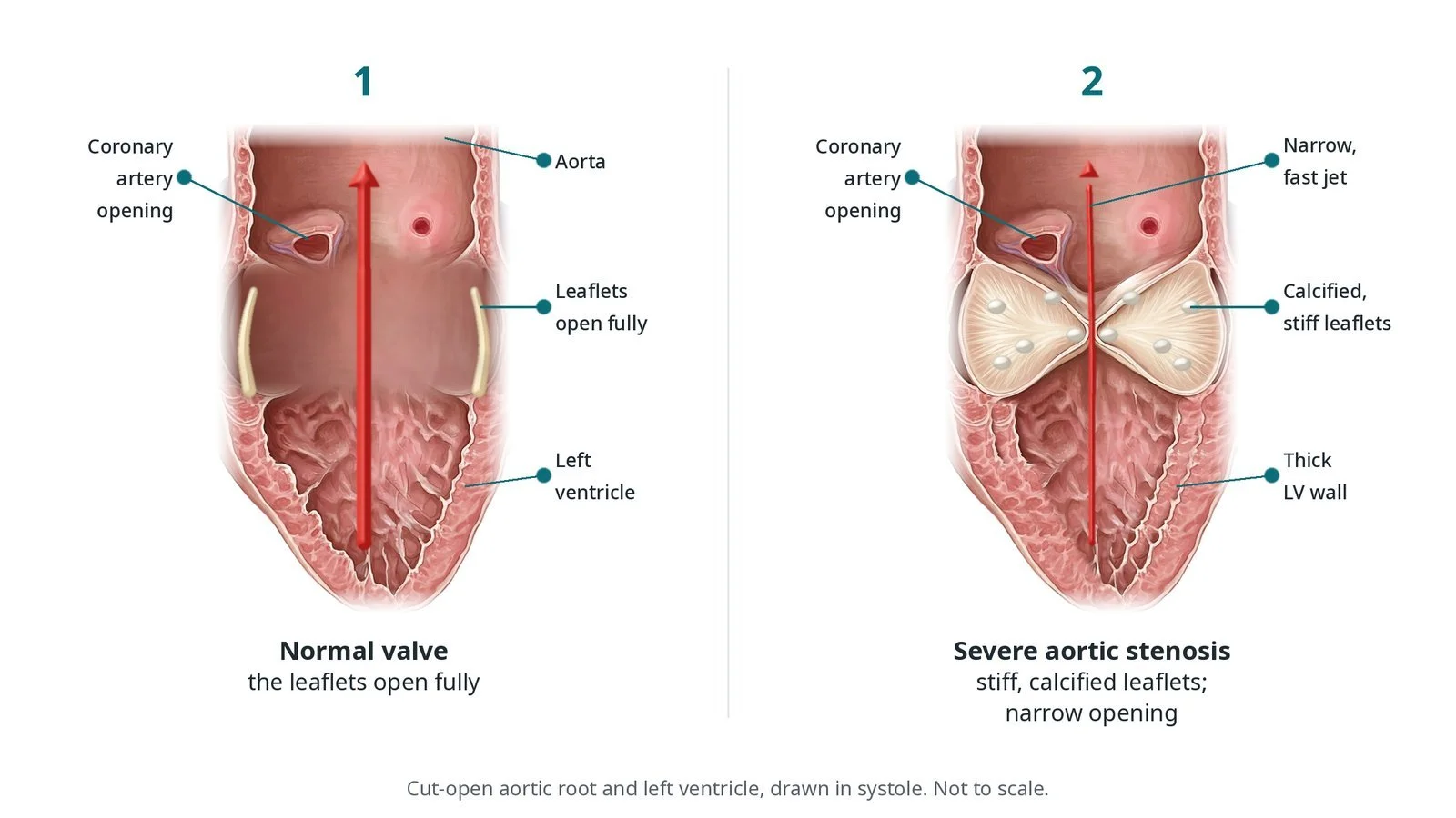
What it is: In aortic regurgitation (AR) the aortic valve leaks, so blood falls back into the left ventricle. Standard TAVI valves rely on calcium to grip, and a leaking valve often has little. Dedicated valves clip onto the native leaflets so they can anchor without it.
Who it is for: Adults with severe, symptomatic aortic regurgitation who are at high risk from open-heart surgery, as decided by a heart team. Surgery remains the standard treatment for most people.
Main risks: Valve moving out of position, a leak around the valve, need for a pacemaker, stroke, and bleeding or damage at the groin artery. Your team will explain your own risk.
Usually: Under sedation or general anaesthetic, about 1 to 2 hours, home after 1 to 3 days for most.
On this page: What is TAVI for aortic regurgitation? · Who is it for? · When is it done? · Step by step · Recovery · Risks · Kit list · First time in the lab? · FAQ
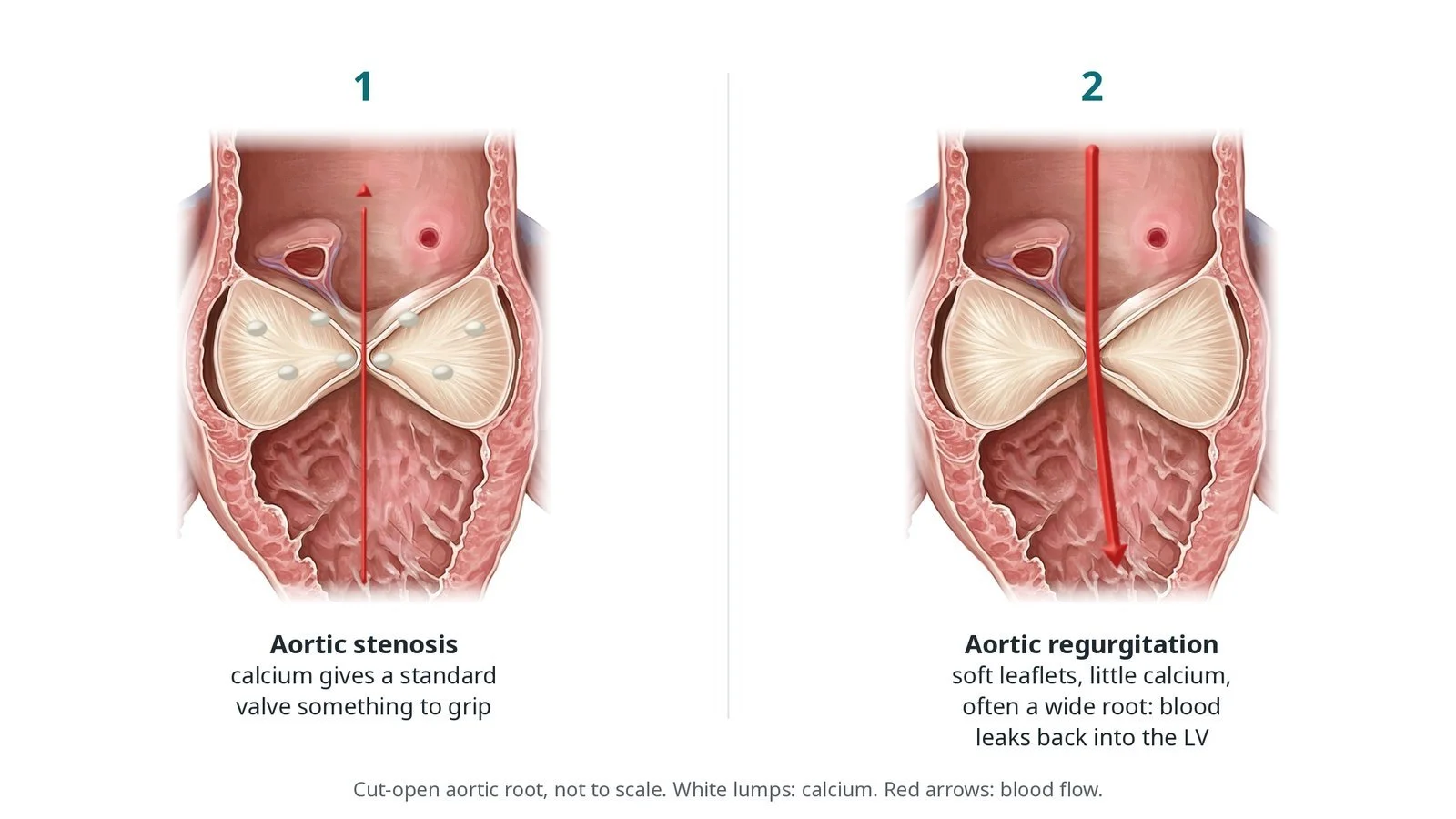
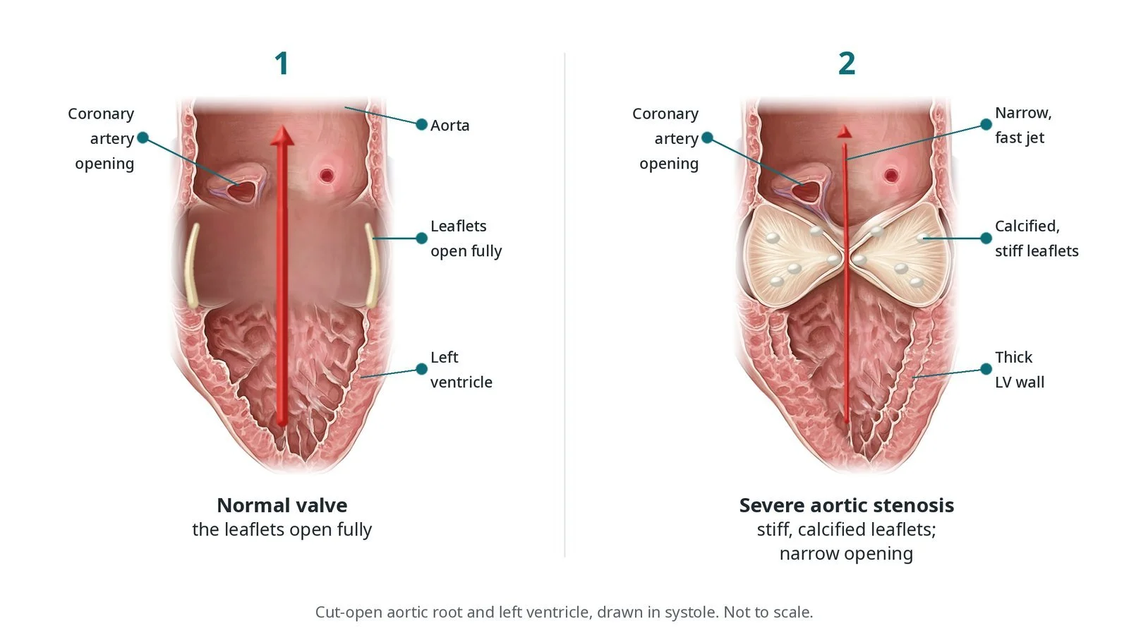
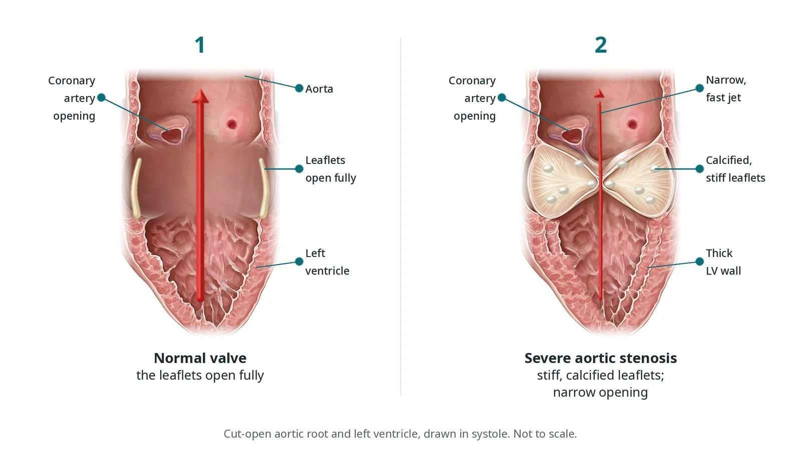
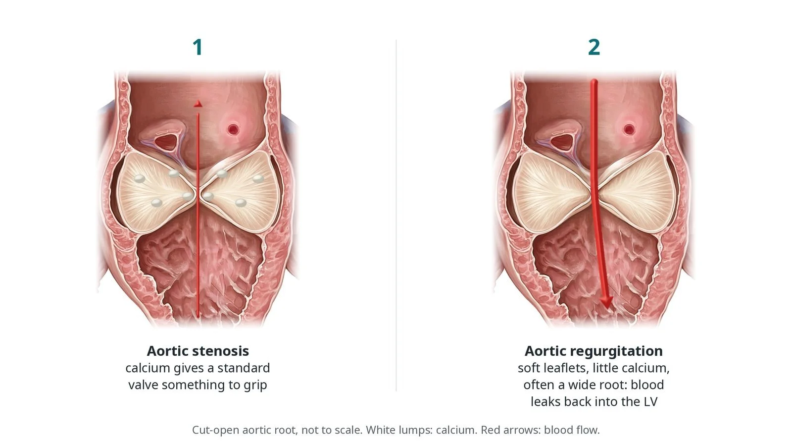
(1) In aortic stenosis, calcium gives a standard valve something to grip. (2) In aortic regurgitation the leaflets are soft with little calcium and the root is often wide, so blood leaks back into the left ventricle.
What is TAVI for aortic regurgitation?
The aortic valve sits between the left ventricle and the aorta. It has three leaflets, and behind each one is a pouch called a sinus. Two of the sinuses give rise to the coronary arteries.
In aortic stenosis the leaflets are thick and calcified. A standard TAVI valve pushes them aside and the calcium helps hold it in place. In pure aortic regurgitation the leaflets are often thin and soft and the root may be wide, so a standard valve has little to grip and can slip (embolise) into the aorta or the left ventricle.
Dedicated AR valves have locating arms that sit in the sinuses behind the native leaflets. The leaflets are then held between the arms and the frame.
New to the acronyms? See the Cath Lab Abbreviations A–Z.
Who is it for?
Adults with severe aortic regurgitation causing symptoms or affecting the left ventricle, who are at high risk from surgery. The heart team considers:
the cause of the leak and the shape of the root on CT
whether the root size fits the available devices
the leg arteries for access
frailty and other illnesses
Who it may not suit: People suitable for surgery, those with a root too large for the device, a bicuspid valve or an aortic aneurysm needing surgery, and people with active infection of the valve (endocarditis).
The decision is made by your heart team, who will talk through the reasons, the alternatives and the risks with you.
When is it done?
It is a planned procedure in specialist centres, after a heart team meeting with a surgical opinion. CT sizing is essential.
gives you a chance to ask questions and give consent
arranges blood tests, including kidney function and a blood count, and an ECG
Step by step
1. Getting ready
You lie on the X-ray table. Sedation or a general anaesthetic is given, as per centre protocol. Monitoring includes ECG, invasive blood pressure and oxygen levels; echo is used throughout.
2. Access
A large sheath is placed in the femoral artery under ultrasound, with closure sutures placed first if used. Sizes are per device and operator. A second artery or vein is used for a pigtail catheter and a temporary pacing wire. Heparin is given as per centre protocol.
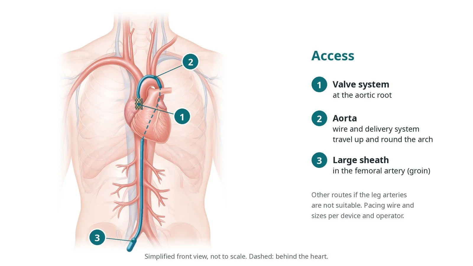
Access: (1) valve system at the aortic root; (2) aorta, round the arch; (3) large sheath in the femoral artery. Other routes are used if the leg arteries are not suitable.
3. Placing the valve
A. Cross. The valve is crossed and a pre-shaped stiff wire is looped in the left ventricle.
B. Locate. The delivery system is advanced and the locating arms are opened into the sinuses behind the native leaflets, checked on X-ray and echo.
C. Deploy. The valve is released so the native leaflets are held between the arms and the frame. Pacing is used as per device and operator.
D. Check. Echo and an aortogram look for leaks, valve position and coronary flow.
Watch: A sudden drop in blood pressure or the valve moving on the screen: possible embolisation or a severe leak. Call out, and have the bail-out plan and surgical team ready.
4. Finishing
The pacing wire may be left in if there is concern about the heart rhythm. The sheaths are removed and the arteries closed with closure devices or pressure, per operator.
5. After the valve
An echo the next day checks the valve and the left ventricle. An ECG looks for new heart block, which can need a pacemaker.
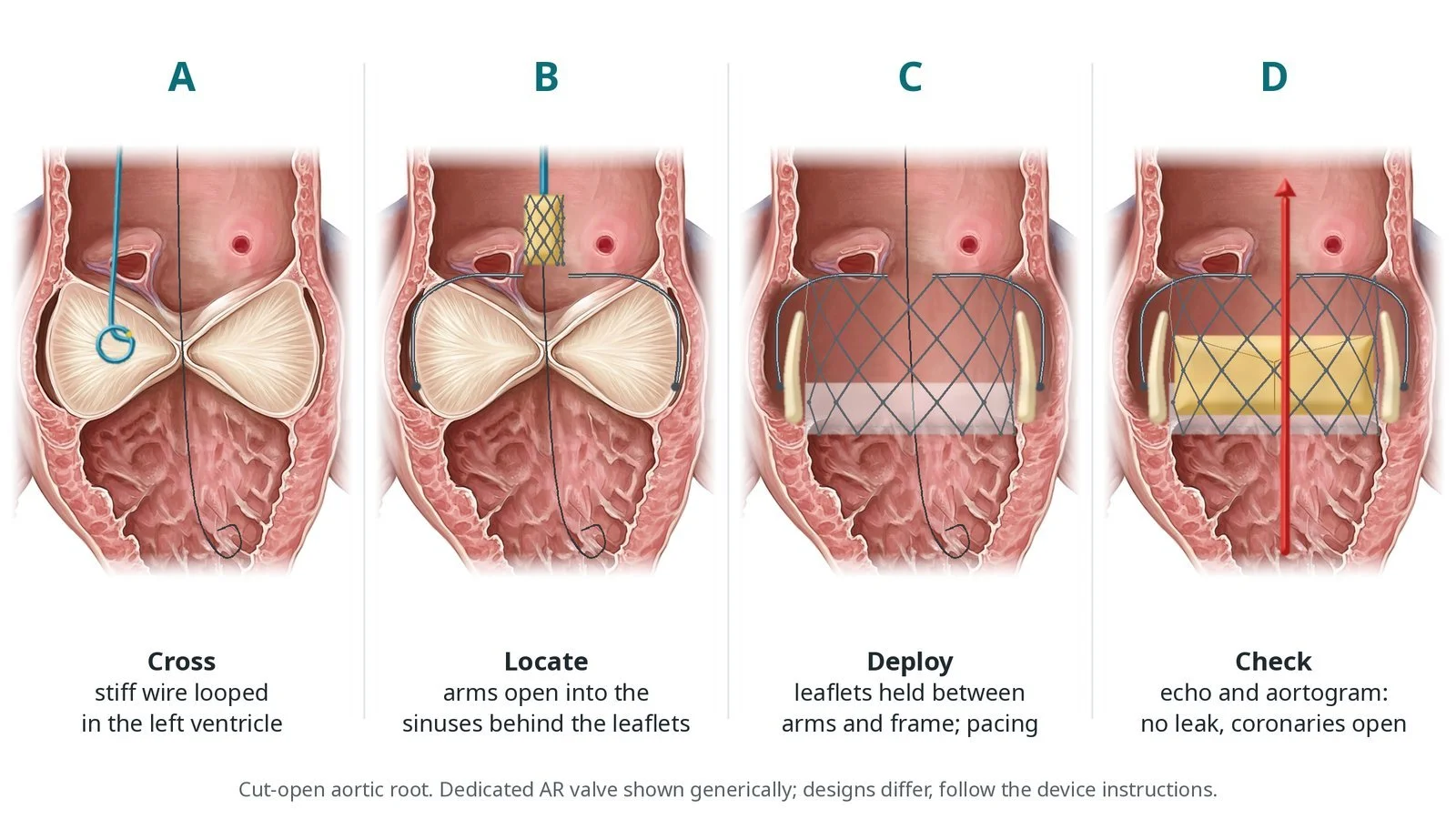
A: cross with a stiff wire looped in the left ventricle. B: arms open into the sinuses behind the leaflets. C: deploy, holding the native leaflets between the arms and the frame. D: check with echo and an aortogram.
Recovery and aftercare
First hours: bed rest with checks of the groin, leg pulses, heart rhythm and blood pressure, as per centre protocol.
Going home: after 1 to 3 days for most.
Medicines: an antiplatelet or anticoagulant, as your team advises.
Activity: walk the next day; avoid heavy lifting for about a week, or as advised.
Driving: do not drive for at least a week, or as your team advises; check DVLA rules, especially if you have a new pacemaker.
Follow-up: echo and clinic review, then regular checks.
Call 999 if you have chest pain, sudden breathlessness, fainting, signs of a stroke, or heavy bleeding from the groin.
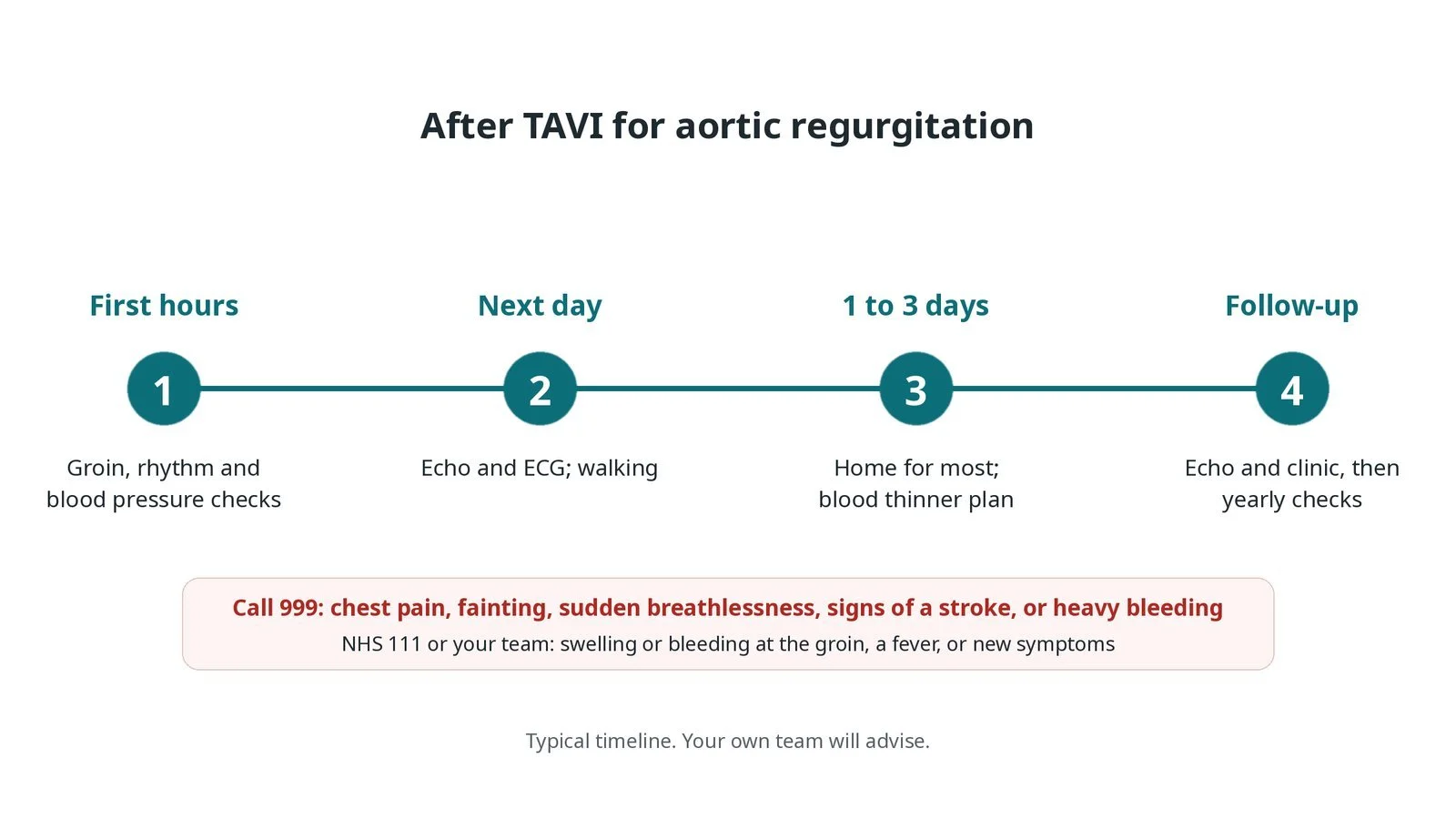
After TAVI for aortic regurgitation: (1) groin, rhythm and blood pressure checks; (2) echo, ECG and walking the next day; (3) home for most in 1 to 3 days; (4) echo and clinic follow-up.
Risks and complications
Your team will explain the risks that apply to you. Possible problems include:
Valve moving out of position (embolisation) (uncommon)
Leak around the valve
Heart block needing a pacemaker
Stroke (uncommon)
Bleeding or damage at the groin artery
Blocked coronary artery (rare)
Kidney strain from contrast
Cath lab kit list (printable)
Barts example kit list
An example from one UK centre, written generically. Every lab has its own protocol, and sizes and doses vary.
large-bore femoral arterial sheath and closure devices, per operator
pre-shaped stiff left ventricular guidewire
pigtail catheter for aortograms
dedicated aortic regurgitation valve and delivery system, per device
temporary pacing wire and pacing box
echo: TOE or transthoracic, per operator
contrast; heparin and anaesthetic medicines, as per centre protocol
snares and bail-out kit for a moved valve
CT planning report and device sizing chart
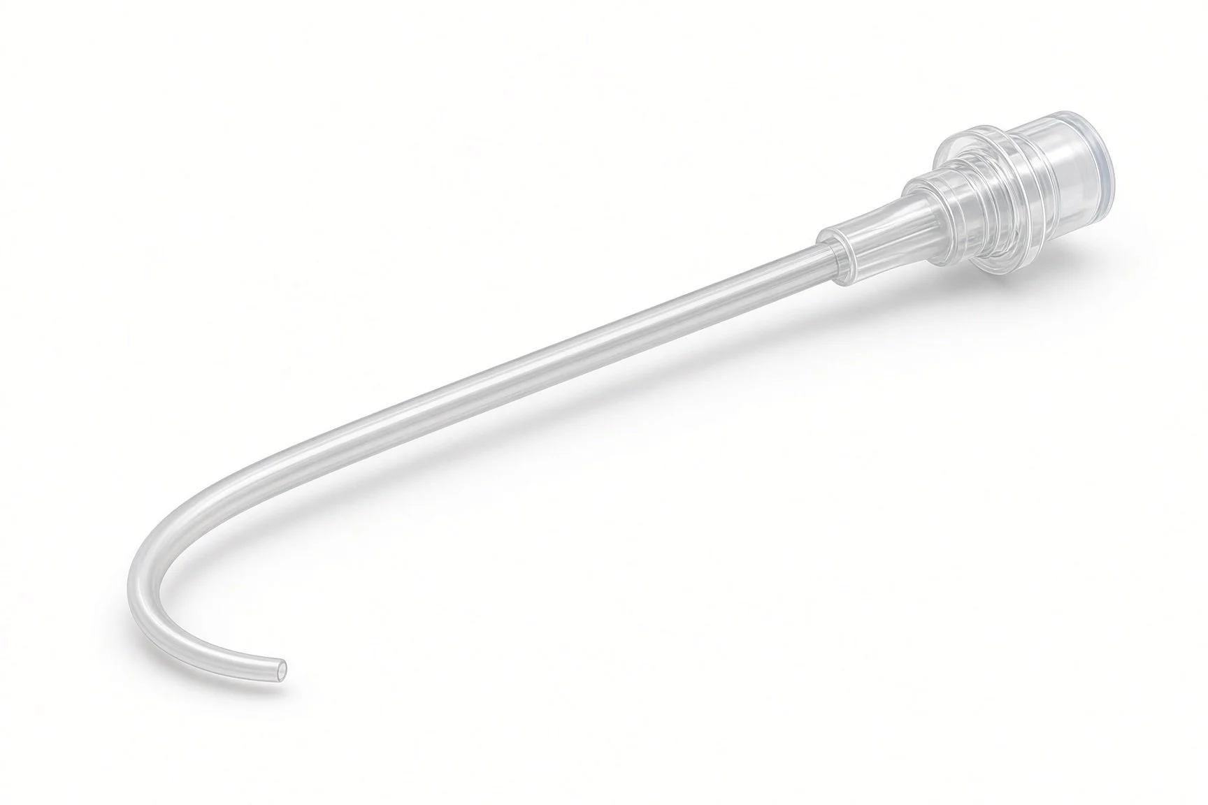
Pigtail catheter.
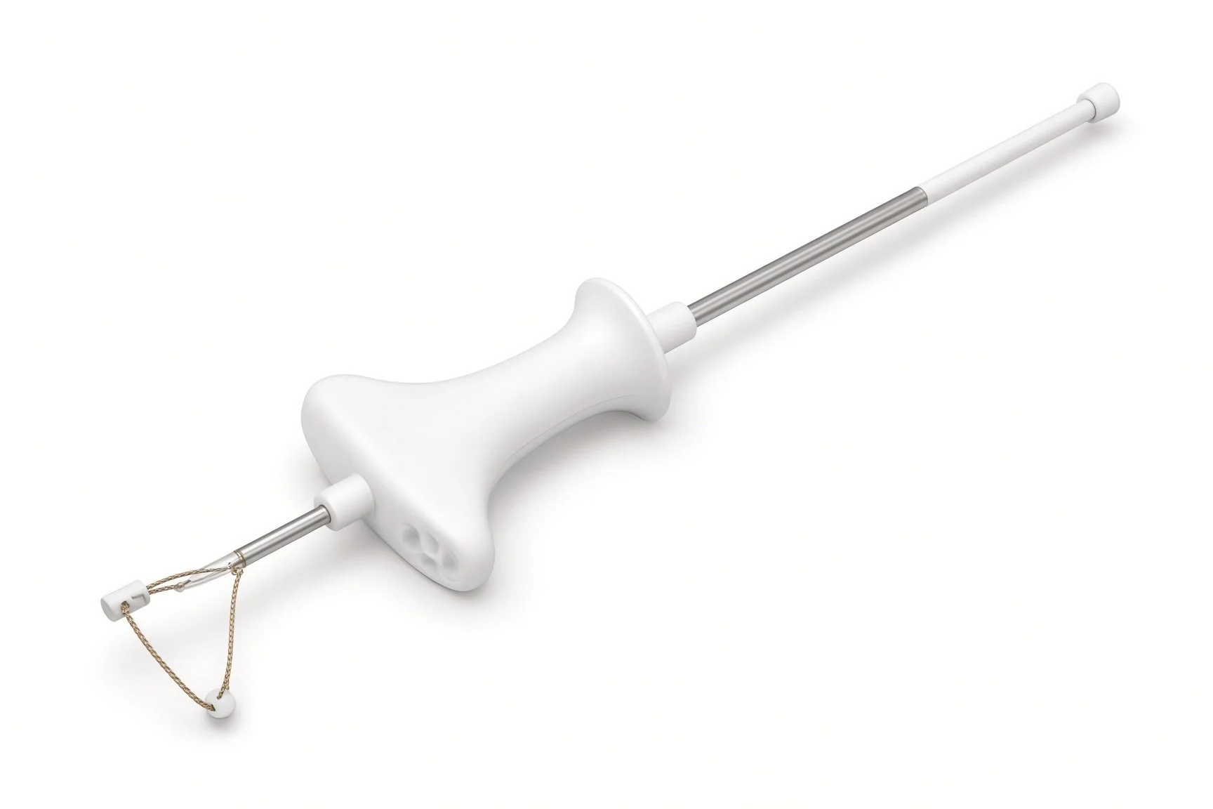
Femoral closure devices.
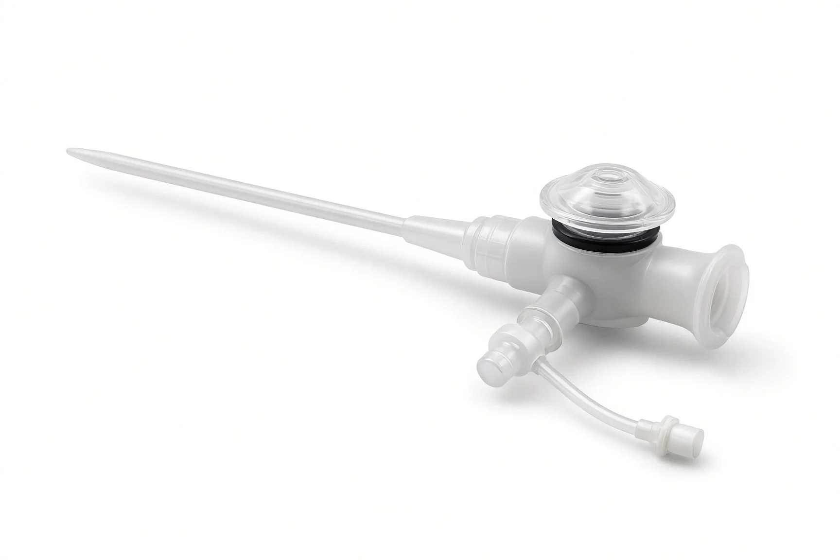
Large-bore femoral sheath.
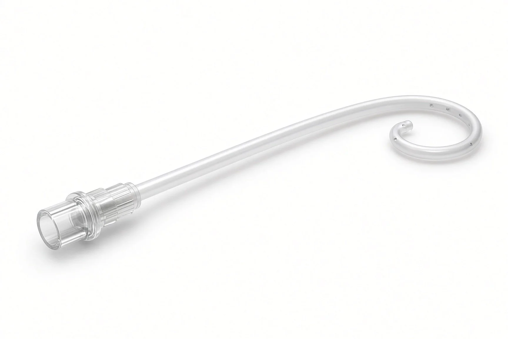
Pigtail catheter.
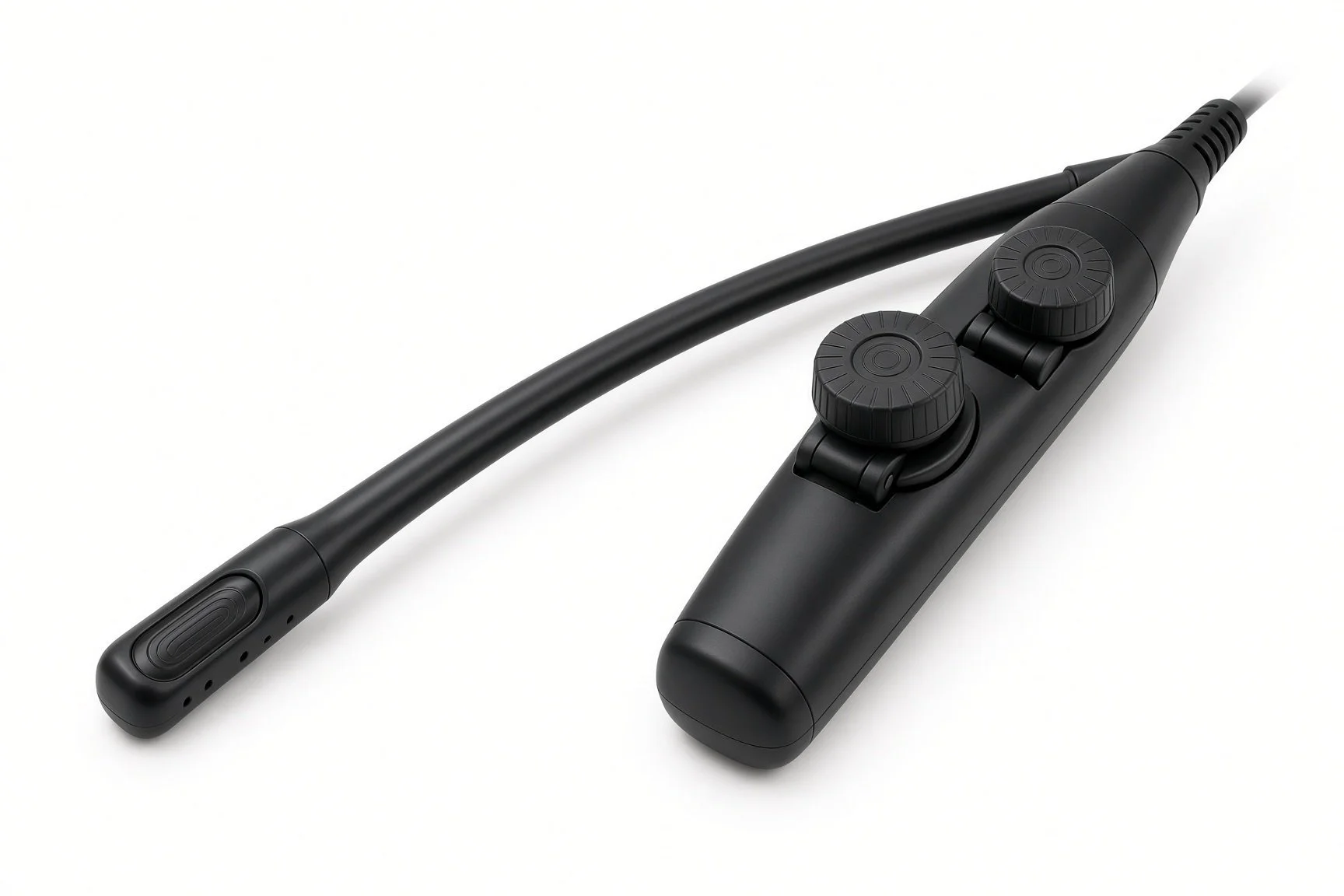
TOE or transthoracic echo.
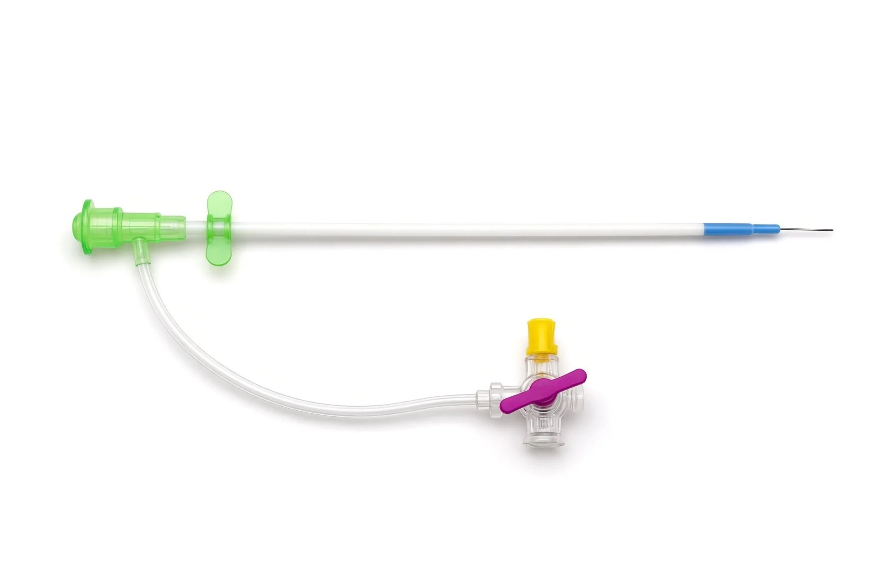
Large-bore femoral sheath.
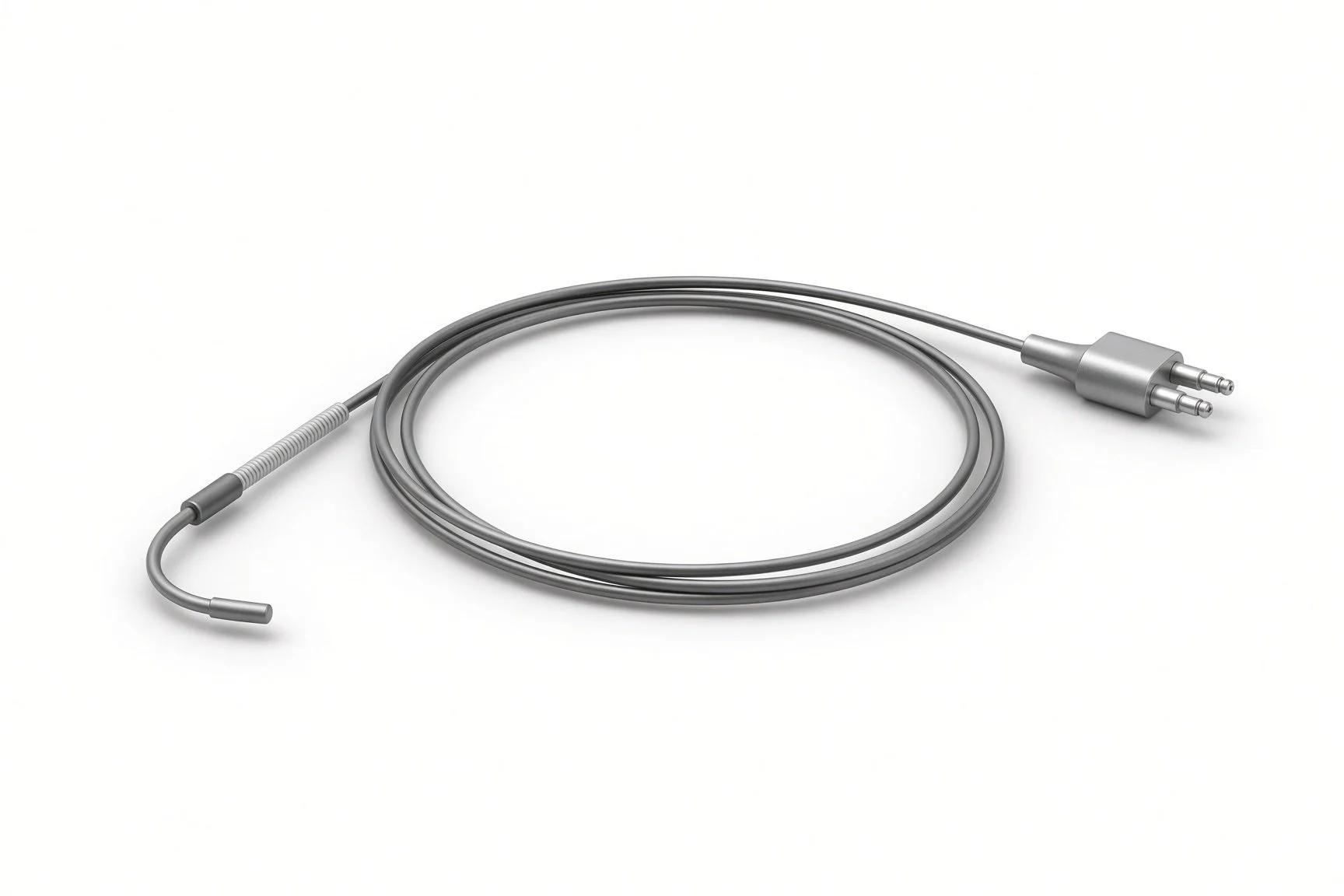
Temporary pacing wire.
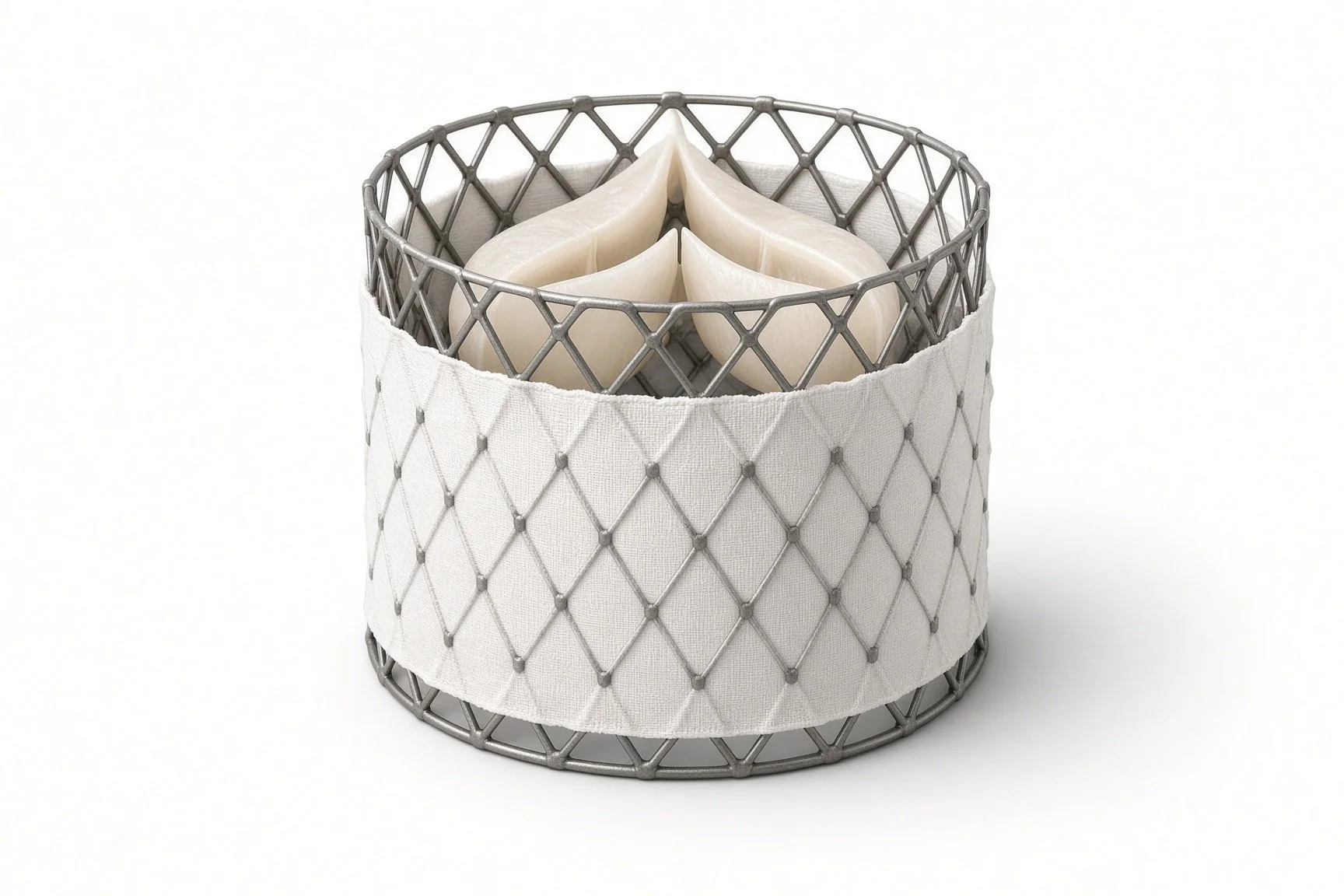
Dedicated AR valve and delivery system.

Echo.
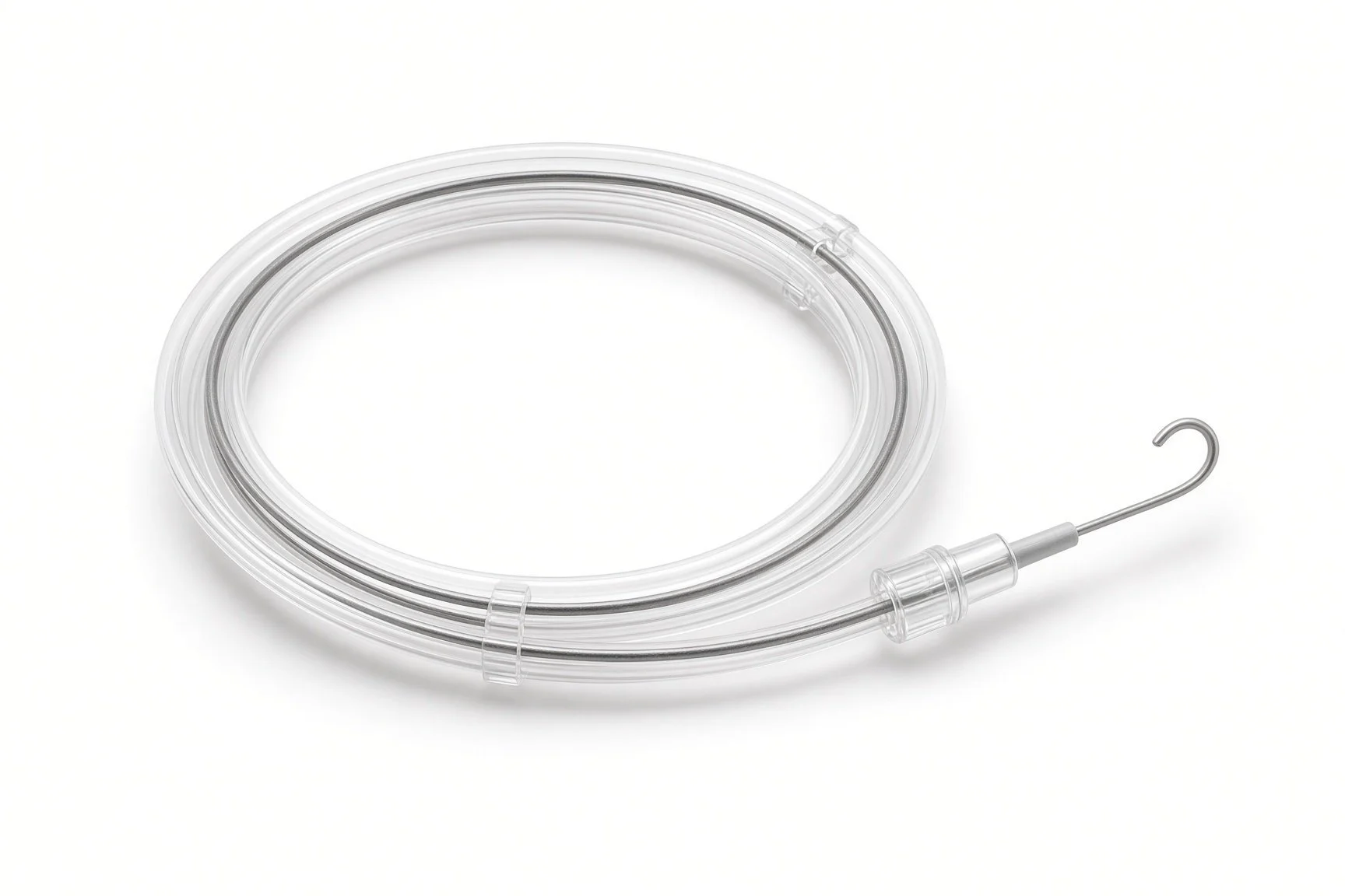
Pre-shaped stiff LV wire.
Procedural steps (Barts example)
Getting ready
Access
Placing the valve
Finishing
After the valve
First time in the cath lab?
AR is not AS. There is little calcium, so the valve has less to grip; the team watches position very closely.
Know the device steps. Dedicated AR valves have extra steps for the locating arms; read the device instructions with the team.
Be ready for bail-out. Know where the snares, extra sheaths and surgical team are before the valve goes in.
Watch the ECG after. New heart block can appear hours later.
Confirm the CT plan, the device size and the planned viewing angle are on the screen before the patient arrives.
Pacing needs may differ from standard TAVI. Follow the device instructions and the operator's call.
FAQ
Why not use a normal TAVI valve?
A leaking valve often has little calcium for a standard valve to grip, so it can slip. Dedicated valves clip onto the leaflets instead.
Is surgery better?
For most people with AR, open-heart surgery is still the standard. This option is for people at high risk from surgery.
Is it widely available?
It is offered in selected specialist centres.
Will I need a pacemaker?
Some people do. Your team will check your ECG before you go home.
Related Cath Lab 101 guides
Redo TAVI (TAV-in-TAV) and coronary access after TAVI (coming soon)
Cerebral embolic protection during TAVI (coming soon)
Sources
All checked on 12 November 2026. Each page's own date is shown where it gives one.
Praz F, et al. 2025 ESC/EACTS Guidelines for the management of valvular heart disease. Eur Heart J 2025
US Food and Drug Administration. JenaValve Trilogy Heart Valve System approval for severe aortic regurgitation in high surgical risk patients, March 2026. fda.gov
StatPearls (NCBI Bookshelf), search: aortic regurgitation. ncbi.nlm.nih.gov/books
Disclaimer
Education only. This page is for general education for healthcare students, cath lab staff and curious readers. It isn't medical advice and doesn't replace advice from your own doctor, heart team or local unit protocols. Equipment and medicines are described generically. No product or manufacturer is recommended. If you think someone is having a heart emergency, call 999.
Disclosure: I work in the medical device industry. This page is independent education and isn't sponsored or reviewed by any company.
All diagrams on this page are original illustrations created for rodnieoro.com.