CardioMEMS pulmonary artery pressure sensor: a Cath Lab 101 guide
Reviewed by Rodnie Oro, cath lab charge nurse · Last reviewed: November 2026 · Education only, not medical advice
CardioMEMS pulmonary artery pressure sensor in one minute
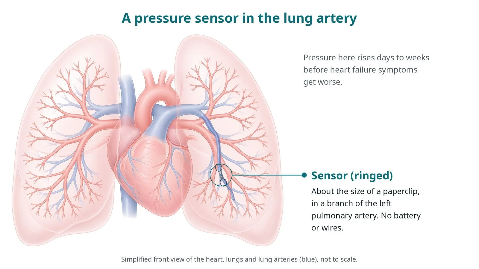
What it is: A small wireless pressure sensor, about the size of a paperclip, is placed in a branch of the left pulmonary artery through a vein in the groin. At home the patient lies on a special pillow for a short daily reading, which goes to the heart failure team.
Who it is for: Selected adults with heart failure who have been in hospital or needed urgent treatment for it, and who can take daily readings, as decided by a specialist heart failure team. NICE recommended it for NHS use in February 2026.
Main risks: Bleeding or bruising at the groin, rhythm upset during the catheter, and rarely injury to the lung artery. Your team will explain your own risk.
Usually: Under local anaesthetic, about an hour, often home the same or next day.
On this page: What is cardioMEMS pulmonary artery pressure sensor? · Who is it for? · When is it done? · Step by step · Recovery · Risks · Kit list · First time in the lab? · FAQ
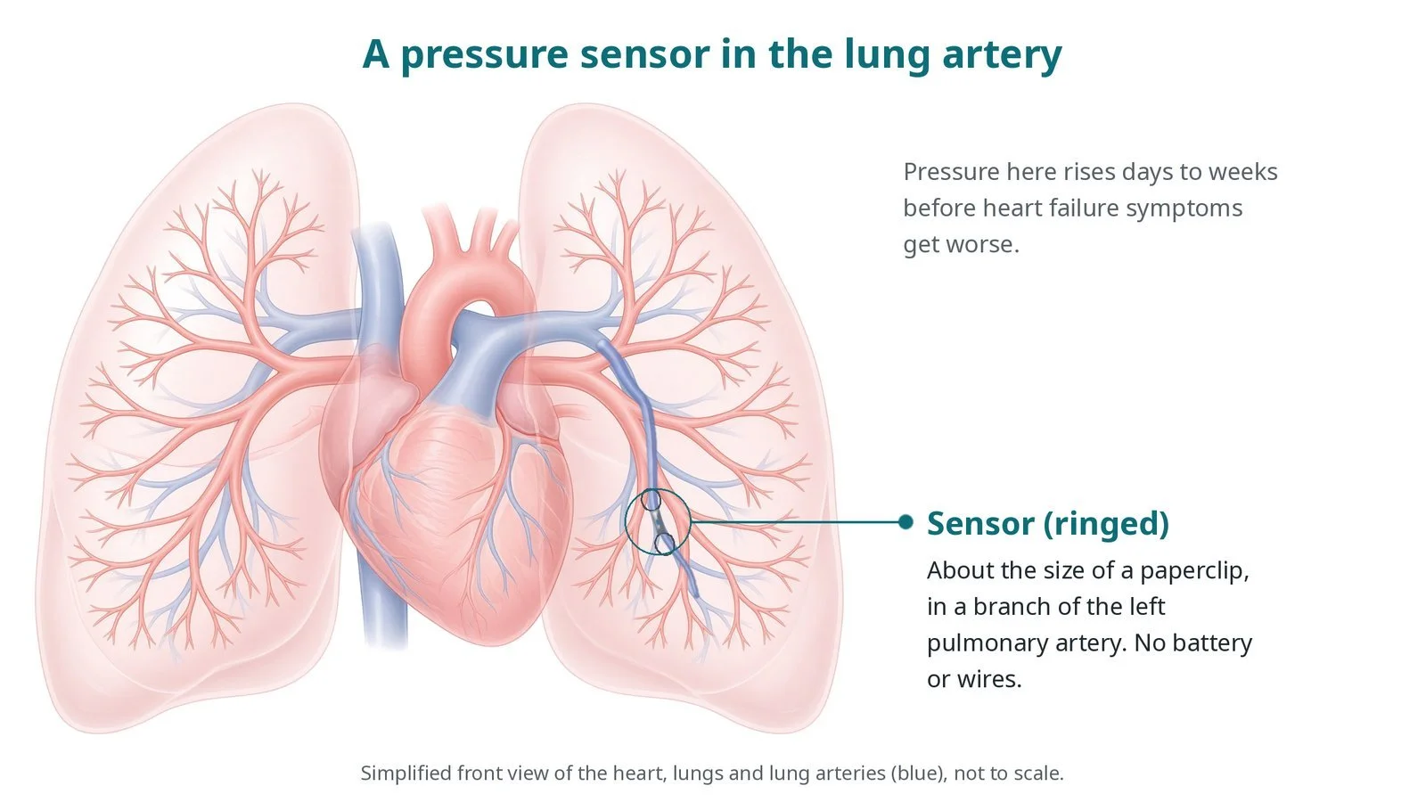
A small sensor (ringed) sits in a branch of the left pulmonary artery. Pressure there rises days to weeks before heart failure symptoms get worse.
What is cardioMEMS pulmonary artery pressure sensor?
In heart failure the heart cannot pump well enough to keep pressures normal. Fluid builds up and pressure rises in the left atrium, and then in the lung arteries, often days to weeks before the person notices more breathlessness or swelling.
The pulmonary artery carries blood from the right ventricle to the lungs. Its pressure reflects the filling pressure on the left side of the heart. A sensor sitting in a lower branch of the left pulmonary artery can measure it every day.
The sensor has no battery and no wires. When the patient lies on the pillow, an antenna powers it for a moment and reads the pressure. The team looks at the trend and adjusts diuretics and other medicines before the patient becomes unwell, aiming to keep them out of hospital.
New to the acronyms? See the Cath Lab Abbreviations A–Z.
Who is it for?
Adults with heart failure chosen by a specialist heart failure team, usually after a hospital stay or urgent treatment for heart failure. The person needs to:
be on the best tolerated heart failure medicines
be able and willing to take a daily reading at home
have suitable lung artery anatomy and body size for the device
be able to take the blood thinning medicines needed after the implant
Who it may not suit: People who cannot take the antiplatelet or anticoagulant medicines advised afterwards, those with an active infection or a recent clot in the lungs, or people who would not be able to take daily readings. The team will discuss alternatives.
The decision is made by your heart team, who will talk through the reasons, the alternatives and the risks with you.
When is it done?
It is a planned procedure in the cath lab. NICE recommended pulmonary artery pressure monitoring with CardioMEMS for NHS use in February 2026, alongside a team that can review readings and act on them.
gives you a chance to ask questions and give consent
arranges blood tests, including kidney function and a blood count, and an ECG
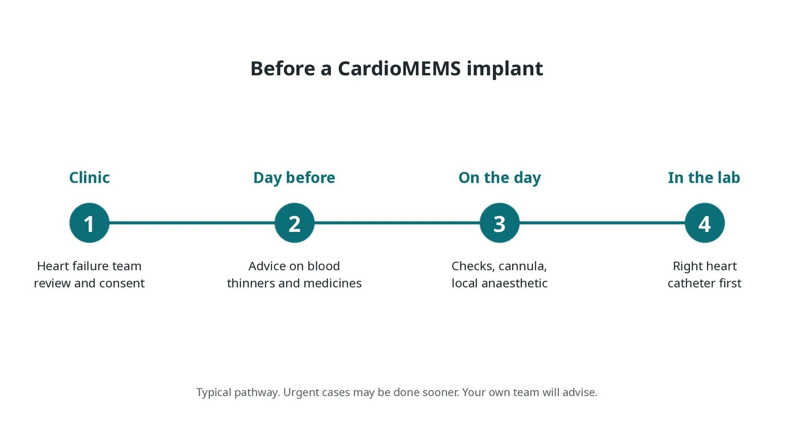
Before a CardioMEMS implant: heart failure team review and consent; advice on blood thinners and medicines; checks and local anaesthetic on the day; a right heart catheter first.
Step by step
1. Getting ready
You lie on the X-ray table and the groin is cleaned and numbed with local anaesthetic. Light sedation may be offered. Monitoring includes ECG, blood pressure and oxygen levels.
2. Right heart catheter
A sheath is placed in the femoral vein under ultrasound. Sizes are per device and operator. A balloon-tipped catheter is floated through the right atrium and right ventricle into the pulmonary artery, and pressures are recorded. Heparin is given as per centre protocol.
Watch: Extra beats or a run of fast rhythm as the catheter crosses the right ventricle: tell the operator, who will usually pull back; have the defibrillator ready.
3. Placing the sensor
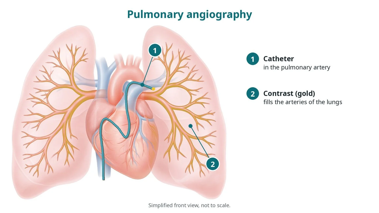
A. Angiogram. Contrast pictures of the left pulmonary artery choose a lower (descending) branch of the right size for the device.
B. Wire. A wire is placed deep in the chosen branch.
C. Deploy. The sensor is advanced on its delivery catheter and released, its wire loops holding it in place.
D. Calibrate. The sensor reading is checked against the pressure measured by the catheter at the same time, and the patient's details are linked to the system.
Watch: Coughing, coughing blood or a drop in oxygen levels: possible injury to the lung artery. Tell the operator at once.
4. Finishing
The catheters and sheath are removed and the vein is closed with pressure or a stitch, per operator. A first reading may be taken on the pillow before going home.
5. Home readings
Each day the patient lies on the pillow for about a minute. The reading goes to a secure website reviewed by the heart failure team, who contact the patient to change medicines when pressures drift up or down.
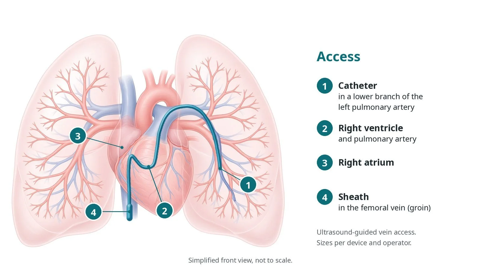
Access: (1) catheter in a lower branch of the left pulmonary artery; (2) right ventricle and pulmonary artery; (3) right atrium; (4) sheath in the femoral vein.
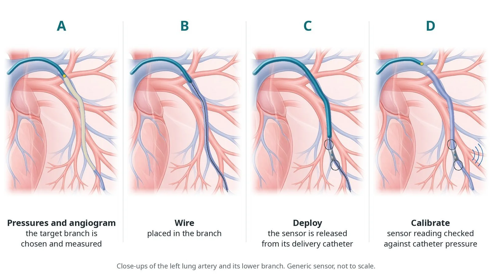
A: pressures and angiogram choose the branch. B: wire in the branch. C: the sensor is released. D: its reading is calibrated against catheter pressure.
Recovery and aftercare
First hours: rest with checks of the groin, heart rhythm, breathing and blood pressure, as per centre protocol.
Going home: often the same or next day.
Medicines: antiplatelet or anticoagulant medicines for a period afterwards, as your team advises; keep taking your heart failure medicines.
Readings: lie on the pillow for a short reading each day; the team will contact you if your medicines need changing.
Driving: ask your team; usually you can drive once the groin has healed and the effects of any sedation have worn off, subject to DVLA rules for your heart condition.
Follow-up: heart failure clinic and remote monitoring.
Call 999 if you have chest pain, sudden severe breathlessness, cough up blood, faint, or have heavy bleeding from the groin.
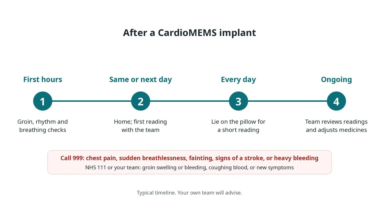
After a CardioMEMS implant: (1) groin, rhythm and breathing checks; (2) home the same or next day; (3) a short reading on the pillow every day; (4) the team reviews readings and adjusts medicines.
Risks and complications
Your team will explain the risks that apply to you. Possible problems include:
Bleeding, bruising or swelling at the groin
Extra beats or a fast rhythm during the catheter: Usually settles when the catheter is moved.
Injury to the lung artery, such as bleeding into the lung (rare)
Reaction to the contrast dye (uncommon)
Sensor moving out of position or not reading (rare)
Bleeding from the blood thinning medicines needed afterwards
Cath lab kit list (printable)
Barts example kit list
An example from one UK centre, written generically. Every lab has its own protocol, and sizes and doses vary.
femoral venous sheath, size per device and operator
balloon-tipped pulmonary artery catheter
guidewires, per operator
pulmonary artery sensor (CardioMEMS) on its delivery catheter
pressure transducer and haemodynamic monitoring system
contrast and syringes for the pulmonary angiogram
home electronics unit with pillow antenna
heparin and local anaesthetic, as per centre protocol
ultrasound for vein access
venous closure: pressure or a figure-of-eight stitch, per operator
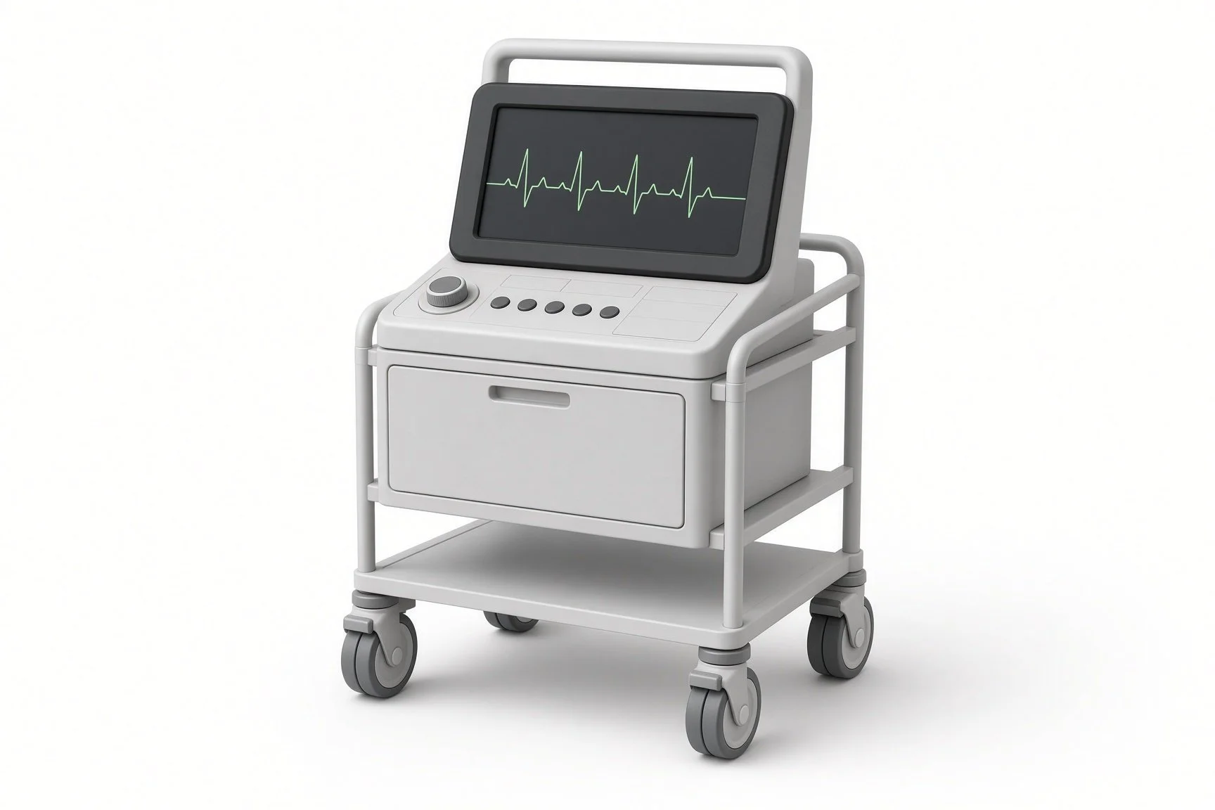
Pressure transducer.
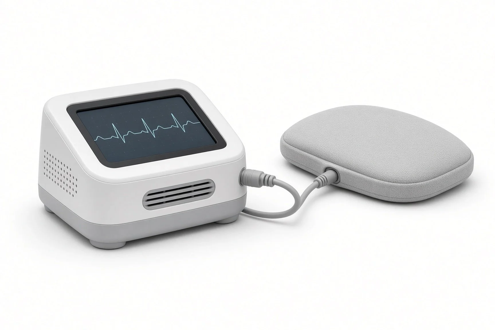
Home reading unit and pillow.
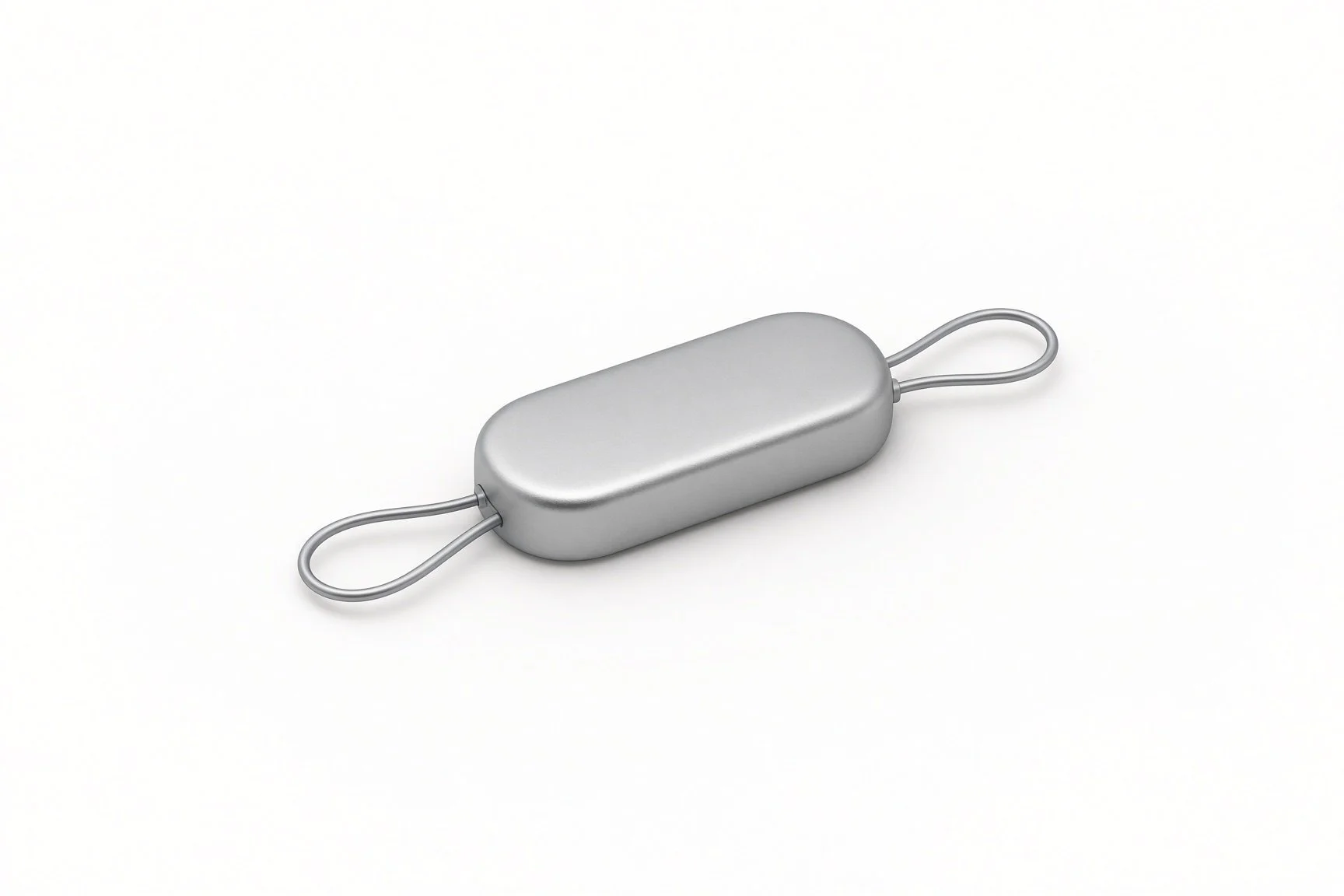
Sensor on its delivery catheter.
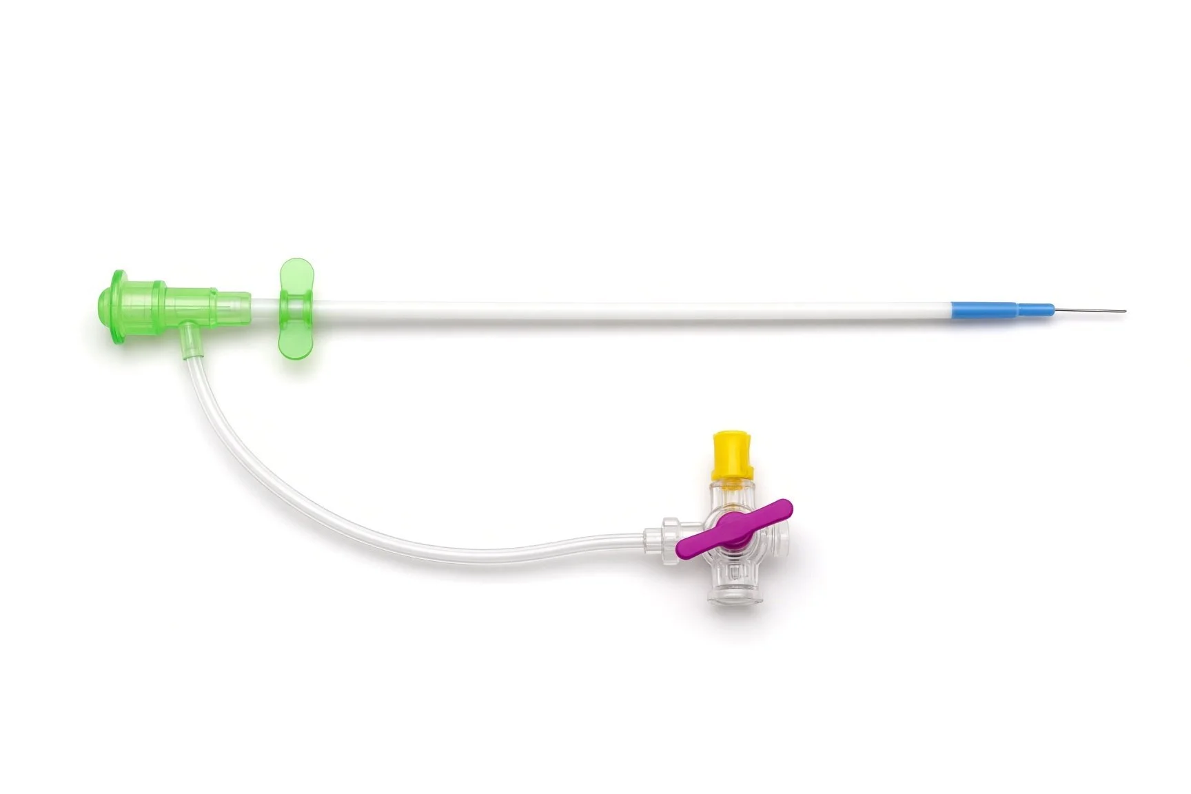
Venous sheath.
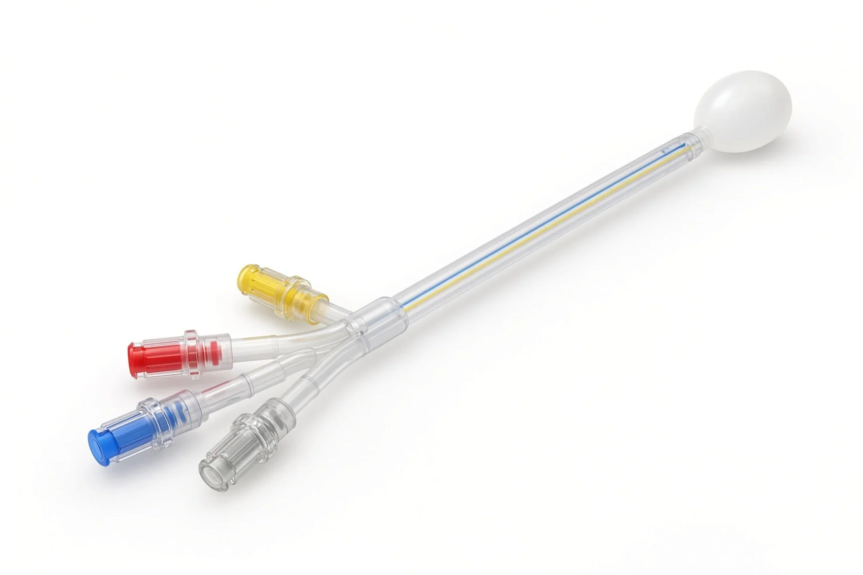
Balloon-tipped pulmonary artery catheter.
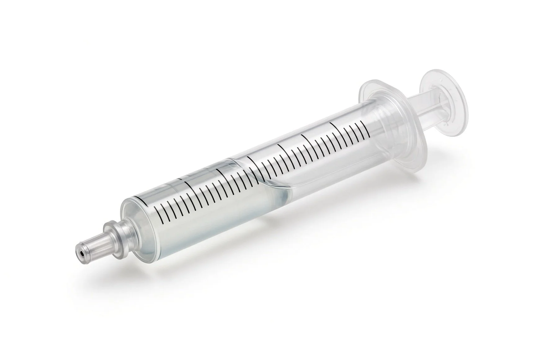
Contrast syringe.
Contrast.
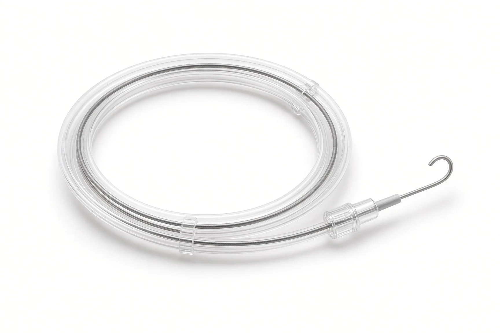
Guidewire.
Procedural steps (Barts example)
Getting ready
Right heart catheter
Placing the sensor
Finishing
Home readings
First time in the cath lab?
Think of it as a right heart catheter first. The set-up, pressures and catheter handling are the same; the sensor comes afterwards.
Zero and level the transducer carefully. Calibration depends on an accurate catheter pressure.
Watch the rhythm. Crossing the right ventricle can cause extra beats; keep the defibrillator nearby.
Teach the pillow. Patients often leave with their home unit, so make sure they know who will call them about readings.
Check the patient's weight and that blood thinners have been managed as per centre protocol. The plan for medicines after the implant should be clear before the case.
The radiographer and physiologist work together for the calibration: the catheter pressure must be recorded at the same moment as the sensor reading.
FAQ
Will I feel the sensor?
No. It is very small, has no battery and sits inside the lung artery.
Does it replace my tablets?
No. It helps your team adjust your heart failure medicines earlier, before you become unwell.
Can I have an MRI scan?
The sensor is MRI conditional: scans are possible under set conditions. Tell the MRI team you have it and carry your implant card.
What if I miss a reading?
Take it when you can. Your team will contact you if readings stop.
Related Cath Lab 101 guides
Cardiac resynchronisation therapy (CRT-P and CRT-D) (coming soon)
Interatrial shunt devices for heart failure (coming soon)
Sources
All checked on 12 November 2026. Each page's own date is shown where it gives one.
NICE. Pulmonary artery pressure monitoring with CardioMEMS for heart failure, recommended for NHS use in February 2026. nice.org.uk
McDonagh TA, et al. 2021 ESC Guidelines for the diagnosis and treatment of acute and chronic heart failure. Eur Heart J 2021;42:3599-3726
StatPearls (NCBI Bookshelf), search: pulmonary artery catheterization. ncbi.nlm.nih.gov/books
Disclaimer
Education only. This page is for general education for healthcare students, cath lab staff and curious readers. It isn't medical advice and doesn't replace advice from your own doctor, heart team or local unit protocols. Equipment and medicines are described generically. No product or manufacturer is recommended. If you think someone is having a heart emergency, call 999.
Disclosure: I work for a company that makes a device used in this procedure. This page is independent education, isn't sponsored or reviewed by any company, and doesn't recommend any product.
All diagrams on this page are original illustrations created for rodnieoro.com.