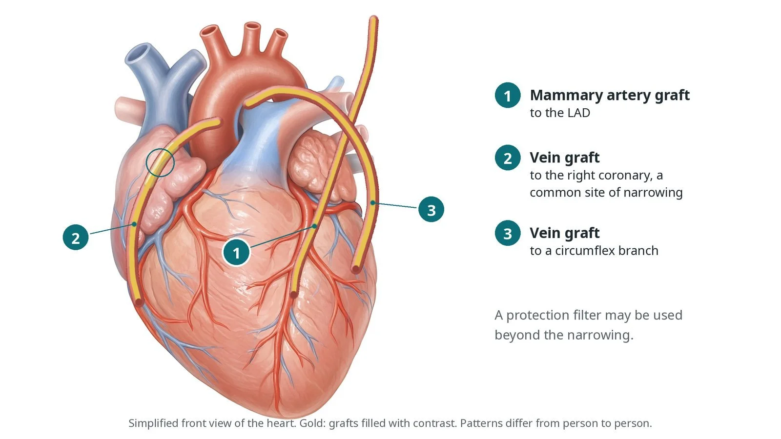
Internal mammary angiography and subclavian stenosis: a Cath Lab 101 guide
Reviewed by Rodnie Oro, cath lab charge nurse · Last reviewed: November 2026 · Education only, not medical advice
Internal mammary angiography and subclavian stenosis in one minute
What it is: An angiogram that places a shaped catheter into the internal mammary artery (LIMA or RIMA), often used as a bypass graft, and checks the subclavian artery it comes from.
Who it is for: People with a mammary artery bypass graft who have symptoms, people being assessed before bypass surgery when a subclavian narrowing is suspected, and people with a blood pressure difference between the arms.
Main risks: Bleeding at the wrist or groin, spasm or a tear in the mammary artery, stroke (rare), and reaction to contrast. Your team will explain your own risk.
Usually: Awake, under local anaesthetic, as part of a coronary angiogram; about 30 to 60 minutes; home the same day for most.
On this page: What is internal mammary angiography and subclavian stenosis? · Who is it for? · When is it done? · Step by step · Recovery · Risks · Kit list · First time in the lab? · FAQ
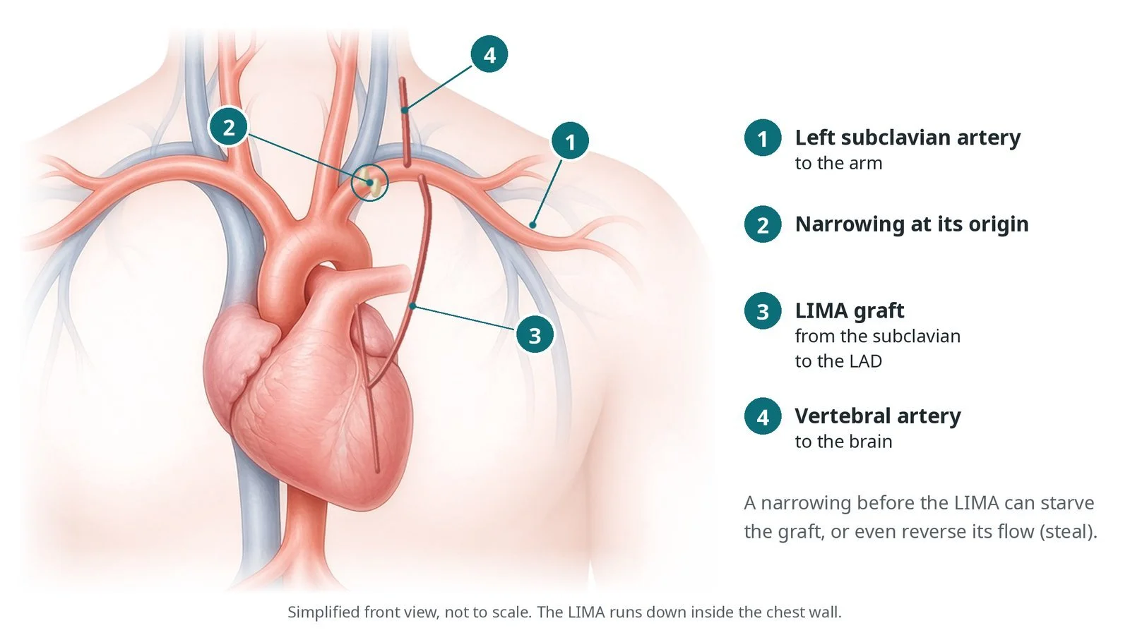
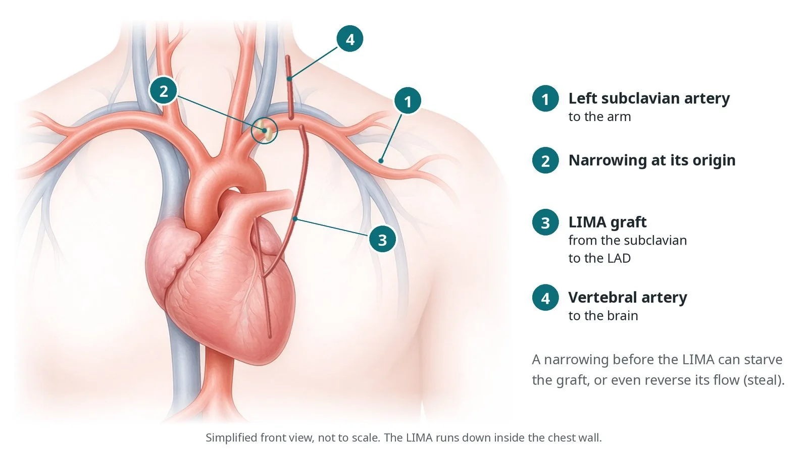
(1) The left subclavian artery runs to the arm; (2) a narrowing at its origin; (3) the LIMA graft runs from the subclavian to the LAD; (4) the vertebral artery runs to the brain. A narrowing before the LIMA can starve the graft or even reverse its flow (steal).
What is internal mammary angiography and subclavian stenosis?
The left and right internal mammary arteries (LIMA and RIMA, also called internal thoracic arteries) branch downwards from the subclavian arteries, just behind the chest wall beside the breastbone. The LIMA is the usual bypass graft to the LAD because it stays open for many years.
The left subclavian artery comes off the aortic arch and runs to the arm. The vertebral artery to the brain and the LIMA branch from it. A narrowing at the start of the subclavian artery, before these branches, can reduce flow to the arm, to the brain, and to a LIMA graft.
In coronary-subclavian steal, the narrowing is so tight that blood flows backwards up the LIMA graft from the heart to supply the arm, causing angina, especially with arm exercise. A difference in blood pressure between the arms (often quoted as more than 15 mmHg) is a warning sign.
New to the acronyms? See the Cath Lab Abbreviations A–Z.
Who is it for?
People with a LIMA or RIMA graft and angina or a positive stress test.
People being assessed for bypass surgery when the mammary artery is to be used and there is a blood pressure difference between the arms or other signs of subclavian narrowing.
People with symptoms of subclavian steal, such as arm pain or dizziness with arm use.
Who it may not suit: The approach is tailored to the anatomy; very tortuous or diseased arteries may need a different route or CT angiography instead.
The decision is made by your heart team, who will talk through the reasons, the alternatives and the risks with you.
When is it done?
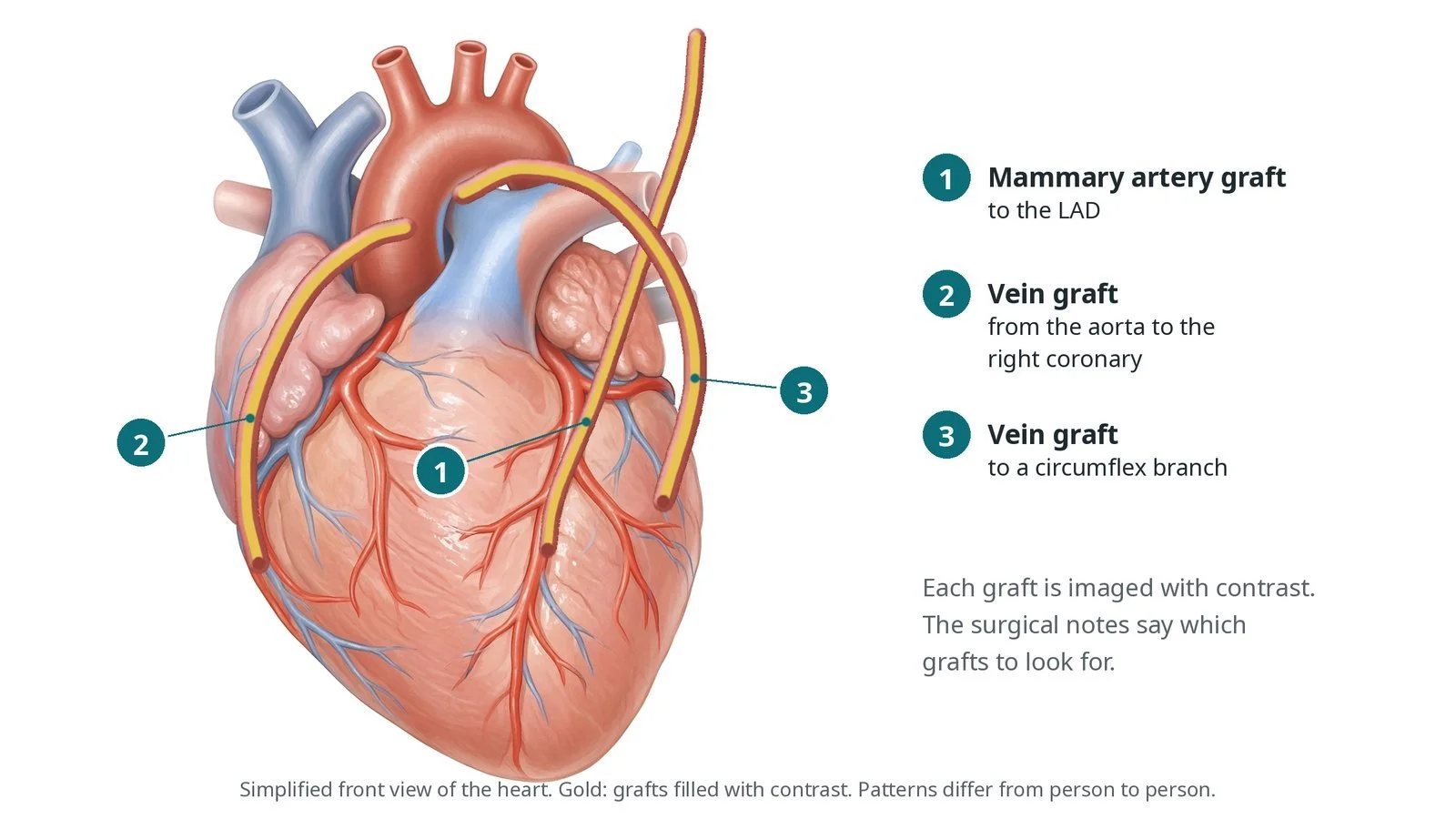
Usually with a coronary angiogram or bypass graft angiogram, as a planned day case. The surgical notes are checked beforehand to know which grafts to look for.
gives you a chance to ask questions and give consent
arranges blood tests, including kidney function and a blood count, and an ECG
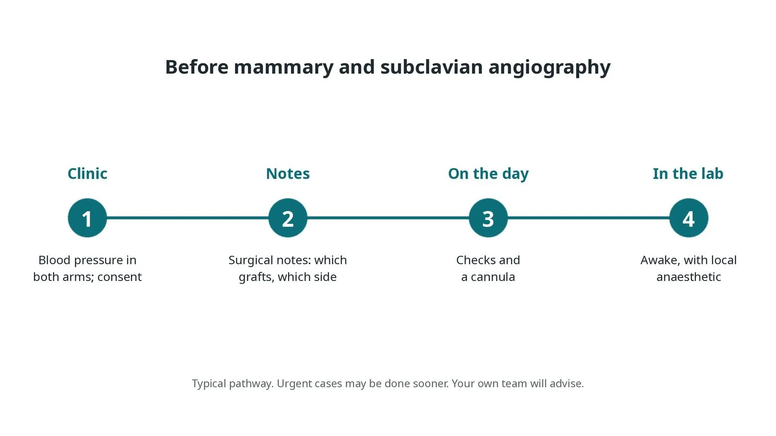
Before mammary and subclavian angiography: blood pressure in both arms and consent; surgical notes on which grafts and which side; checks and a cannula on the day; awake with local anaesthetic.
Step by step
1. Getting ready
Blood pressure is measured in both arms. The surgical notes are reviewed for the number and type of grafts. You lie on the X-ray table with monitoring.
2. Access
For a LIMA, the femoral artery at the groin or the left radial artery at the wrist is used; for a RIMA, the right radial artery is common. A sheath is placed and heparin given as per centre protocol.
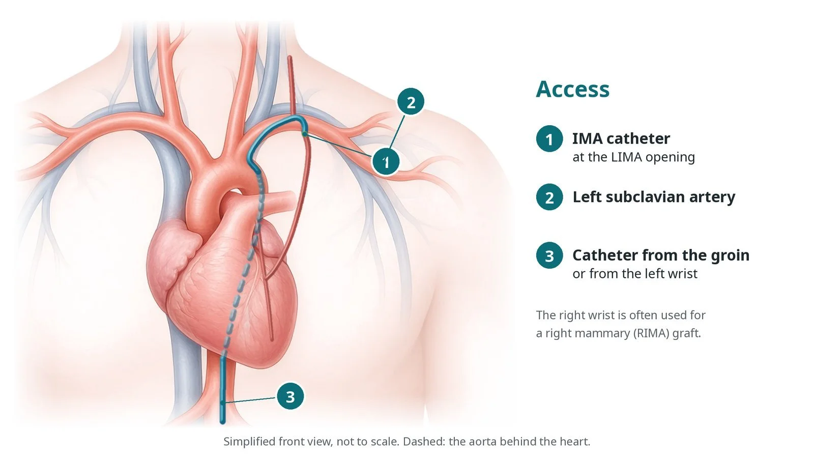
Access: (1) IMA catheter at the LIMA opening; (2) left subclavian artery; (3) catheter from the groin, or from the left wrist. The right wrist is often used for a right mammary (RIMA) graft.
3. Imaging the mammary artery
A. Into the subclavian. A catheter is guided from the aortic arch into the subclavian artery, and a wire is passed beyond the mammary artery opening.
B. Engage the LIMA. A dedicated IMA catheter with a hooked tip is advanced over the wire, then gently pulled back and turned until it drops into the mammary artery opening, which points downwards.
C. Selective injection. Small, gentle injections fill the graft down to where it joins the LAD. Pictures are taken in more than one view to see the join.
D. Subclavian check. If the arm pressures differ or the graft fills poorly, contrast is injected in the subclavian artery, and pressures may be measured across any narrowing.
Watch: A damped or ventricularised pressure in the mammary artery means the catheter is wedged. Tell the operator before any injection.
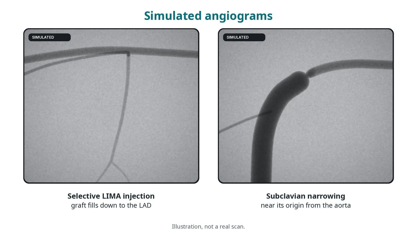
Illustration, not a real scan: (1) selective LIMA injection, the graft filling down to the LAD; (2) a subclavian narrowing near its origin from the aorta.
4. If a narrowing is found
A subclavian narrowing is described and measured. Treatment with a stent is usually done by an interventional radiologist, vascular team or cardiologist as a separate, planned procedure, as per local pathways.
5. Finishing
The catheters are removed. The sheath is taken out and the wrist or groin closed, as per centre protocol.
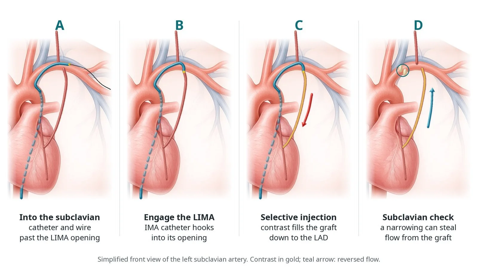
A: catheter and wire into the subclavian, past the LIMA opening. B: the IMA catheter hooks into the LIMA opening. C: selective injection fills the graft down to the LAD. D: subclavian check; a narrowing can steal flow from the graft.
Recovery and aftercare
First hours: wrist or groin, heart rhythm and blood pressure checks, as per centre protocol.
Going home: the same day for most.
Medicines: keep taking your usual medicines unless told otherwise.
Activity: avoid heavy lifting with the wrist or leg used for a few days.
Results: your team explains the findings; a subclavian narrowing may be referred for stenting.
Driving: ask your team; most people can drive after a day or two if the site has healed and they feel well.
Call 999 if you have chest pain that does not settle, breathlessness, fainting, signs of a stroke, or heavy bleeding.
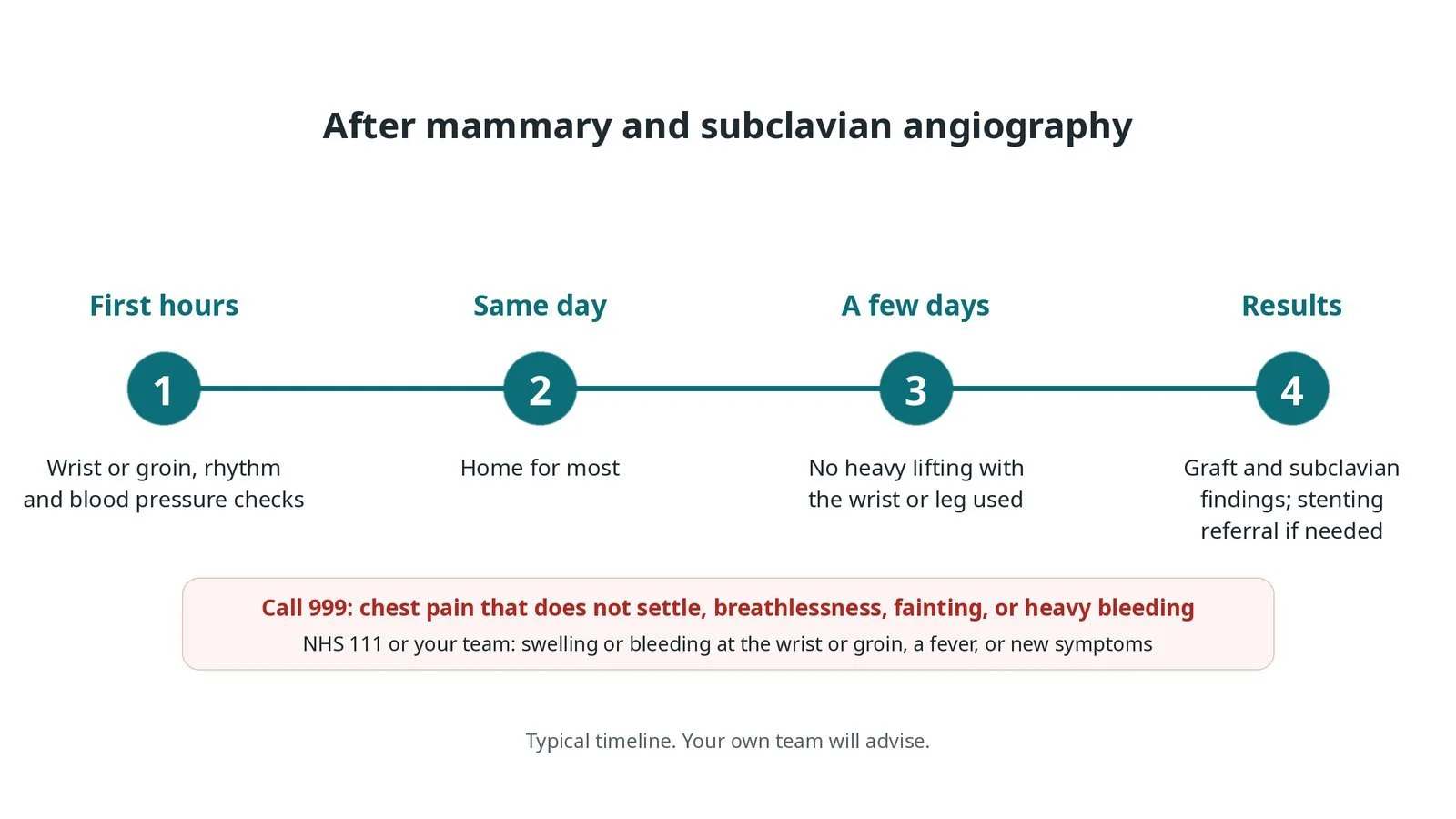
After mammary and subclavian angiography: (1) wrist or groin, rhythm and blood pressure checks; (2) home the same day for most; (3) no heavy lifting with the wrist or leg used for a few days; (4) results explained, with referral for stenting if needed.
Risks and complications
Your team will explain the risks that apply to you. Possible problems include:
Bleeding or bruising at the wrist or groin
Spasm of the mammary artery: Usually settles with GTN.
Tear of the mammary artery or subclavian artery (rare)
Stroke from catheter work near the vertebral artery (rare)
Kidney strain from contrast
Cath lab kit list (printable)
Barts example kit list
An example from one UK centre, written generically. Every lab has its own protocol, and sizes and doses vary.
radial or femoral sheath, size per operator
diagnostic catheters for the aortic arch and subclavian artery
internal mammary (IMA) catheter with a hooked tip
guidewires, including a soft-tipped wire
contrast and heparin, as per centre protocol
GTN for spasm, per operator
pressure monitoring for gradients across a narrowing
surgical notes with the graft details
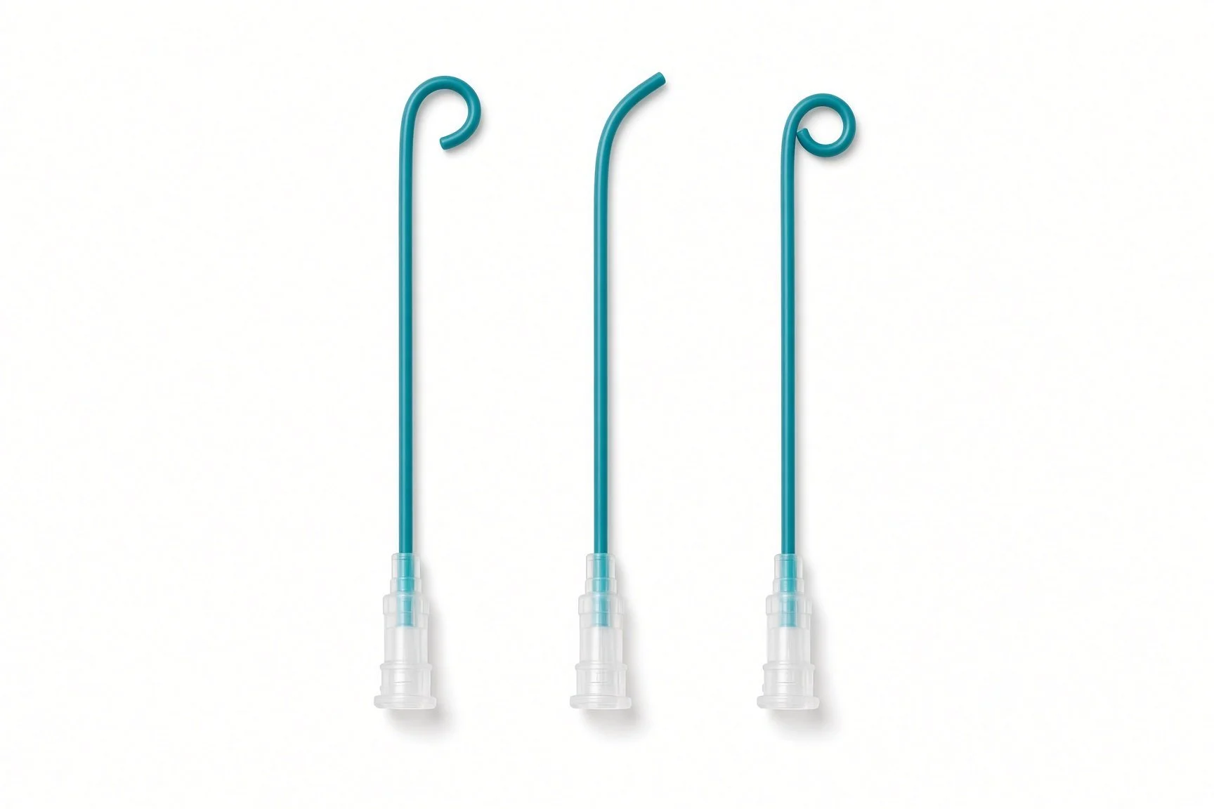
Diagnostic catheters for the aortic arch and subclavian artery.
Procedural steps (Barts example)
Getting ready
Access
Imaging the mammary artery
If a narrowing is found
Finishing
First time in the cath lab?
Measure both arms. A blood pressure difference between the arms is a key clue; record it before the case.
Read the surgical notes. Know how many grafts and which mammary artery was used before the patient is on the table.
Warn about the burning feeling. Mammary injections often cause a warm or burning feeling in the chest wall.
Watch the trace. A damped pressure means the catheter may be wedged in a small artery that is easy to injure.
Write both arm blood pressures on the record. The operator uses them to choose the route.
Warn the patient before each injection: a warm or burning feeling in the chest wall is common.
Watch the pressure trace: damping means the catheter may be wedged in the artery.
FAQ
Why is my blood pressure checked in both arms?
A difference can mean a narrowing in the subclavian artery, which can affect a mammary graft.
What is subclavian steal?
When a narrowing is so tight that blood flows backwards from the heart or brain to supply the arm.
Why do I feel burning in my chest during the injection?
The mammary artery also supplies the chest wall; the contrast causes a brief warm feeling there.
Can the narrowing be treated?
Yes, usually with a stent as a separate planned procedure.
Related Cath Lab 101 guides
Subclavian, vertebral and mesenteric stenting (coming soon)
Sources
All checked on 12 November 2026. Each page's own date is shown where it gives one.
Mazzolai L, et al. 2024 ESC Guidelines for the management of peripheral arterial and aortic diseases. Eur Heart J 2024
StatPearls (NCBI Bookshelf), search: subclavian steal syndrome. ncbi.nlm.nih.gov/books
StatPearls (NCBI Bookshelf), search: internal mammary artery. ncbi.nlm.nih.gov/books
Disclaimer
Education only. This page is for general education for healthcare students, cath lab staff and curious readers. It isn't medical advice and doesn't replace advice from your own doctor, heart team or local unit protocols. Equipment and medicines are described generically. No product or manufacturer is recommended. If you think someone is having a heart emergency, call 999.
Disclosure: I work in the medical device industry. This page is independent education and isn't sponsored or reviewed by any company.
All diagrams on this page are original illustrations created for rodnieoro.com.