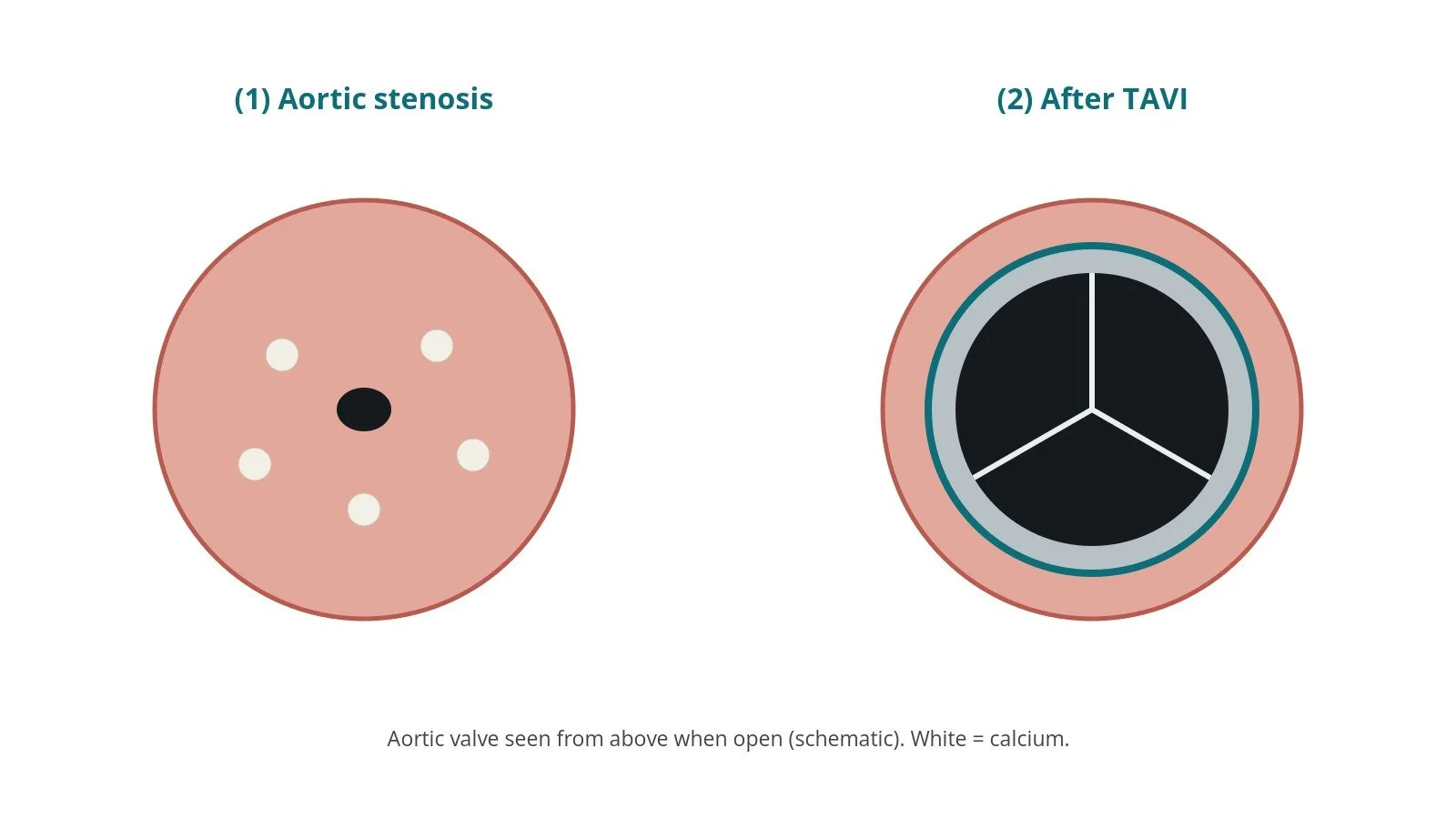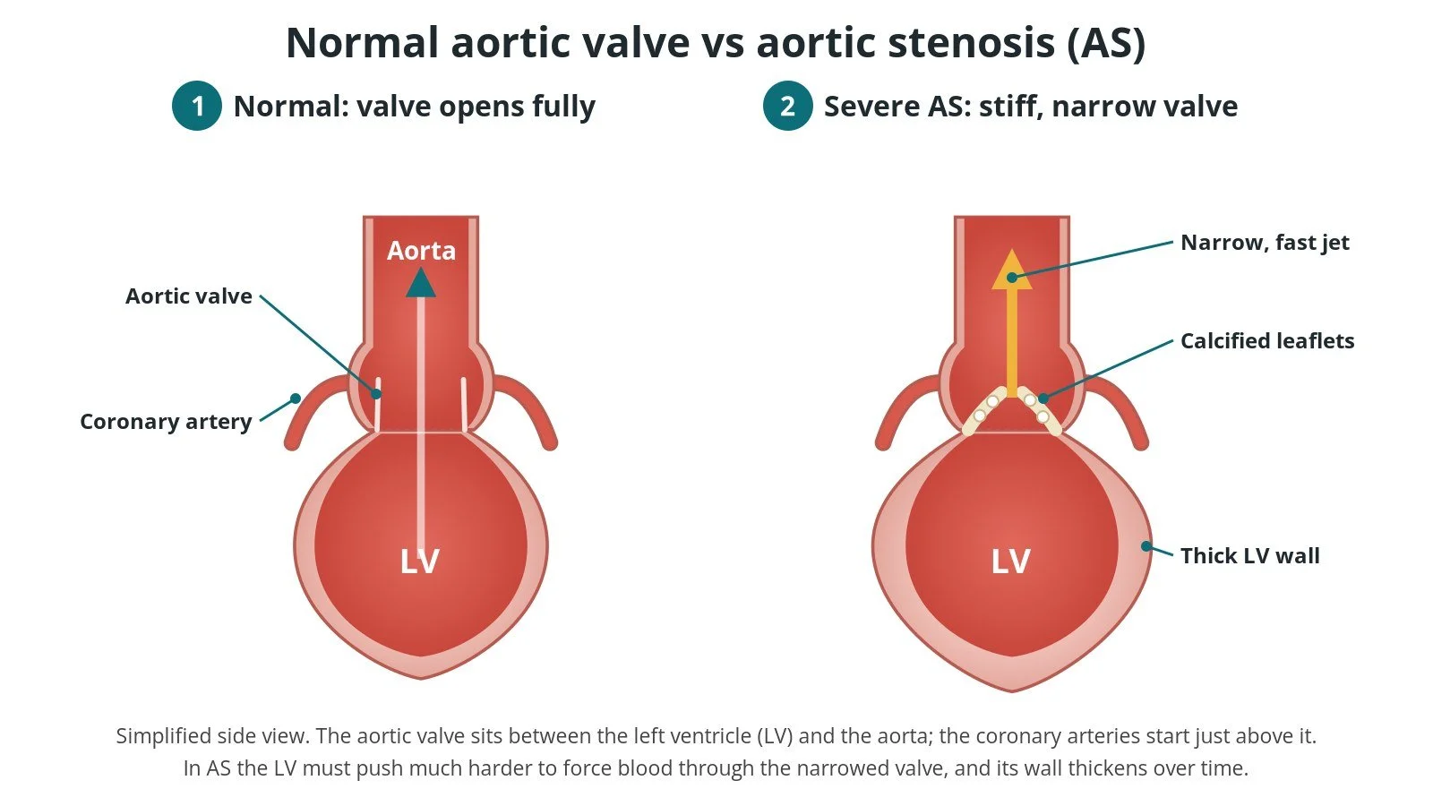
TAVI (transcatheter aortic valve implantation): a Cath Lab 101 guide
Reviewed by Rodnie Oro, cath lab charge nurse · Last reviewed: October 2026 · Education only, not medical advice
TAVI in one minute
What it is: TAVI (transcatheter aortic valve implantation, also called TAVR) is a keyhole (catheter) treatment for a narrowed aortic valve, the valve between the left ventricle and the aorta. A new tissue valve, folded inside a thin tube, is passed up an artery, usually from the groin, and opened inside the old valve, which is pushed aside.
Who it is for: adults with severe aortic stenosis who have symptoms, such as breathlessness, chest pain, dizziness or blackouts, or who have weakening heart muscle. A specialist heart team decides whether TAVI, open surgery or another plan is best.
Main risks: bleeding or damage to the artery, needing a permanent pacemaker, a leak around the new valve, and stroke. Your heart team will explain your own risk.
Usually: under local anaesthetic with sedation (sometimes a general anaesthetic), about 1 to 2 hours, then home after 1 to 2 days for many people.
On this page: What is TAVI? · Who is it for? · When is it done? · Step by step · Recovery · Risks · Kit list · First time in the lab? · FAQ
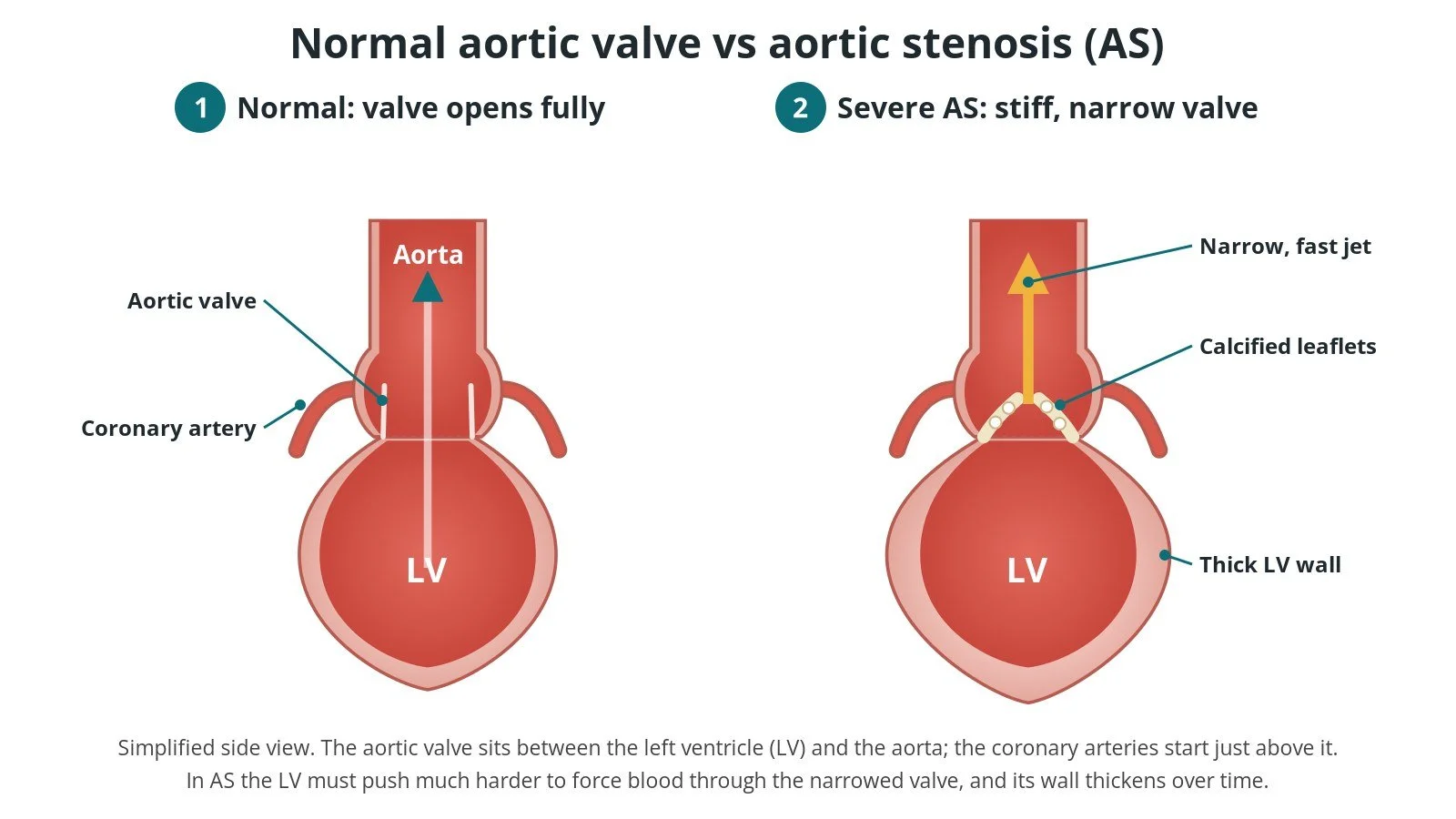
Normal vs aortic stenosis (AS). (1) Normal: the valve opens fully. (2) Severe AS: calcified leaflets, a narrow jet and a thick LV wall. The coronary arteries start just above the valve.
What is TAVI?
The aortic valve sits between the left ventricle (LV), the main pumping chamber, and the aorta, the large artery that carries blood to the body. It usually has three thin leaflets that open fully with each beat and close to stop blood falling back. The two coronary arteries, which feed the heart muscle, start from the aorta just above the valve.
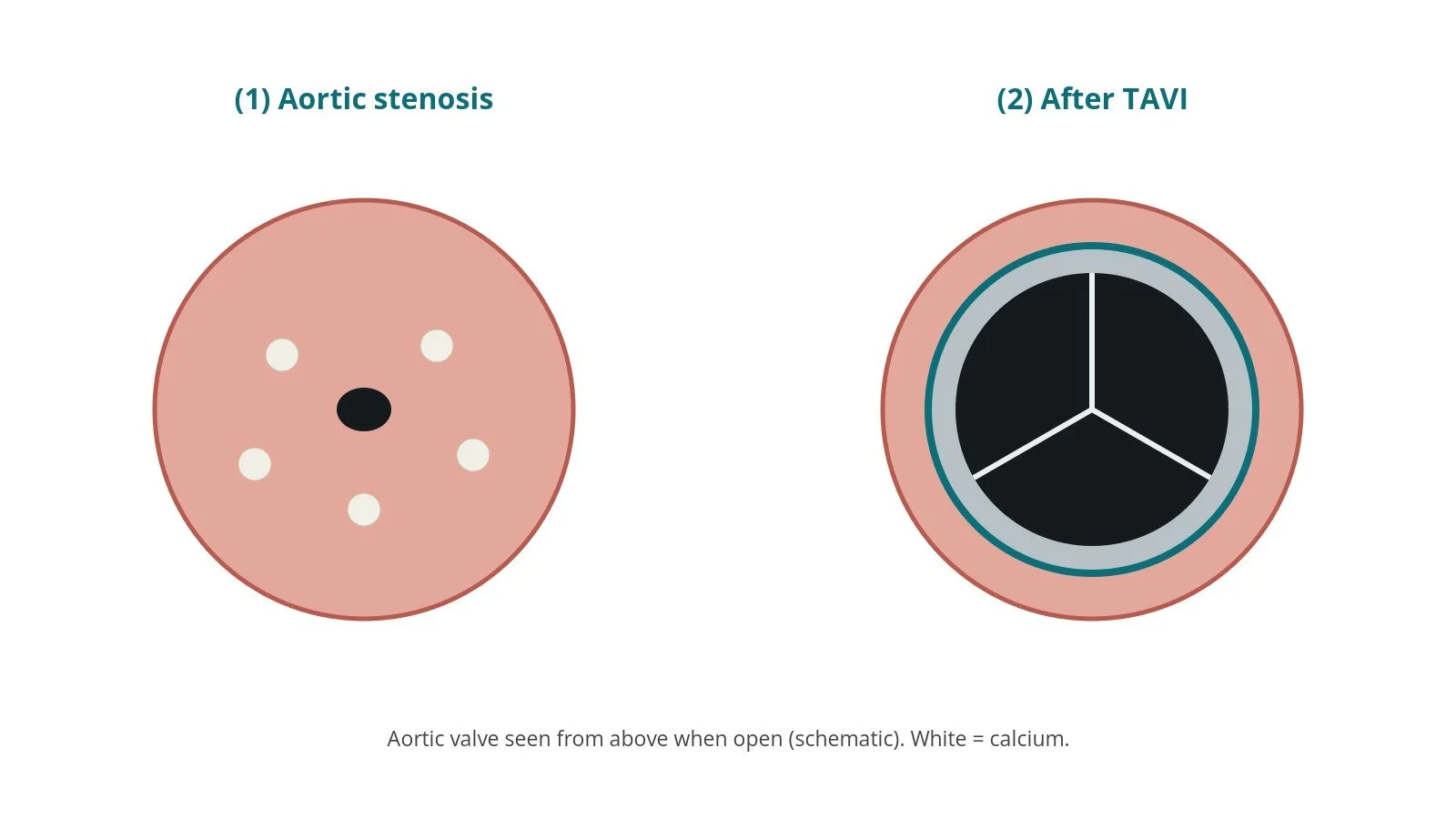
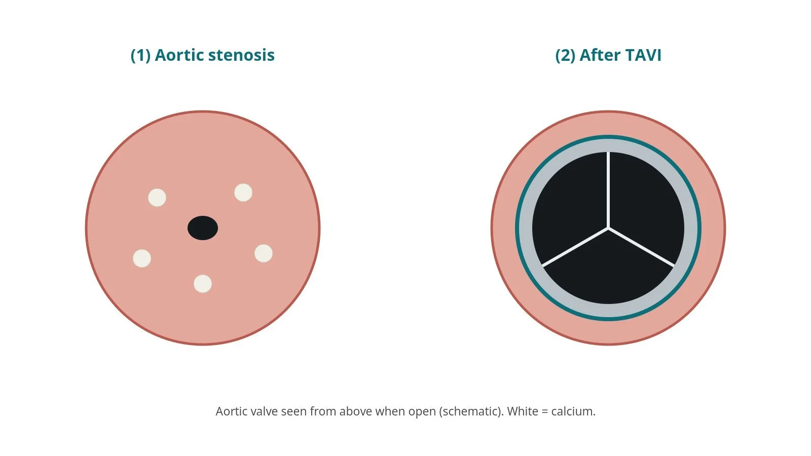
In aortic stenosis (AS) the leaflets become thick, stiff and calcified, so the valve no longer opens fully. The LV has to push much harder to get blood out, and its wall thickens. Most AS in older adults is caused by age-related calcium build-up. It can also happen earlier in people born with a two-leaflet (bicuspid) valve. Once severe AS causes symptoms, such as breathlessness, chest tightness, dizziness or blackouts, it tends to get worse, and medicines cannot open the valve.
TAVI replaces the valve's job without open-heart surgery. A new valve made of animal tissue, mounted on a metal frame, is crimped down onto a delivery catheter. It is passed through an artery to the heart, positioned inside the diseased valve and expanded. The old leaflets are pressed against the wall and the new leaflets take over straight away.
There are two broad types of TAVI valve:
balloon-expandable: the valve is opened by inflating a balloon inside it
self-expanding: the frame opens by itself as a cover is pulled back
The heart team chooses the valve and size for each person's anatomy. Open surgical aortic valve replacement (SAVR) is still the right choice for many people.
New to the acronyms? See the Cath Lab Abbreviations A–Z.
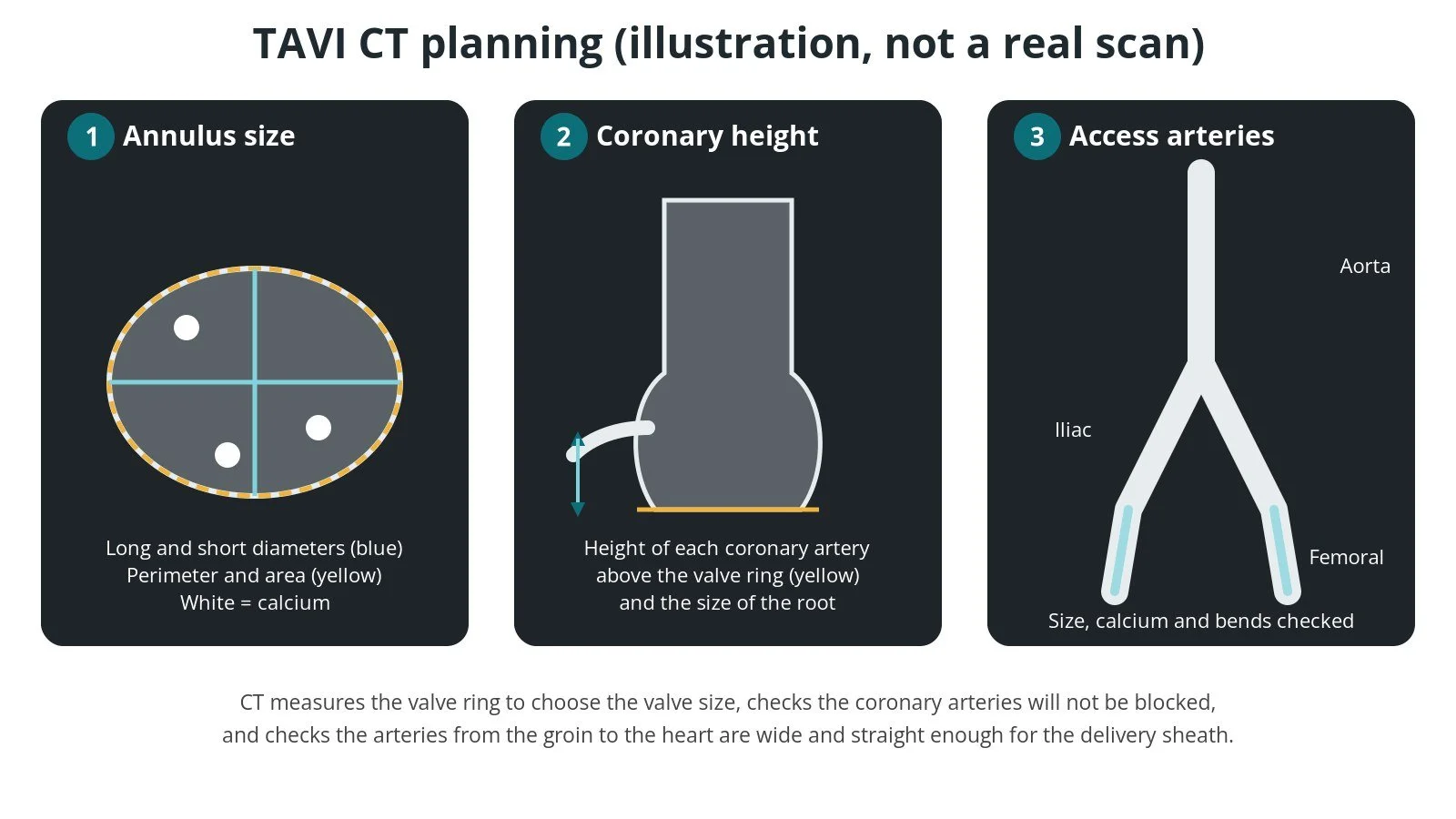
Illustrated CT, not a real scan. (1) Valve ring (annulus) size; (2) coronary height above the valve; (3) access arteries from the aorta to the groin.
Who is it for?
The decision is made by a specialist heart team, usually including interventional cardiologists, cardiac surgeons, imaging specialists and others, after a full assessment. UK (NICE NG208 and the NICE TAVI procedures guidance), European (ESC/EACTS 2025) and American (ACC/AHA 2020) guidance broadly agree that:
treatment is considered for severe aortic stenosis with symptoms, and for some people without symptoms whose LV is starting to weaken
the choice between TAVI and surgery takes into account age, surgical risk, frailty, the valve and aortic root anatomy, the access arteries and the person's own wishes
TAVI is the usual choice for people who are older or at high risk from surgery, while surgery is often preferred in younger people at lower risk, especially with a bicuspid valve
treatment is done in a heart valve centre with a heart team and cardiac surgery on site
NICE NG208 recommends offering TAVI to adults with severe aortic stenosis at high surgical risk or when surgery is unsuitable, and surgery first-line for those at low or intermediate risk. NHS England has its own commissioning position on lower-risk patients. NICE also asks centres to enter every TAVI into the UK TAVI registry.
TAVI may not be suitable if the access arteries are too small or diseased and no alternative route is safe, if the valve anatomy is unsuitable, or if other illness means the person is unlikely to benefit.
When is it done?
TAVI is usually a planned procedure, though some people are treated while in hospital. Before it, the team usually:
confirms the severity of AS with an echo (TTE), and sometimes a TOE or other tests
does a CT scan (TAVI CT) of the heart, aorta and leg arteries to measure the valve ring (annulus), check the height of the coronary arteries above the valve, look at the calcium, and check that the femoral and iliac arteries are large enough for the delivery sheath
checks the coronary arteries, often with an angiogram or the CT, and treats significant narrowings if needed
reviews the ECG for conduction problems that raise the chance of needing a pacemaker
assesses frailty, other illnesses, dental health, kidney function and bloods, and reviews medicines including blood thinners, as per centre protocol
talks through the options and the person's goals before consent
Step by step
1. Anaesthetic and set-up
Most transfemoral TAVIs are done under local anaesthetic with light sedation, with an anaesthetist present. Some centres or patients need a general anaesthetic. Monitoring, a urinary catheter if needed and a defibrillator are set up. The procedure is guided by X-ray (fluoroscopy) and echo.
2. Access
Main access: usually the femoral artery in the groin, entered under ultrasound guidance. Suture-based closure devices are often pre-placed before the artery is enlarged. A large delivery sheath is then inserted. Sheath sizes are per device and operator.
Second artery: a smaller sheath, in the radial artery at the wrist or the other femoral artery, for a pigtail catheter in the aortic root. This is used for aortograms, pressure monitoring and checking the groin at the end.
Alternative access: if the leg arteries are unsuitable, the valve can be delivered through another artery, such as the subclavian/axillary or carotid, or rarely directly through the chest. See the TransAxillary TAVI guide.
Anticoagulation is given as per centre protocol, and reversed at the end if needed.
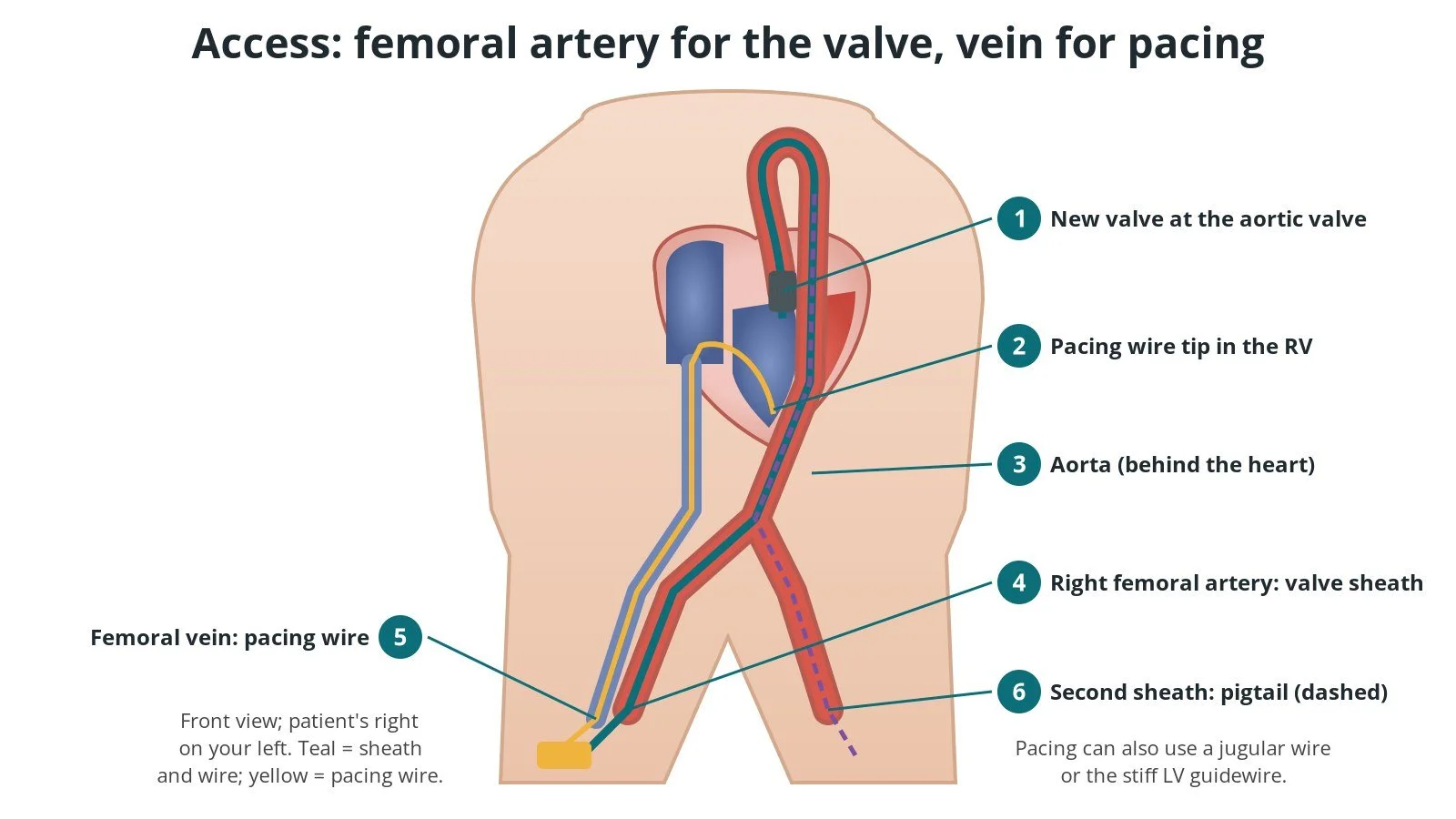
Access: (1) new valve at the aortic valve; (2) pacing wire tip in the RV; (3) aorta; (4) right femoral artery with the valve sheath; (5) femoral vein with the pacing wire; (6) second sheath for the pigtail.
3. Pacing
The heart needs to be paced during TAVI, for two reasons:
Rapid pacing briefly makes the heart beat very fast, so it pumps out very little. This drops the blood pressure and steadies the valve while it is opened or a balloon is inflated. The rate and timing are per operator and centre protocol.
Back-up pacing protects against slow heart rhythms (heart block) if the new valve presses on the heart's conduction system.
Pacing can be done through a temporary pacing wire placed in the right ventricle through a vein (femoral or jugular), or through the stiff guidewire in the LV, with a skin electrode as the return. Many centres use a vein wire when the ECG already shows a conduction problem, such as a broad QRS, because the risk of heart block afterwards is higher.
4. Crossing, preparing, deploying and assessing
A. Crossing the valve. A catheter and straight wire are used to cross the narrowed valve into the LV. The wire is exchanged for a pre-shaped extra-stiff wire that sits safely in the LV apex and supports the delivery system.
B. Balloon valvuloplasty, if needed. Some valves are first stretched with a balloon (BAV) under rapid pacing. Many cases go straight to the valve.
C. Positioning and deploying. The new valve is advanced across the old one and positioned at the right height using the aortogram and fluoroscopy. A balloon-expandable valve is opened by inflating its balloon under rapid pacing. A self-expanding valve is opened gradually as its cover is pulled back, often with pacing to steady it, and some can be recaptured and repositioned before full release.
D. Final check. The team looks at the valve position and shape, the pressure gradient, any leak around the valve (paravalvular leak, PVL), the coronary arteries, the heart rhythm and the blood pressure. If needed, the valve can be expanded further with a balloon (post-dilation).
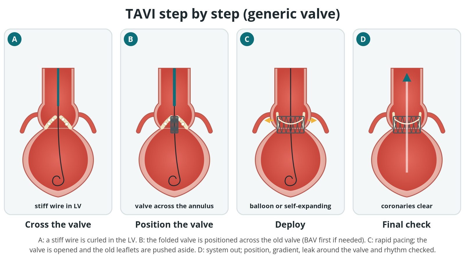
A: a stiff wire is placed in the LV. B: the folded valve is positioned across the old valve. C: the valve is opened (balloon-expandable or self-expanding). D: the result is checked.
5. Closing and finishing
The delivery system and wire are removed. The femoral artery is closed with the pre-placed suture devices, sometimes with an extra plug-based device, and an angiogram from the second sheath checks the groin artery for bleeding or narrowing. The small sheath is removed and the wrist is closed with a compression band. The pacing wire is either removed or left in for a period, depending on the ECG.
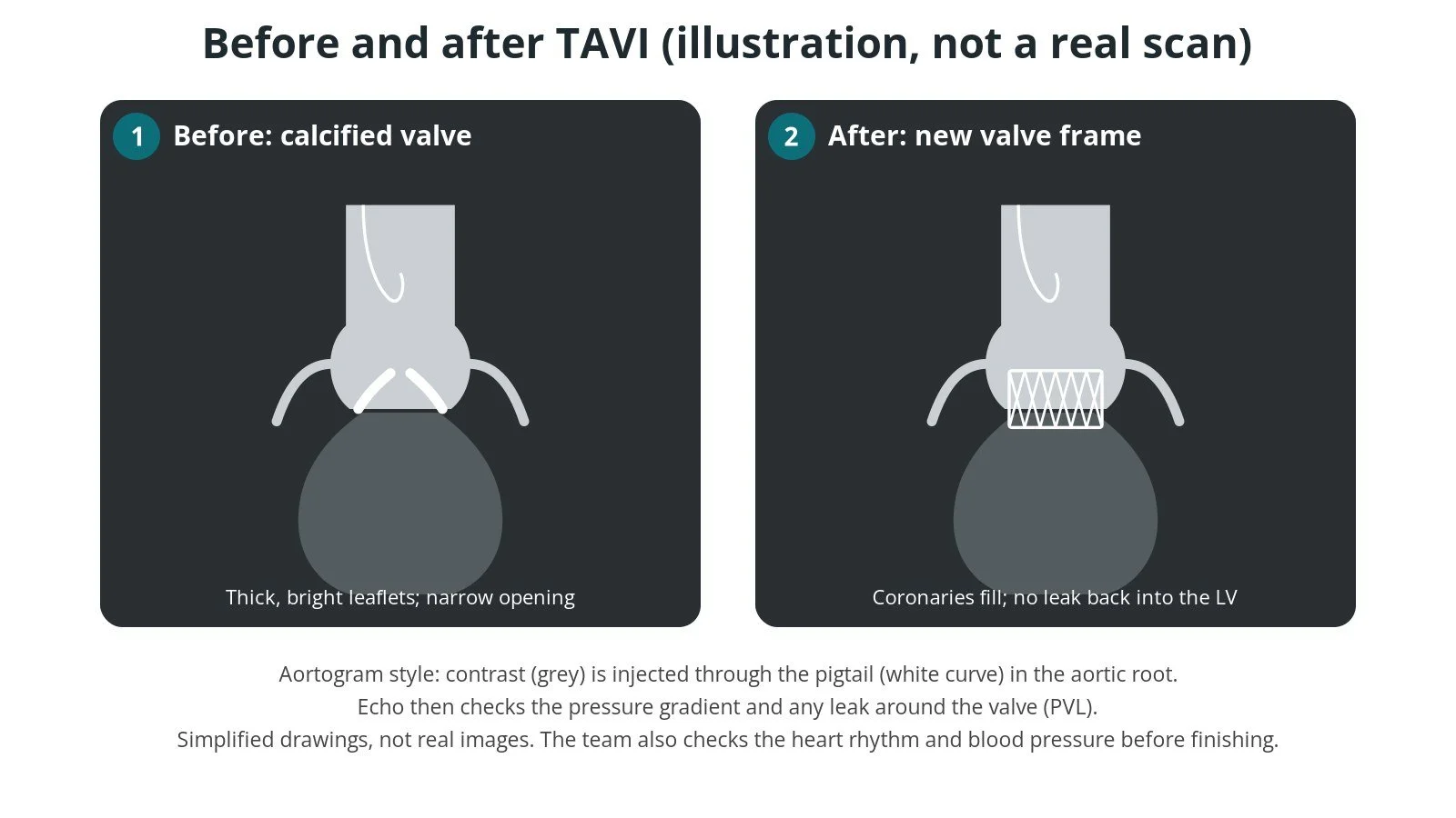
Illustrated aortogram, not a real scan. Before: calcified leaflets. After: the new valve frame, coronaries filling, no leak back. Echo then checks the gradient and any leak around the valve.
Recovery and aftercare
First hours. Bed rest and lying flat for a few hours, with checks of the groin, wrist, blood pressure, heart rhythm and pulses in the feet, as per centre protocol.
Heart rhythm. The ECG is watched closely, because heart block can develop in the first days. A temporary wire may stay in, and some people need a permanent pacemaker before going home.
Echo. A scan, usually before discharge, checks the new valve and any leak.
Going home. Many people go home after 1 to 2 days, some the next day and some later.
Groin. Avoid heavy lifting and strenuous exercise for about a week, or as your team advises. Keep the site clean and dry.
Medicines. Your team will tell you which blood-thinning medicine to take and for how long.
Follow-up. A clinic review and echo, as per centre protocol, then regular echo checks of the valve. Tell your dentist about your valve, and look after your teeth to lower the risk of valve infection.
Getting back to normal. Many people feel better within weeks, and the BHF says full recovery usually takes 6 to 10 weeks.
Driving (UK). DVLA guidance says car and motorcycle drivers must not drive for 2 weeks after a percutaneous valve procedure and need not tell the DVLA. Bus and lorry drivers must not drive for 3 months and must tell the DVLA. Different rules apply if a pacemaker is fitted. Check the current GOV.UK guidance.
Contact your team or NHS 111 if the groin or wrist swells, bleeds or becomes painful, or if you have a fever. Call 999 for chest pain, fainting, severe breathlessness, or signs of stroke such as face drooping, arm weakness or slurred speech.
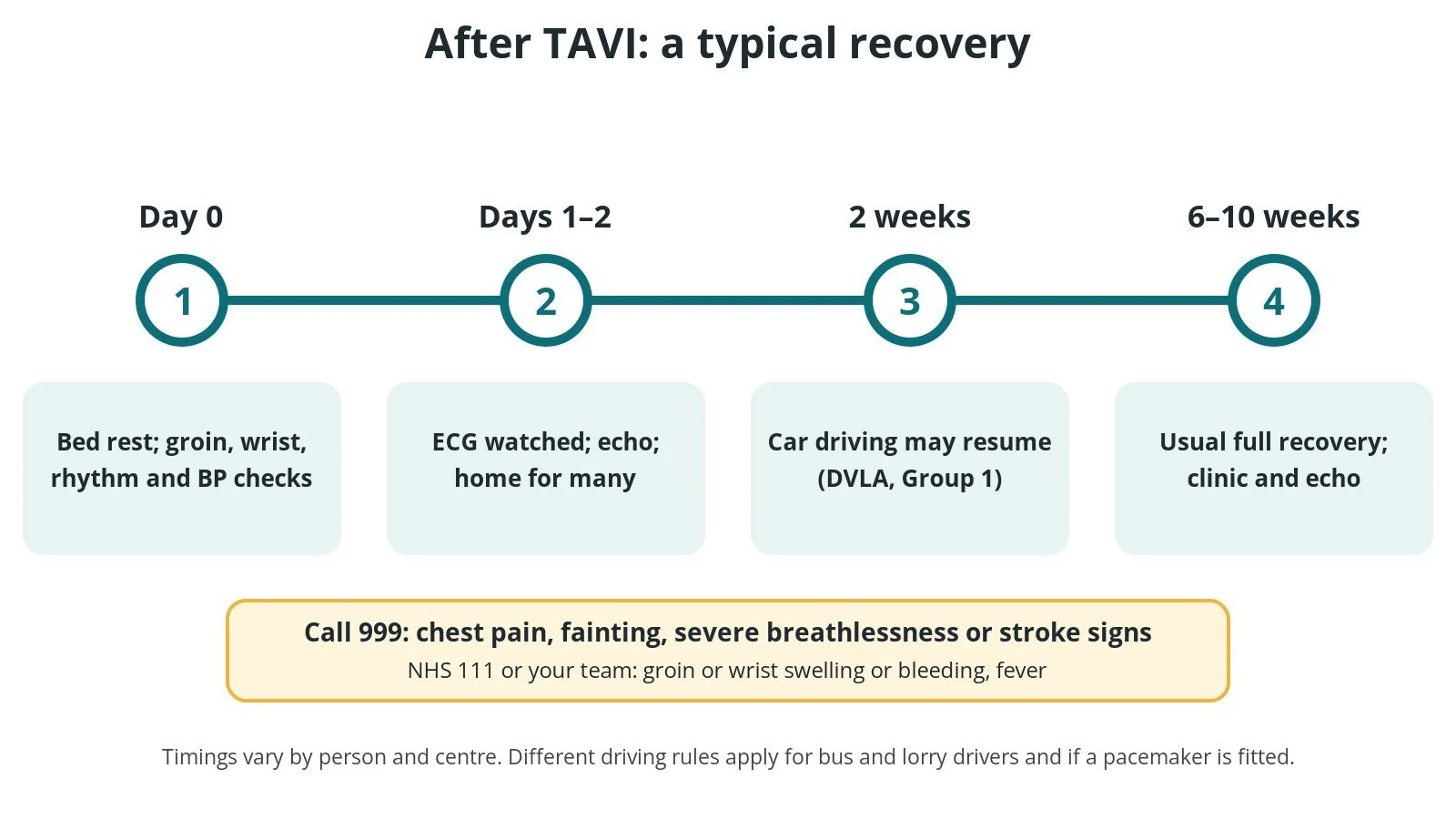
After TAVI: (1) bed rest and checks; (2) ECG watched, echo, home for many after 1 to 2 days; (3) car driving after 2 weeks (DVLA Group 1); (4) usual full recovery 6 to 10 weeks.
Risks and complications
TAVI avoids open-heart surgery, but it is still a major procedure. Possible problems include:
Bleeding or damage to the access artery (vascular complications), sometimes needing a stent, surgery or a transfusion
Needing a permanent pacemaker because of heart block. The chance depends on the person's existing ECG, their anatomy and the valve used
A leak around the new valve (paravalvular leak, PVL), usually small. A larger leak may need a balloon or another valve
Stroke or mini-stroke, from debris or clot released during the procedure
Blockage of a coronary artery by the old valve leaflets (rare, but serious, and planned for on CT)
Kidney injury from the contrast dye
Damage to the aortic root or valve ring, or fluid around the heart (tamponade) (rare)
The valve moving out of position, which may need a second valve
Infection of the new valve (endocarditis) later on
Death is uncommon
Your heart team will explain how these apply to you.
Cath lab kit list (printable)
Drugs, doses and dilutions: always as per centre protocol. Equipment, sizes and choices also vary by unit and operator. Device and product names below are generic. Always check the sheath, wire and closure required in each product's instructions for use.
Example: Barts Heart Centre kit list — always follow your own centre protocol
Typical setup for transfemoral TAVI under local anaesthetic and sedation, shared as one real-world example. It is not a protocol for your unit.
Scrub trolley
structural heart procedure pack and angiography pack
small femoral sheath for initial access, and a radial sheath for the pigtail, per operator
intermediate femoral sheath for pre-closure and exchange, per device and operator
valve delivery sheath, size per device and operator
pigtail catheters (long length for the radial route), per operator
Amplatz left (AL) shaped diagnostic catheter, or another catheter for crossing, per operator
standard and long exchange guidewires, straight-tip wire for crossing
pre-shaped extra-stiff LV guidewire
suture-based vascular closure device(s), with a plug-based closure device as back-up, per operator
radial compression band
syringes, and a contrast and saline mix for balloon inflation, as per centre protocol
valvuloplasty balloon, size per operator, if needed
Large back table
heparinised saline on a pressure bag with a drip stand, giving set and extension, as per centre protocol
haemostasis valve with Y-connector and extension tubing
large bowls for valve rinsing
chilled saline, if the valve's instructions for use require it
local anaesthetic, anticoagulation and emergency drugs, as per centre protocol
Valve preparation table
3-way taps and Luer-lock syringes for flushing and de-airing
rinse bowls of saline: number of rinses and timing per the valve's instructions for use
scissors and forceps
crimping or loading tools supplied with the valve
balloon-expandable valves: contrast and saline mix for the delivery balloon, as per centre protocol
self-expanding valves: loading per the instructions for use
Pacing
temporary pacing wire and venous sheath (femoral or jugular), or an external pacing lead for pacing through the LV guidewire, per operator
temporary pacing box, checked before the case
Contrast injector: flow rates and volumes per operator and centre protocol.
Alternative closure: large-bore closure device sized to the sheath, per device and operator, with its own wire and introducer.
Extra for some self-expanding systems or alternative access: longer large-bore sheaths, high-pressure injector lines, extra rinse bowls and warm saline on standby, per device and operator.
Procedural steps (Barts example)
Pacing plan agreed from the ECG: pacing through the LV wire, or a venous temporary wire if the QRS is broad
Small sheath to the right femoral artery; radial (or other femoral) sheath for the pigtail
Pigtail positioned in the aortic root for aortograms and pressure monitoring
Femoral angiogram to check the puncture and artery
Suture-based closure pre-placed; intermediate sheath inserted
Anticoagulation as per centre protocol
Stiff wire placed in the aorta; delivery sheath inserted under fluoroscopy
Valve crossed with a diagnostic catheter and straight wire; exchanged for a pre-shaped extra-stiff LV wire
Balloon valvuloplasty under rapid pacing if needed
Valve delivery system advanced; position checked on aortogram
Valve deployed with pacing as per operator; result checked (position, gradient, PVL, rhythm)
Delivery system removed; femoral artery closed; angiogram from the pigtail to check the groin
Radial sheath removed with a compression band; pacing wire removed or left in per ECG
End of case; transfer to recovery
First time in the cath lab?
💡 First-time tip
Know the pacing plan. Check before the case whether pacing is through the LV wire or a venous wire, test the pacing box, and know who calls the rapid-pacing start and stop. Watch the blood pressure during rapid pacing. It falls on purpose. Be ready for it to be slow to come back. Respect the valve preparation. Rinsing, crimping and loading are exact steps. Follow the instructions for use and don't rush. Keep the wire still. The stiff wire in the LV supports everything. Avoid knocking the table, and tell the operator if the wire moves. Watch the groin and the ECG at the end. Large-bore closure and new heart block are the most common things to catch early.
FAQ
What is the difference between TAVI and TAVR?
None. TAVI (implantation) is the usual term in the UK and Europe, and TAVR (replacement) in the US.
Is the old valve taken out?
No. The new valve is opened inside the old one, which is pressed back against the wall of the aorta.
Am I awake for it?
Often yes, with sedation and local anaesthetic, so you may feel drowsy but not pain. Some people have a general anaesthetic.
Why might I need a pacemaker?
The valve sits close to the heart's electrical wiring. Pressure from the new valve can slow the signals (heart block). Your team will check your ECG and fit a pacemaker if needed. See the Pacemaker guide.
How long does a TAVI valve last?
TAVI is still a newer treatment, and research on how long the valves last is ongoing. You will have regular echo checks. If a valve wears out, a second valve can sometimes be placed inside it.
Can I have an MRI scan afterwards?
Most TAVI valves are labelled as MR conditional, which means a scan is possible under set conditions. Always tell the scanning team about your valve and carry your implant card.
Related Cath Lab 101 guides
Sources
All checked on 10 October 2026. Each page's own date is shown where it gives one.
NICE. Heart valve disease presenting in adults: investigation and management (NG208). nice.org.uk/guidance/ng208
NICE. Transcatheter aortic valve implantation for aortic stenosis (IPG586). nice.org.uk/guidance/ipg586 (published 26 Jul 2017)
Praz F, et al. 2025 ESC/EACTS Guidelines for the management of valvular heart disease. Eur Heart J 2025;46(44):4635–4736. PubMed 40878295
Otto CM, et al. 2020 ACC/AHA Guideline for the Management of Patients With Valvular Heart Disease. J Am Coll Cardiol 2021;77(4):e25–e197. PubMed 33342586
NHS. Heart valve replacement. nhs.uk/conditions/aortic-valve-replacement
NHS. Heart valve disease. nhs.uk/conditions/heart-valve-disease
British Heart Foundation. TAVI (transcatheter aortic valve implantation) procedure. bhf.org.uk
DVLA / GOV.UK. Cardiovascular disorders: assessing fitness to drive. gov.uk (last updated 7 Nov 2025)
Disclaimer
Education only. This page is for general education for healthcare students, cath lab staff and curious readers. It isn't medical advice and doesn't replace advice from your own doctor, heart team or local unit protocols. Equipment and medicines are described generically. No product or manufacturer is recommended. If you think someone is having a heart emergency, call 999.
Disclosure: I work for a company that makes a device used in this procedure. This page is independent education, isn't sponsored or reviewed by any company, and doesn't recommend any product.
All diagrams on this page are original illustrations created for rodnieoro.com.