Tricuspid TEER (transcatheter edge-to-edge repair): a Cath Lab 101 guide
Reviewed by Rodnie Oro, cath lab charge nurse · Last reviewed: October 2026 · Education only, not medical advice
Tricuspid TEER in one minute
What it is: tricuspid TEER (transcatheter edge-to-edge repair) is a keyhole (catheter) treatment for a leaking tricuspid valve, the valve between the right atrium and right ventricle. A small clip is passed up a vein from the groin and used to hold the edges of two valve leaflets together, so the valve closes better and less blood leaks backwards.
Who it is for: adults with severe tricuspid regurgitation (TR) who still have symptoms, such as swollen legs and tummy, breathlessness or tiredness, despite medicines, and who are at high risk from open-heart surgery. A specialist heart team decides who is suitable.
Main risks: bleeding at the groin, a clip coming loose from one leaflet, damage to the valve, fluid around the heart, and the leak not being reduced enough. Your heart team will explain your own risk.
Usually: under general anaesthetic or deep sedation, with a heart scan through the food pipe (TOE) guiding each step, about 1 to 3 hours, then home after 1 to 3 days.
On this page: What is tricuspid TEER? · Who is it for? · When is it done? · Step by step · Recovery · Risks · Kit list · First time in the lab? · FAQ
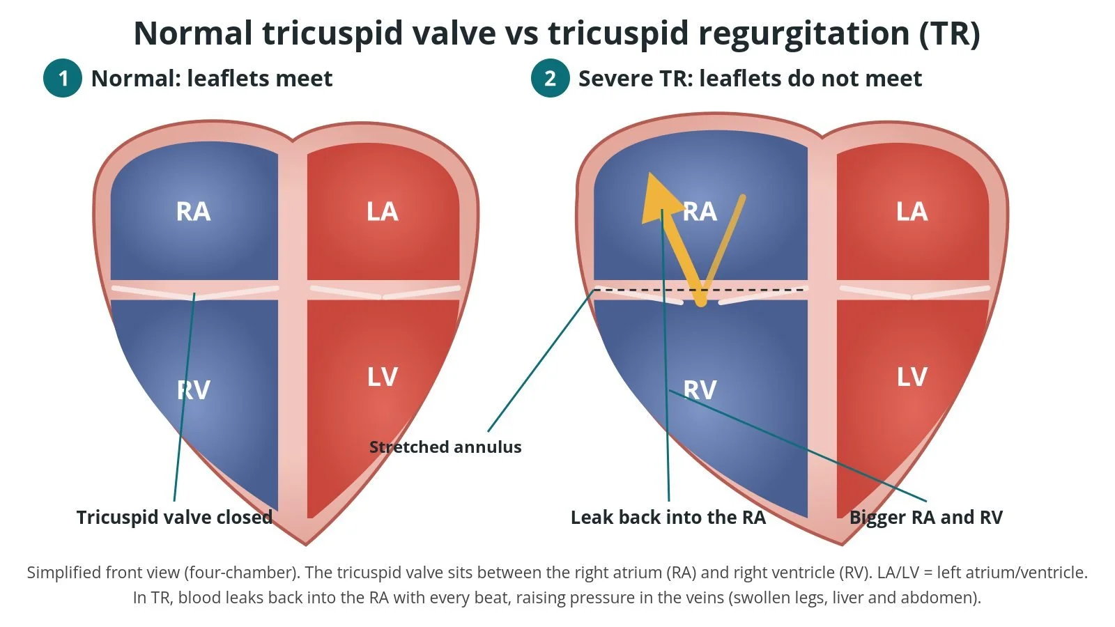
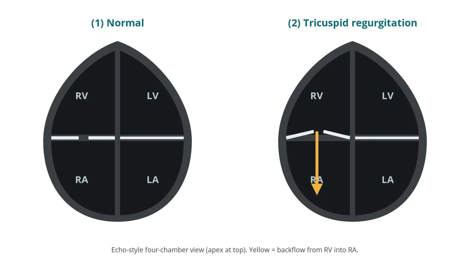
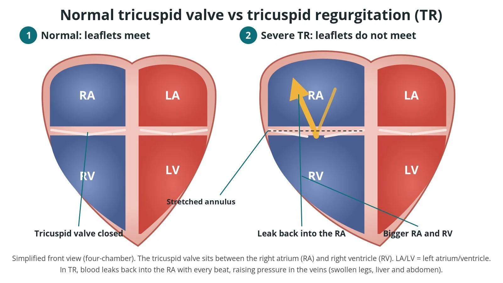
Normal vs tricuspid regurgitation (TR). (1) Normal: the leaflets meet. (2) Severe TR: the ring is stretched, the leaflets don't meet, and blood leaks back into the right atrium (RA).
What is tricuspid TEER?
The tricuspid valve sits between the right atrium (RA), which collects blood returning from the body, and the right ventricle (RV), which pumps it to the lungs. It usually has three leaflets: anterior, septal and posterior. When the RV squeezes, the leaflets meet and stop blood going backwards.
In tricuspid regurgitation (TR) the leaflets no longer meet properly, so blood leaks back into the right atrium with every beat. Most TR is secondary (functional): the valve itself is normal, but the ring it sits in (the annulus) and the right heart have stretched. Common causes are long-standing atrial fibrillation, left-sided heart or valve disease, and high pressure in the lungs. Less often the leaflets themselves are damaged, or a pacemaker or defibrillator lead crossing the valve gets in the way of it closing.
Severe TR raises the pressure in the veins that drain into the right heart. Fluid builds up in the legs, abdomen and liver, and people often feel tired and breathless. Water tablets (diuretics) ease the fluid but do not fix the leak.
Tricuspid TEER repairs the valve from the inside. A steerable tube is passed up the femoral vein, through the inferior vena cava (IVC) and into the right atrium. A clip on the end of a delivery system is lined up over the leak, its arms grab the edges of two leaflets, and it closes them together. One or more clips can be placed. The valve then works as a double (or triple) opening with a much smaller leak.
Other options include valve surgery, and other catheter treatments such as a transcatheter tricuspid valve replacement. The heart team chooses based on the valve's anatomy and the person's overall health.
New to the acronyms? See the Cath Lab Abbreviations A–Z.
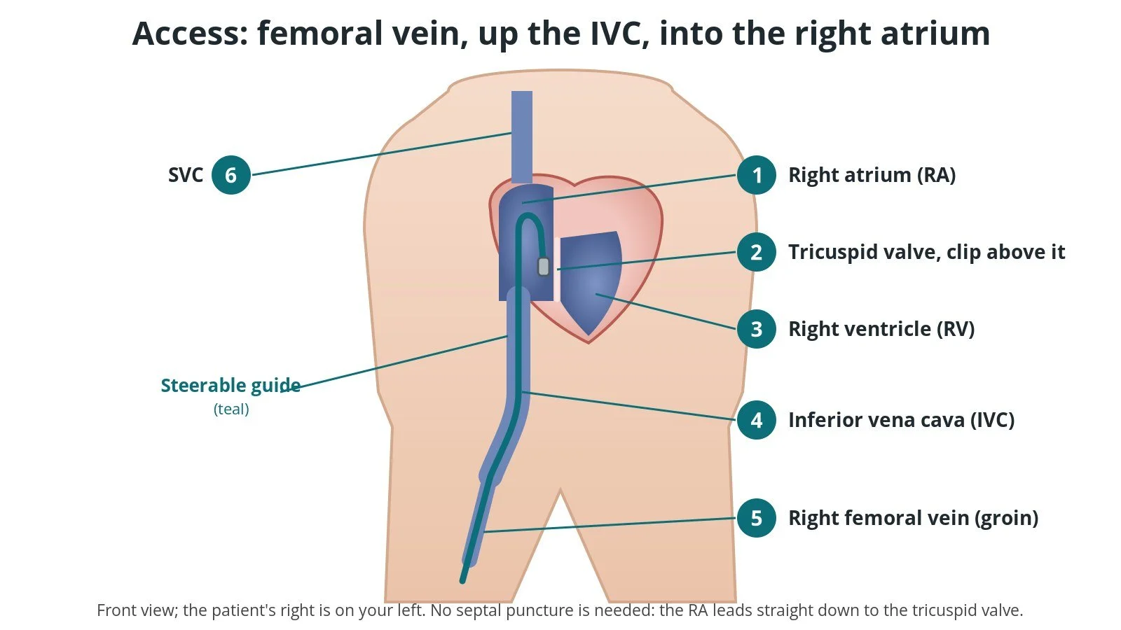
Access: (1) right atrium; (2) the clip above the tricuspid valve; (3) right ventricle; (4) inferior vena cava (IVC); (5) right femoral vein; (6) superior vena cava (SVC). No septal puncture is needed.
Who is it for?
The decision is made by a specialist heart team after a full assessment. European (ESC/EACTS 2025, updating 2021) and American (AHA/ACC 2020) guidelines broadly agree that:
intervention is considered for severe TR with symptoms despite medicines, after the team has looked at the cause of the leak, the right ventricle's function, the lung pressures and the risk of surgery
a transcatheter treatment can be considered in people at high risk from surgery, provided the right ventricle is not severely weakened and there is no severe pre-capillary pulmonary hypertension (ESC/EACTS 2025)
the treatment is done in a heart valve centre with an experienced team
It may be less suitable when the leaflet gap is very wide, the leaflets are short or thickened, the echo pictures of the valve are poor, or a device lead is pinning a leaflet.
NICE (IPG731, now HTG632) says that for severe, symptomatic TR, transcatheter tricuspid leaflet repair should only be used with special arrangements for clinical governance, consent and audit or research, because evidence on how well it works is still limited and there are serious but well-recognised complications. For mild or moderate TR it should only be used in research.
When is it done?
Tricuspid TEER is a planned procedure. Before it, the team usually:
images the valve with a heart scan through the chest (TTE) and through the food pipe (TOE) to check the cause of the leak, the leaflets, the gap between them and whether good pictures are possible during the procedure
may do heart CT or MRI and a right heart catheter to measure the lung pressures
reviews the ECG and any pacemaker or ICD, and checks the leads
checks bloods, kidney and liver function and medicines, including blood thinners, as per centre protocol
optimises fluid with diuretics where possible
Step by step
1. Anaesthetic and imaging set-up
The patient is usually under general anaesthetic so that a TOE probe can stay in place throughout. Some centres use deep sedation, and some use a scan from inside the heart (intracardiac echo, ICE) as well or instead. A urinary catheter and arterial line are often placed, as per centre protocol. Clear echo pictures are essential: the procedure is guided mainly by echo, with X-ray (fluoroscopy) in support.
2. Femoral vein access
The operator puts a needle into the femoral vein in the groin, usually the right, guided by ultrasound. A suture-based closure device may be pre-placed. The vein is then stretched with dilators so it can take the large steerable guide. Sheath and dilator sizes are per device and operator. Anticoagulation is given as per centre protocol.
3. Steering into the right atrium
A stiff wire is passed up the IVC into the right atrium or the superior vena cava, and the steerable guide is advanced over it into the right atrium. There is no need to cross the septum, unlike mitral TEER. The clip delivery system goes through the guide, and the operator uses its dials to bend and steer the clip down towards the tricuspid valve, from above.
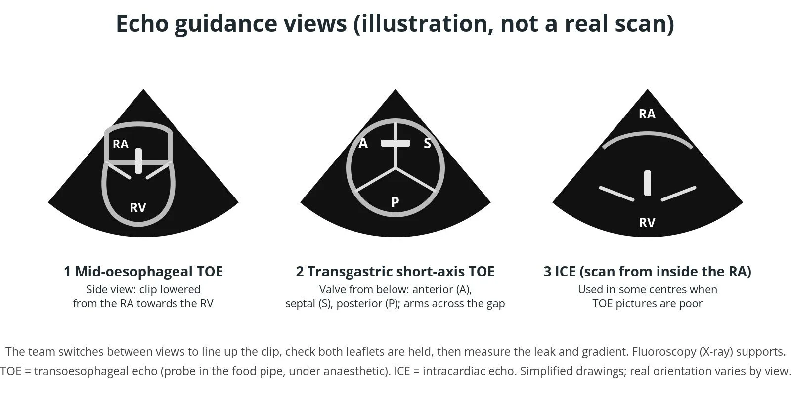
Illustrated echo, not real scans. (1) Mid-oesophageal TOE side view; (2) transgastric short-axis view of the anterior (A), septal (S) and posterior (P) leaflets; (3) intracardiac echo (ICE), used in some centres.
4. Aligning, grasping and closing
Using several echo views, the team:
A. Steers the clip above the leak, often near where the anterior and septal leaflets meet
B. Opens the arms and turns the clip so they sit at right angles to the line where the leaflets meet
C. Advances the clip into the RV, pulls it back gently and grasps the edges of two leaflets, checking on echo that both are held well
D. Closes the clip, checks the leak and the pressure across the valve, and releases it if the result is good
If the grasp is poor, the clip can be opened and repositioned before release. Often a second or third clip is added beside the first until the leak is small enough without narrowing the valve too much.
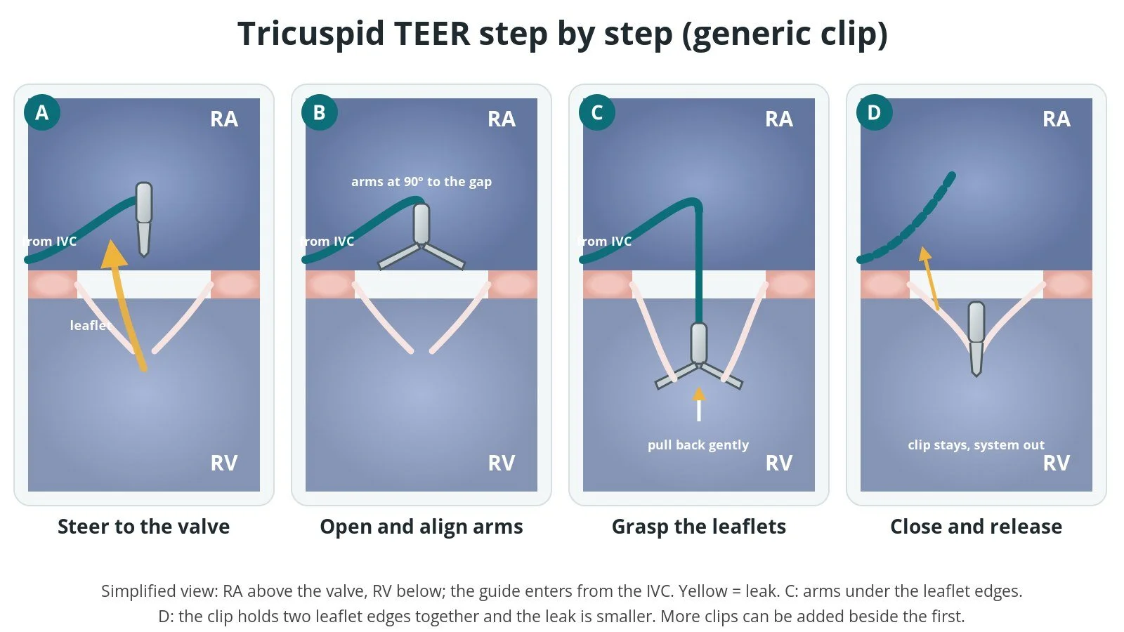
A: the clip is steered above the leak. B: the arms open at right angles to the gap. C: the clip goes into the RV and grasps two leaflet edges. D: the clip is closed and released; the leak is smaller.
5. Checking the result and finishing
Echo shows the size of the remaining leak and the pressure difference (gradient) across the valve, to make sure it has not been narrowed too much. The delivery system and guide are removed, and the groin vein is closed with a pre-placed suture device or a figure-of-eight stitch, per operator. The patient wakes up in recovery or goes to a monitored bed.
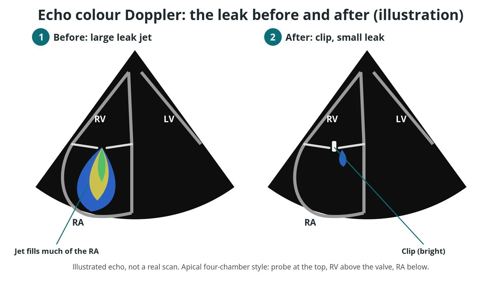
Illustrated echo, not a real scan. Before: a large colour jet of blood leaking back into the RA. After: the clip holds the leaflets and the jet is much smaller.
Recovery and aftercare
First hours. Bed rest, with checks of the groin, blood pressure and heart rhythm, as per centre protocol.
Echo. A scan, usually the next day, checks the clip or clips and the leak before going home.
Going home. Many people go home after 1 to 3 days.
Groin. Avoid heavy lifting and strenuous exercise for about a week, or as your team advises. Keep the site clean and dry.
Medicines. Your team will tell you which blood thinners to take and for how long. Diuretics are often reviewed and sometimes reduced as the swelling settles.
Follow-up. A clinic review and echo, often at about a month and then yearly, as per centre protocol. Improvement in symptoms and energy can build over weeks to months.
Driving (UK). Ask your team when you can drive. Check the current GOV.UK guidance.
Contact your team or NHS 111 if the groin swells, bleeds or becomes painful, or if your ankle swelling or breathlessness gets worse. Call 999 for chest pain, fainting or severe breathlessness.
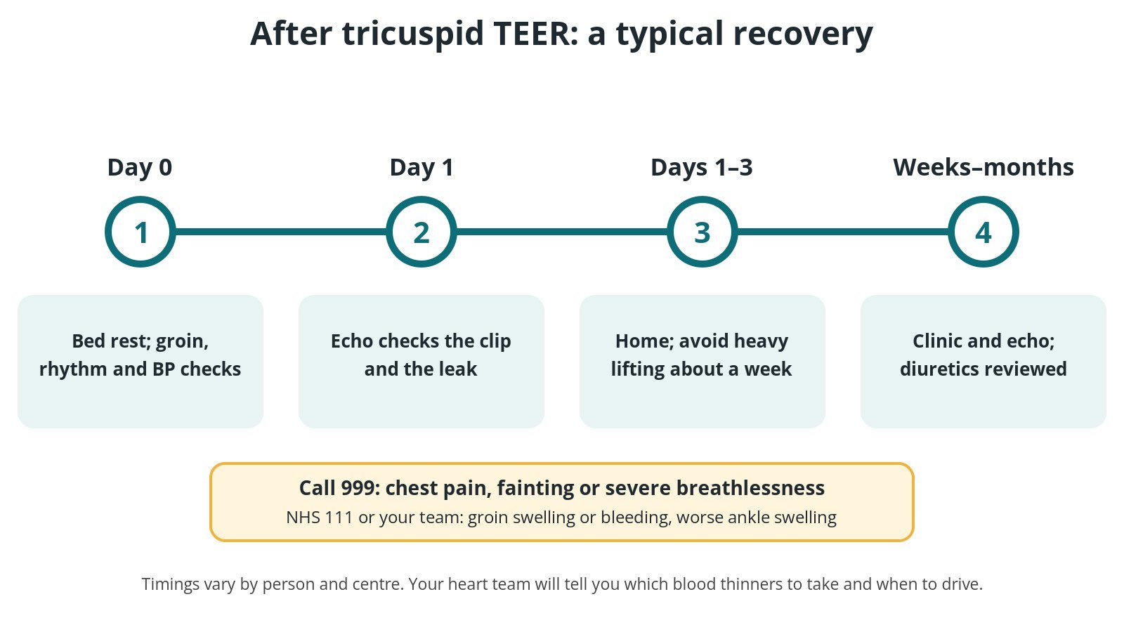
After tricuspid TEER: (1) bed rest and checks; (2) echo the next day; (3) home after 1 to 3 days; (4) clinic and echo, with diuretics reviewed.
Risks and complications
Tricuspid TEER avoids open-heart surgery, but it has risks. NICE describes them as serious but well recognised. Possible problems include:
Bleeding or bruising at the groin, sometimes needing a transfusion
A clip coming loose from one leaflet (single leaflet device attachment), which may need another clip or other treatment
Leaflet damage or tears
Narrowing of the valve (tricuspid stenosis) if too much of the opening is closed
The leak not being reduced enough, or coming back over time
Fluid around the heart (tamponade) or injury to the heart or veins
Heart rhythm problems, including slow rhythms (uncommon)
Interference with a pacemaker or defibrillator lead
Stroke or death are rare
Your heart team will explain how these apply to you.
Cath lab kit list (printable)
Drugs, doses and dilutions: always as per centre protocol. Equipment, sizes and choices also vary by unit and operator. Device and product names below are generic. Always check the sheath, wire and closure required in each product's instructions for use.
Example: Barts Heart Centre kit list — always follow your own centre protocol
Typical setup for tricuspid TEER under general anaesthetic with TOE guidance, shared as one real-world example. It is not a protocol for your unit.
Device and set-up
steerable guide catheter
clip delivery system (clip size chosen for the anatomy, per operator)
device stabiliser, support plate and height-adjustable stool or booster
distance from mid-chest to the stool measured before set-up (85 cm in this example)
large back table for device preparation
Flush and preparation
3-way taps and needle-free Luer access devices
heparinised saline flush bags, prepared as per centre protocol, with giving sets and pressure bags
large Luer-lock syringes and a drip stand
sterile water for device preparation, as per the instructions for use
Scrub trolley
structural heart procedure pack
femoral access needle
femoral venous sheaths for access and pre-dilation, per device and operator
chlorhexidine skin preparation applicators
pressure lines
diagnostic catheter (right Judkins shape in this example), per operator
extra-stiff exchange guidewire, tip shape per device and operator
sterile ultrasound probe cover and saline for ultrasound-guided access
suture-based vascular closure devices (two in this example), or a figure-of-eight suture, per operator
sharps and needle-count block
Medications (scrub nurse): anticoagulation and local anaesthetic, as per centre protocol. Anaesthetic drugs are managed by the anaesthetic team.
Procedural steps (Barts example)
General anaesthetic and TOE probe placed; baseline echo of the valve and leak
Ultrasound-guided femoral vein access; closure sutures pre-placed per operator
Anticoagulation as per centre protocol
Stiff wire to the right atrium or SVC; vein dilated; steerable guide advanced to the right atrium
Clip delivery system prepared, flushed and introduced; stabiliser set at the measured height
Clip steered over the leak; arms opened and aligned at right angles to the leaflet edges
Clip advanced into the RV, pulled back to grasp two leaflets; grasp checked on echo
Clip closed; leak and gradient checked; clip released if the result is good
Repeat for further clips if needed
System and guide removed; groin closed; patient woken and transferred
First time in the cath lab?
💡 First-time tip
Watch the echo screen. Tricuspid TEER is guided mostly by echo. Knowing which view is on screen helps you follow each step. Respect the device preparation. Flushing and de-airing the guide and delivery system is slow and exact. Follow the instructions for use and don't rush it. Keep the stabiliser steady. Once the system is on the stabiliser, avoid knocking the table or the plate. Small movements change the clip's position in the heart. Check for device leads. If the patient has a pacemaker or ICD, know where the leads are and whether the device team needs to be on hand. Be ready for bleeding. The venous sheath is large. Have closure devices, pressure and a plan for the groin ready before the guide comes out.
FAQ
What does TEER stand for?
Transcatheter edge-to-edge repair: a catheter treatment that holds the edges of two valve leaflets together with a clip. It is used for both the mitral and tricuspid valves.
Is it the same as mitral TEER?
The idea is the same, but the route is different. For the mitral valve the device has to cross the wall between the two upper chambers (a transseptal puncture). For the tricuspid valve it goes straight from the IVC into the right atrium, with no septal puncture. See the mitral TEER guide.
Am I awake for it?
Usually not. Most centres use a general anaesthetic because the TOE probe stays in the food pipe for the whole procedure.
Will I feel better straight away?
Some people notice less swelling and more energy within weeks, as fluid comes off. For others it is more gradual. The leak is reduced, not always removed completely.
Can I still have a pacemaker or ICD?
Yes. Existing leads are checked before the procedure. If a new device is needed later, the team will plan how the leads should cross the repaired valve. See the Pacemaker guide.
Can I have an MRI scan afterwards?
Many valve clips are labelled as MR conditional, which means a scan is possible under set conditions. Always tell the scanning team about your clip and carry your implant card.
Related Cath Lab 101 guides
Sources
All checked on 10 October 2026. Each page's own date is shown where it gives one.
NICE. Transcatheter tricuspid valve leaflet repair for tricuspid regurgitation (HTG632, formerly interventional procedures guidance IPG731). nice.org.uk/guidance/htg632 (published 27 Jul 2022; moved to HealthTech guidance with the recommendations unchanged)
Praz F, et al. 2025 ESC/EACTS Guidelines for the management of valvular heart disease. Eur Heart J 2025;46(44):4635–4736. PubMed 40878295
Vahanian A, et al. 2021 ESC/EACTS Guidelines for the management of valvular heart disease. Eur Heart J 2022;43(7):561–632. PubMed 34453165
Otto CM, et al. 2020 ACC/AHA Guideline for the Management of Patients With Valvular Heart Disease. J Am Coll Cardiol 2021;77(4):e25–e197. PubMed 33342586
NHS. Heart valve disease. nhs.uk/conditions/heart-valve-disease (page last reviewed 22 Jan 2025)
British Heart Foundation. Heart valve disease. bhf.org.uk (published 5 Mar 2024)
GOV.UK. Health conditions and driving. gov.uk/health-conditions-and-driving
Disclaimer
Education only. This page is for general education for healthcare students, cath lab staff and curious readers. It isn't medical advice and doesn't replace advice from your own doctor, heart team or local unit protocols. Equipment and medicines are described generically. No product or manufacturer is recommended. If you think someone is having a heart emergency, call 999.
Disclosure: I work for a company that makes a device used in this procedure. This page is independent education, isn't sponsored or reviewed by any company, and doesn't recommend any product.
All diagrams on this page are original illustrations created for rodnieoro.com.