Catheter-directed therapy for pulmonary embolism: a Cath Lab 101 guide
Reviewed by Rodnie Oro, cath lab charge nurse · Last reviewed: November 2026 · Education only, not medical advice
Catheter-directed therapy for pulmonary embolism in one minute
What it is: A pulmonary embolism (PE) is a blood clot blocking the lung arteries. Catheter-directed therapy treats the clot from inside: thin catheters from a vein in the groin either drip a clot-busting drug straight into the clot, or a wider catheter sucks the clot out.
Who it is for: Selected adults with a large PE that is straining the right side of the heart (intermediate-high risk) or causing low blood pressure (high risk), especially when a full dose of clot-busting drug into a vein is too risky. A PE response team decides.
Main risks: Bleeding at the groin or elsewhere (rarely in the brain), heart rhythm problems, damage to the lung artery, and blood loss with suction devices. Your team will explain your own risk.
Usually: Under local anaesthetic, with light sedation only if needed, about 1 to 2 hours in the lab. With an infusion, the catheters stay in for a set time, as per centre protocol, on a coronary care or high-dependency unit.
On this page: What is catheter-directed therapy for pulmonary embolism? · Who is it for? · When is it done? · Step by step · Recovery · Risks · Kit list · First time in the lab? · FAQ
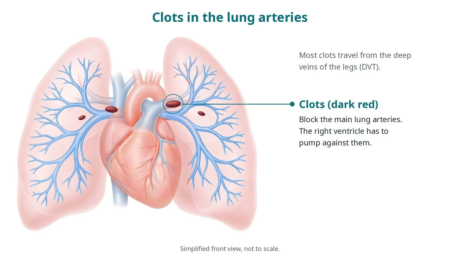
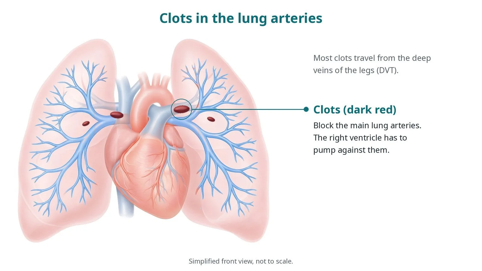
Clots (dark red) block the main lung arteries, and the right ventricle has to pump against them. Most clots travel from the deep veins of the legs.
What is catheter-directed therapy for pulmonary embolism?
Blood returning from the body passes through the right atrium and right ventricle into the pulmonary artery, which divides into right and left branches to the lungs. Most pulmonary emboli start as a clot in the deep veins of the leg or pelvis (DVT) that breaks off, travels through the right heart and lodges in the lung arteries.
A large clot raises the pressure the right ventricle has to pump against. The right ventricle is thin-walled and copes badly with a sudden rise: it stretches, weakens and can fail, and the blood pressure can fall. This right ventricular strain, seen on CT or echo and in heart blood tests (troponin, BNP), is what makes a PE intermediate-high or high risk.
Blood thinners (anticoagulation) stop the clot growing while the body breaks it down. Catheter-directed therapy aims to clear the clot faster and relieve the right ventricle. There are two main approaches: catheter-directed thrombolysis, where a low dose of clot-busting drug (such as alteplase) runs through a catheter with many side holes lying in the clot, sometimes with ultrasound to help the drug in; and mechanical aspiration thrombectomy, where a large-bore catheter pulls the clot out, often with no clot-busting drug at all.
New to the acronyms? See the Cath Lab Abbreviations A–Z.
Who is it for?
The decision is made by a PE response team, which may include acute or respiratory physicians, cardiologists, interventional radiologists, intensivists and haematologists. It may be considered for:
intermediate-high-risk PE (right ventricle strain on CT or echo and a raised troponin), especially if the patient is getting worse on blood thinners
high-risk PE (low blood pressure or shock) when a full dose of clot-busting drug into a vein is not suitable or has not worked
people at higher risk of bleeding with a full-dose clot-busting drug
Who it may not suit: low-risk PE, which is treated with blood thinners alone, often at home people with recent bleeding in the brain, brain surgery or major trauma, for whom any clot-busting drug is too risky (aspiration without a drug may still be possible) old, firm clot, or anatomy the catheters cannot reach safely
The decision is made by your heart team, who will talk through the reasons, the alternatives and the risks with you.
When is it done?
gives you a chance to ask questions and give consent
arranges blood tests, including kidney function and a blood count, and an ECG
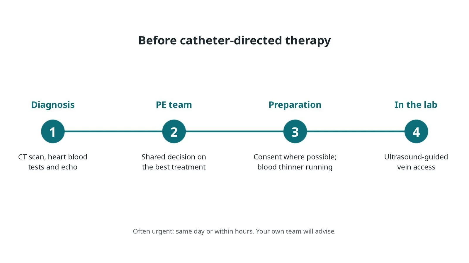
Before catheter-directed therapy: CT scan, heart blood tests and echo; a PE team decision; consent where possible with a blood thinner running; ultrasound-guided vein access.
Step by step
1. Getting ready
The patient is often unwell: breathless, with a fast heart rate and low oxygen levels, so the team works calmly and quickly. Monitoring includes ECG, blood pressure (often an arterial line) and oxygen levels, with defibrillator pads on. Oxygen is given. Sedation is kept light, because it can drop the blood pressure when the right ventricle is strained.
Watch: Falling blood pressure or oxygen levels, or a slowing heart rate: tell the operator at once, as the right ventricle may be failing, and call for senior and anaesthetic help as per centre protocol.
2. Access
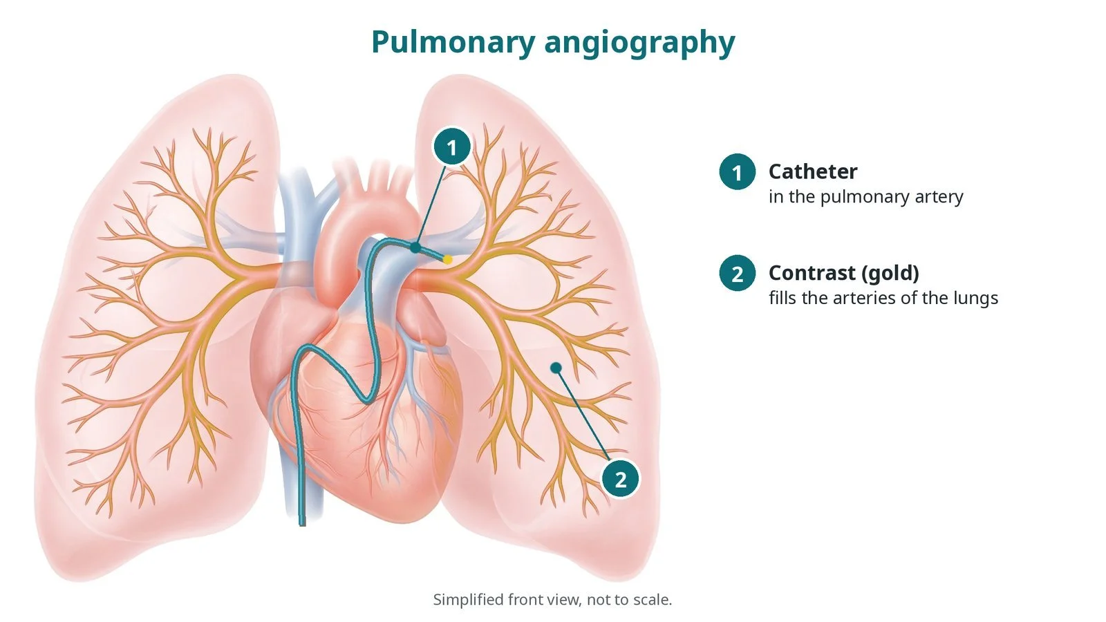
A sheath is placed in the femoral vein in the groin under ultrasound; sometimes the jugular vein in the neck is used. Sizes are per device and operator. Heparin is given, as per centre protocol. A catheter is passed through the right atrium and right ventricle into the pulmonary artery. Pressures are measured and a pulmonary angiogram may be taken to show the clot.
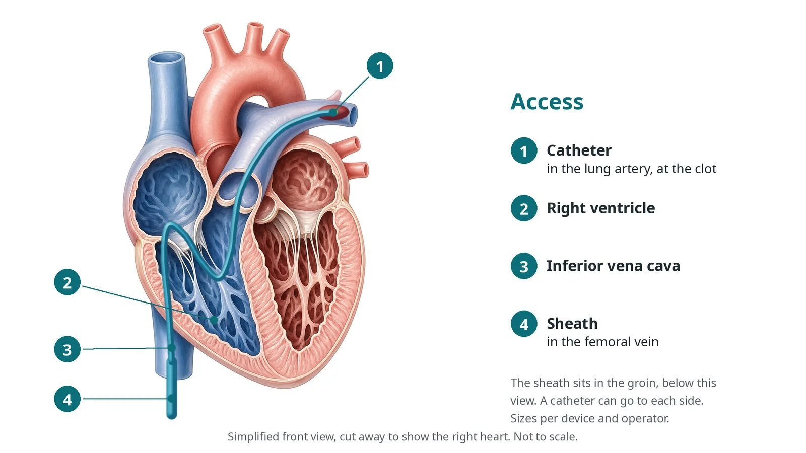
Access: (1) catheter in the lung artery, at the clot; (2) right ventricle; (3) inferior vena cava; (4) sheath in the femoral vein.
3. Treating the clot
A. To the clot. A wire and catheter are steered into the right and/or left pulmonary artery, into the clot.
B. Clot-busting infusion. For catheter-directed thrombolysis, an infusion catheter with many side holes is left lying in the clot, one in each lung if both sides are affected. A low dose of clot-busting drug, such as alteplase, runs slowly through it; dose and duration are as per centre protocol. Ultrasound-facilitated catheters add an ultrasound core to help the drug into the clot.
C. Or suction. For mechanical thrombectomy, a large-bore catheter is placed against the clot and suction pulls it out into a syringe or canister. Several passes may be needed. Blood comes out with the clot, so the amount is measured; some systems return filtered blood.
D. Check. Pulmonary artery pressures, oxygen levels and heart rate are checked again, and a final angiogram may be taken.
Watch: A sudden drop in blood pressure or a slow heart rate during aspiration or wire work: say so straight away; the operator may pause, and fluids, medicines to support the blood pressure or resuscitation may be needed, as per centre protocol.
4. Finishing
After aspiration, the sheath is removed and the vein is closed with pressure or a stitch, per operator. After setting up thrombolysis, the infusion catheters are secured and connected to labelled infusion pumps, and the patient goes to a coronary care or high-dependency unit with the catheters in place.
5. Infusion care and catheter removal
During the infusion, nurses check the access site, signs of bleeding (gums, urine, stools, puncture sites), neurological status and observations, as per centre protocol, and blood tests may be taken. Heparin usually continues alongside. When the infusion finishes, the catheters and sheath are removed and firm pressure is applied. Blood thinners then continue.
Watch: A new headache, confusion, weakness or drowsiness during the infusion: stop the clot-busting drug and get urgent medical review, as per centre protocol.
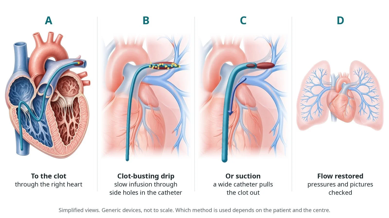
A: catheter to the clot through the right heart. B: a slow clot-busting infusion through side holes. C: or a wide catheter sucks the clot out. D: flow restored and checked.
Recovery and aftercare
First hours: care on a coronary care, high-dependency or intensive care unit, with checks of the access site, bleeding, oxygen and blood pressure, as per centre protocol.
Infusion: if a clot-busting infusion is running, you rest in bed with the catheters in place until it finishes; they are then removed and the site pressed.
Going home: usually after a few days, once breathing, oxygen levels and the access site have settled.
Medicines: blood thinners continue for at least 3 months, often longer depending on the cause; your team will explain which one and for how long.
Activity and driving: build up gently, as breathlessness and tiredness can take weeks to improve; ask your team before driving.
Follow-up: clinic review, often with an echo; ongoing breathlessness is checked for raised lung pressures.
Call 999 if you have worsening breathlessness, chest pain, fainting, coughing up blood, heavy bleeding, or signs of a stroke.
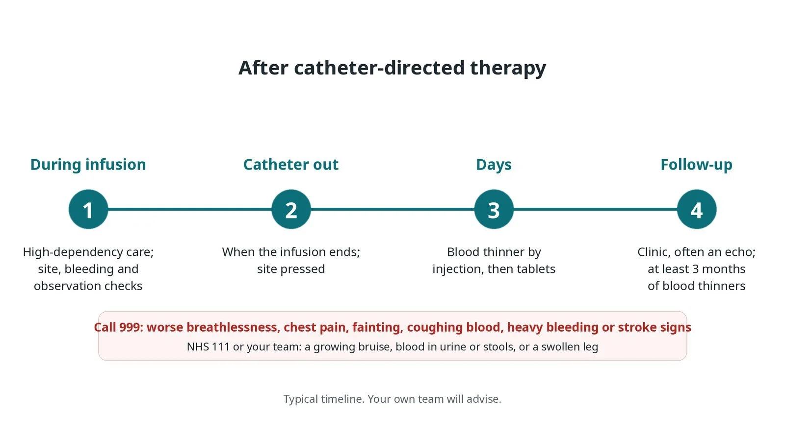
After catheter-directed therapy: (1) high-dependency care during the infusion; (2) catheter out when it ends; (3) blood thinner by injection, then tablets; (4) clinic follow-up, often an echo.
Risks and complications
Your team will explain the risks that apply to you. Possible problems include:
Bleeding at the access site or elsewhere (uncommon): More likely with clot-busting drugs, so it is checked closely during the infusion.
Bleeding in the brain (rare): The most serious risk of clot-busting drugs.
Heart rhythm problems as catheters cross the right heart
Blood loss with suction devices, sometimes needing a transfusion
Low blood pressure or collapse during the procedure (uncommon)
Damage to the pulmonary artery, such as a tear (rare)
Kidney strain or a reaction to the contrast dye
Cath lab kit list (printable)
Barts example kit list
An example from one UK centre, written generically. Every lab has its own protocol, and sizes and doses vary.
ultrasound machine and sterile probe cover for vein access
venous sheaths, size per device and operator
guidewires, including exchange-length and stiff wires, per operator
balloon-tipped or angled catheter for pressures and pulmonary angiography
multi-side-hole infusion catheters for thrombolysis, length per patient and operator
ultrasound-facilitated thrombolysis catheter and control unit, if used
large-bore aspiration thrombectomy catheter with syringe or vacuum canister, per device
pressure transducer, contrast and arterial line
clot-busting drug, heparin and infusion pumps, as per centre protocol
defibrillator pads and resuscitation equipment
Pressure transducer.
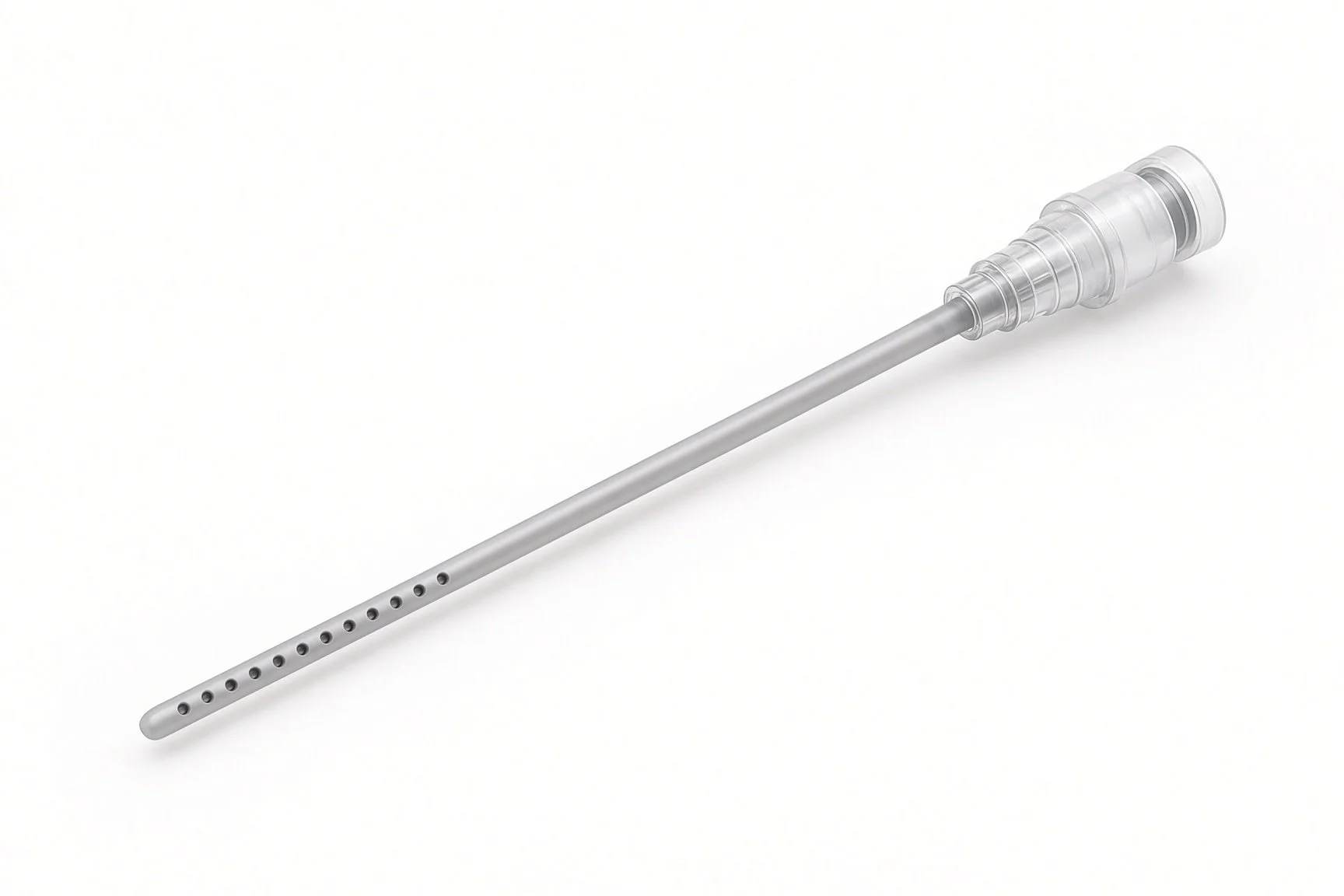
Multi-side-hole infusion catheter.
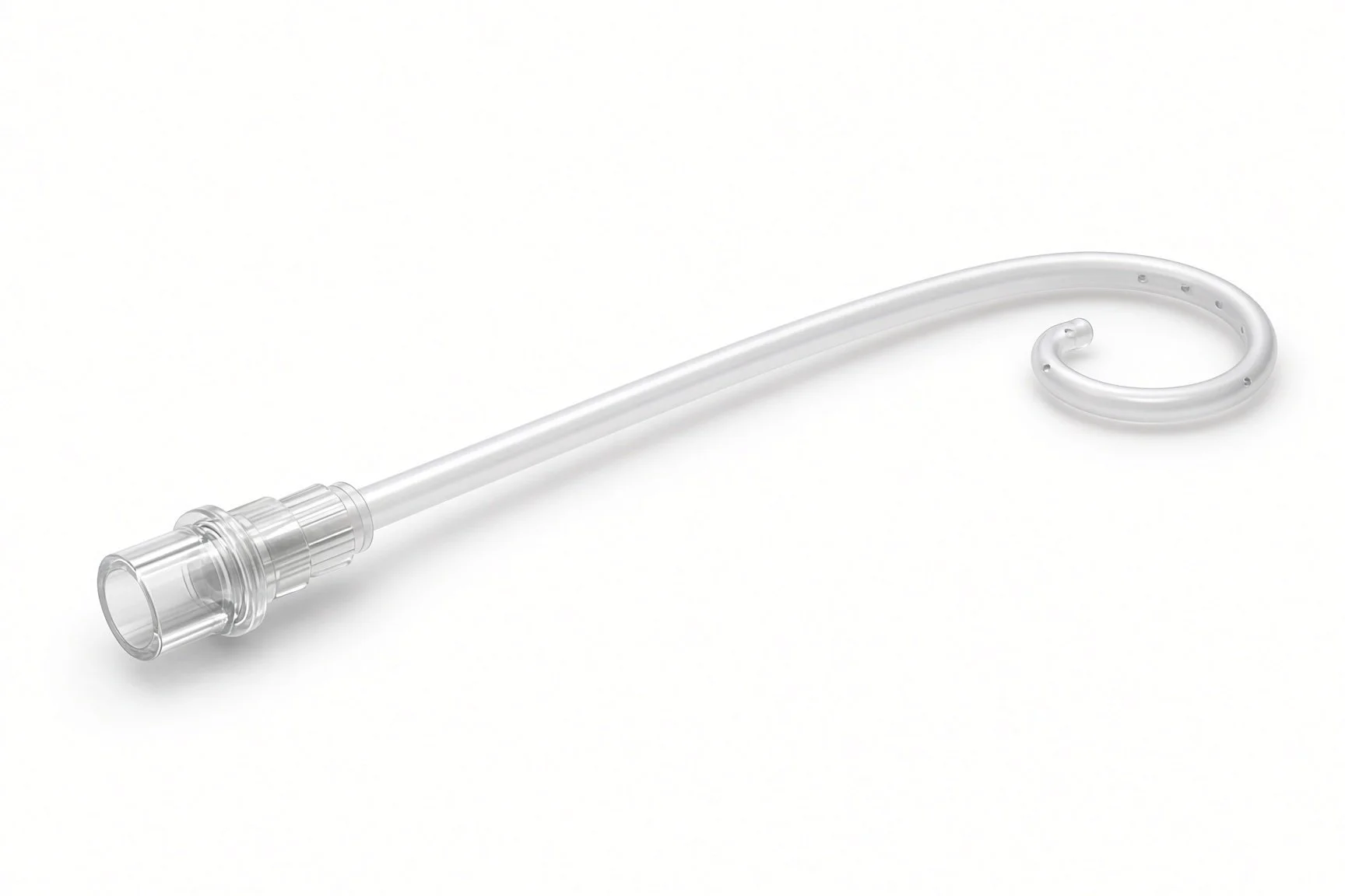
Balloon-tipped or pigtail catheter.
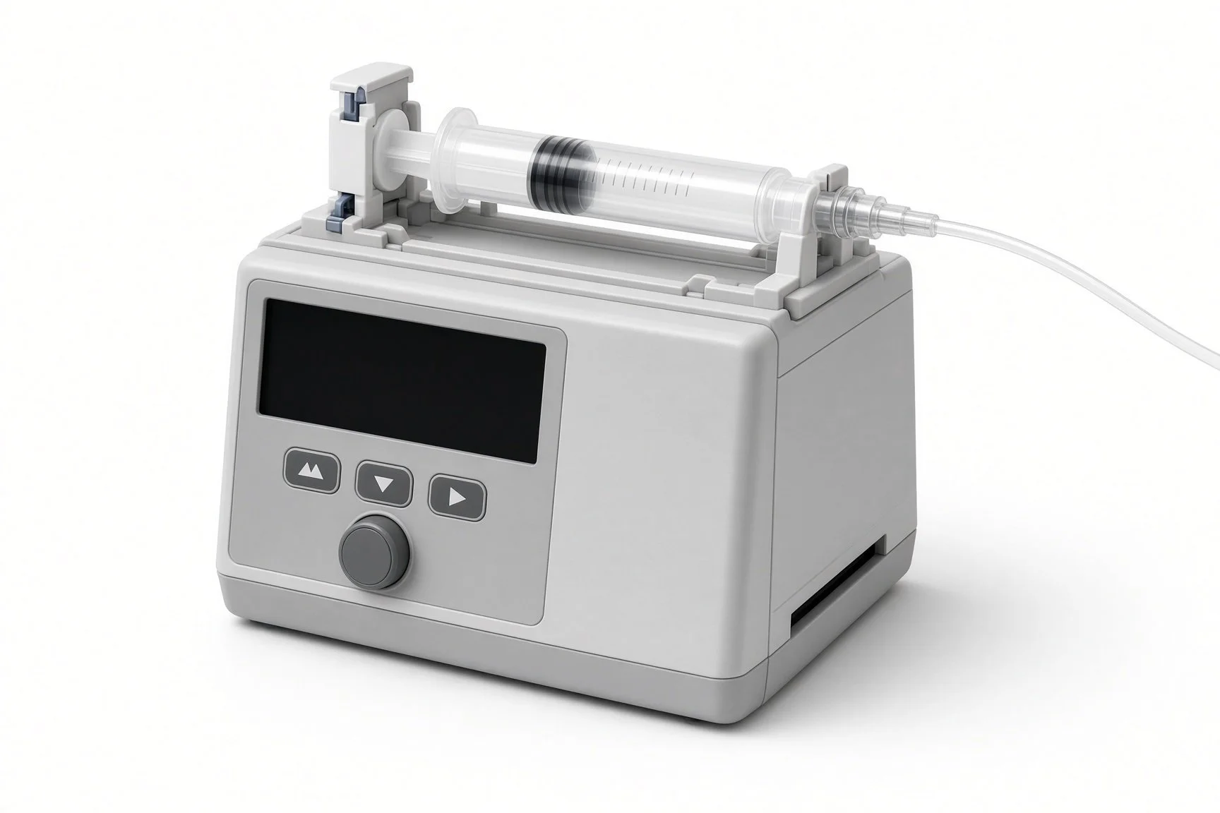
Infusion pumps.
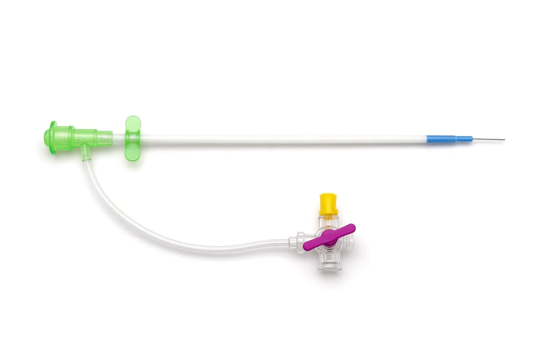
Venous sheath and ultrasound.
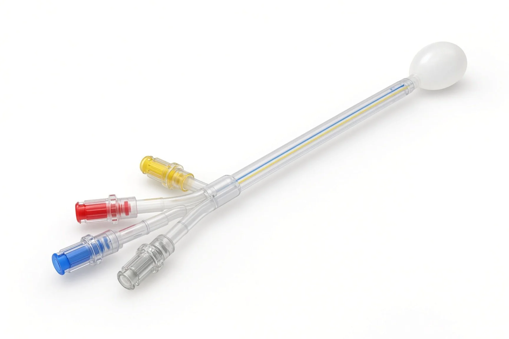
Balloon-tipped or pigtail catheter.
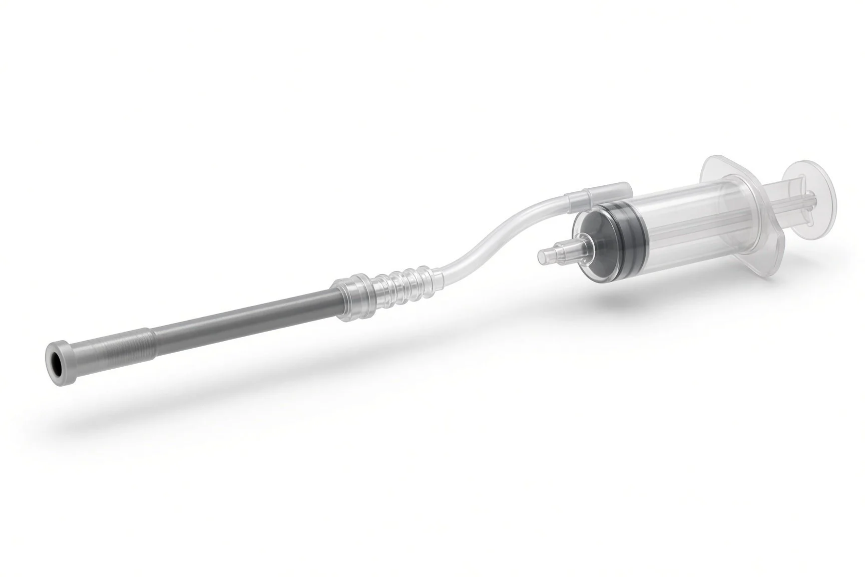
Large-bore aspiration catheter.
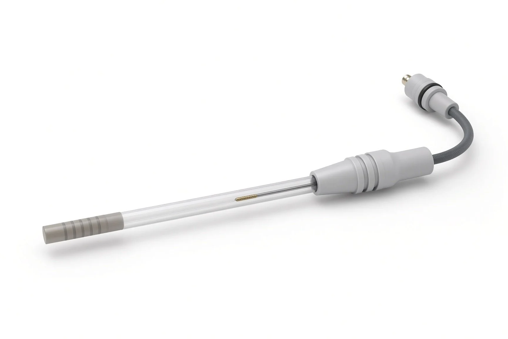
Ultrasound-facilitated lysis catheter.
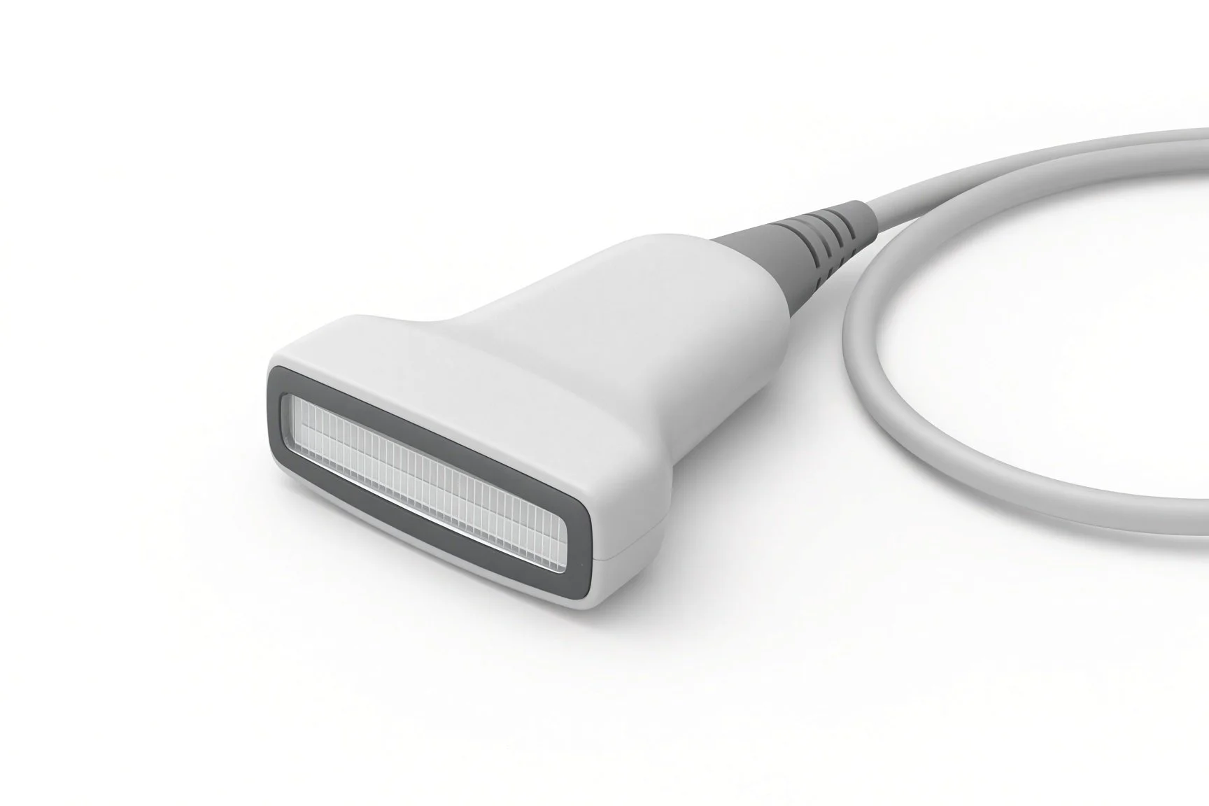
Venous sheath and ultrasound.
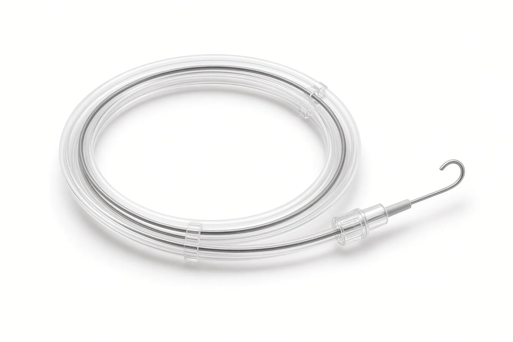
Guidewires.
Procedural steps (Barts example)
Getting ready
Access
Treating the clot
Finishing
Infusion care and catheter removal
First time in the cath lab?
Know the plan before the patient arrives. Ask whether it is thrombolysis, aspiration or both, so the right kit, pumps and bed are ready.
Respect the right ventricle. These patients can collapse with sedation, lying flat or a small drop in blood pressure. Keep the defibrillator and resuscitation drugs close.
Track the blood. In aspiration cases, keep a running total of the blood removed and know how to get blood products quickly.
Hand over every line. At the end, hand over each catheter, its position, what is running through it and when it stops.
Keep the anaesthetic or critical care team close. A fall in blood pressure on sedation or lying flat is a warning sign in this group.
A single front-wall puncture under ultrasound matters more than usual: these patients may receive a clot-busting drug.
Count the blood. With aspiration, record how much blood is removed on each pass and tell the operator the running total.
Label every line. The clot-busting drug, heparin and any flush must be clearly labelled at the patient end and at the pump.
FAQ
Why not just use blood thinners?
Blood thinners stop the clot growing and are the main treatment for every PE. For a few people the clot strains the heart badly, and clearing it faster may help.
Is it better than a clot-busting drip into the arm?
The catheter puts a much smaller dose straight into the clot, aiming for the benefit with less bleeding. Trials are still working out who gains most.
Will I be awake?
Usually yes, with local anaesthetic and light sedation if needed. Heavy sedation is avoided because it can lower the blood pressure.
How long will I need blood thinners?
At least 3 months, sometimes for life, depending on what caused the clot.
Related Cath Lab 101 guides
IVC filter insertion and retrieval (coming soon)
Sources
All checked on 12 November 2026. Each page's own date is shown where it gives one.
Konstantinides SV, et al. 2019 ESC Guidelines for the diagnosis and management of acute pulmonary embolism. Eur Heart J 2020;41(4):543–603.
NICE. Venous thromboembolic diseases: diagnosis, management and thrombophilia testing (NG158). nice.org.uk/guidance/ng158
HI-PEITHO trial (ultrasound-facilitated catheter-directed thrombolysis vs anticoagulation alone, intermediate-high-risk PE, 2026) and PEERLESS trial (large-bore aspiration thrombectomy vs catheter-directed thrombolysis).
StatPearls (NCBI Bookshelf), search: pulmonary embolism. ncbi.nlm.nih.gov/books
Disclaimer
Education only. This page is for general education for healthcare students, cath lab staff and curious readers. It isn't medical advice and doesn't replace advice from your own doctor, heart team or local unit protocols. Equipment and medicines are described generically. No product or manufacturer is recommended. If you think someone is having a heart emergency, call 999.
Disclosure: I work in the medical device industry. This page is independent education and isn't sponsored or reviewed by any company.
All diagrams on this page are original illustrations created for rodnieoro.com.