Peripheral arterial intervention (leg arteries): a Cath Lab 101 guide
Reviewed by Rodnie Oro, cath lab charge nurse · Last reviewed: November 2026 · Education only, not medical advice
Peripheral arterial intervention in one minute
What it is: Peripheral arterial disease is narrowing of the leg arteries by fatty, often calcified plaque. Peripheral intervention (angioplasty) opens the artery from inside: a wire crosses the narrowing, then balloons, drug-coated balloons, stents and sometimes plaque-modifying devices restore flow.
Who it is for: People with leg pain on walking that still limits daily life despite exercise and medicines, and people with chronic limb-threatening ischaemia: pain at rest, ulcers or gangrene. A vascular team decides.
Main risks: Bleeding or a bruise at the access site, a torn or blocked artery, clot or debris travelling down the leg (distal embolisation) and kidney strain from the contrast. Your team will explain your own risk.
Usually: Under local anaesthetic, about 1 to 2 hours (longer for complex blockages), often home the same or next day.
On this page: What is peripheral arterial intervention? · Who is it for? · When is it done? · Step by step · Recovery · Risks · Kit list · First time in the lab? · FAQ
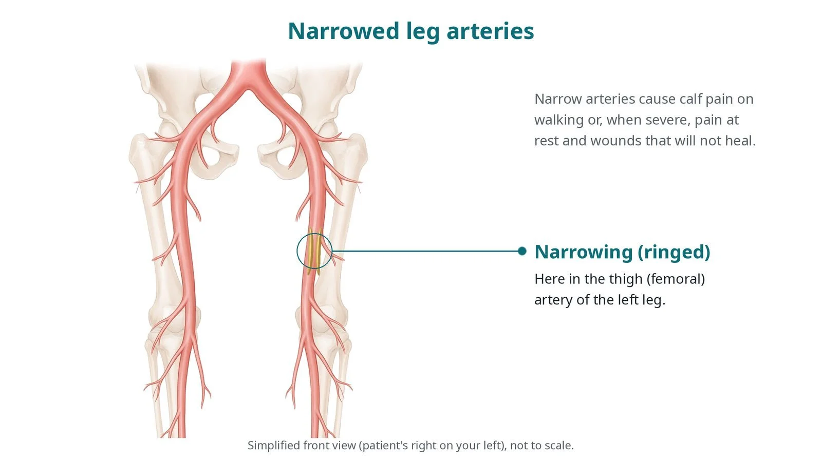
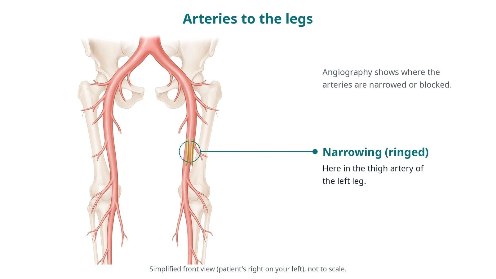
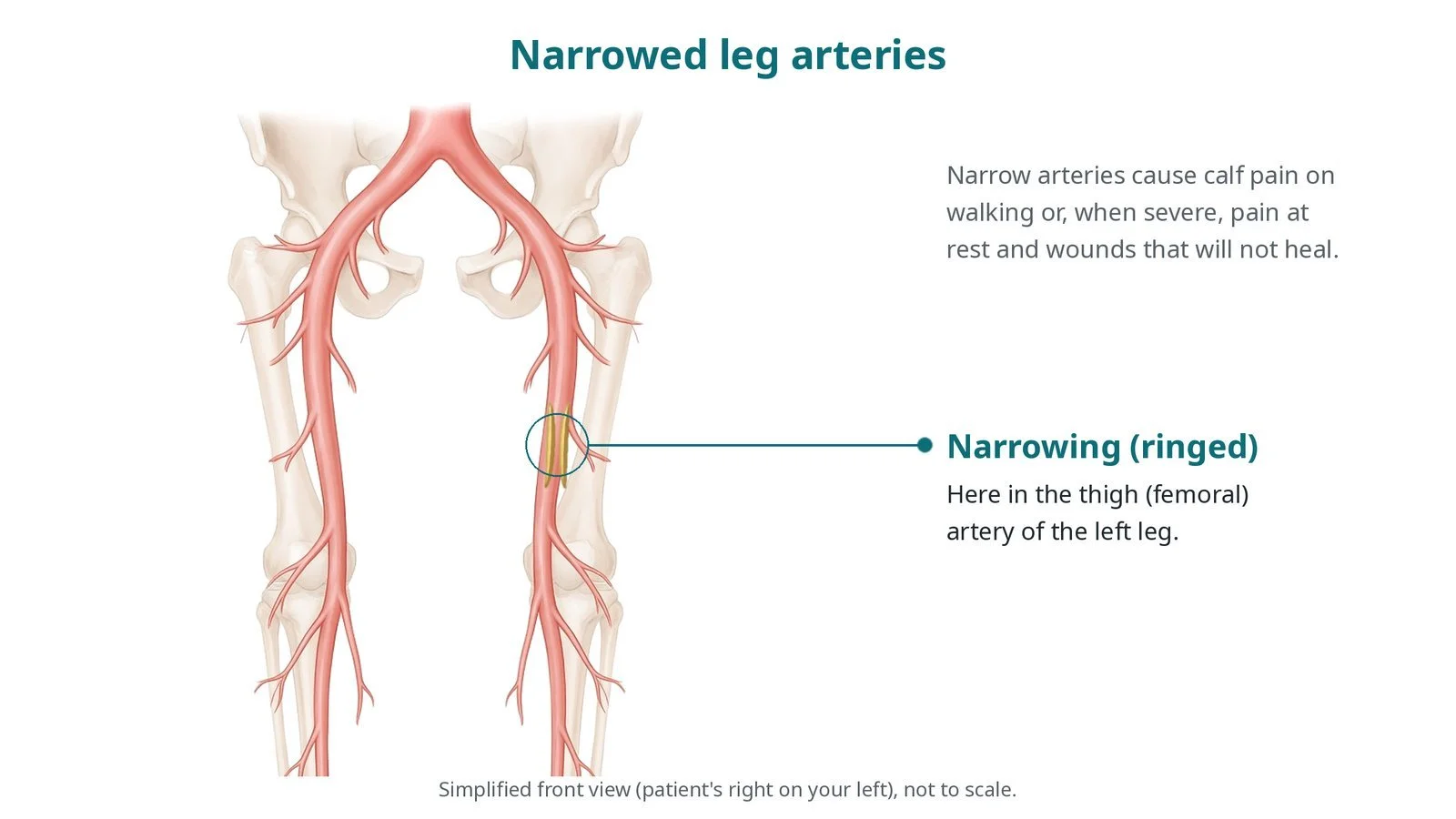
Narrowed leg arteries: a narrowing (ringed) in the thigh (femoral) artery of the left leg. Narrow arteries cause calf pain on walking or, when severe, rest pain and wounds that will not heal.
What is peripheral arterial intervention?
The aorta divides at about the level of the belly button into the two common iliac arteries. Each becomes the external iliac artery, then the common femoral artery in the groin. Below the groin, the superficial femoral artery runs down the thigh and becomes the popliteal artery behind the knee, which divides into three smaller below-the-knee arteries to the foot.
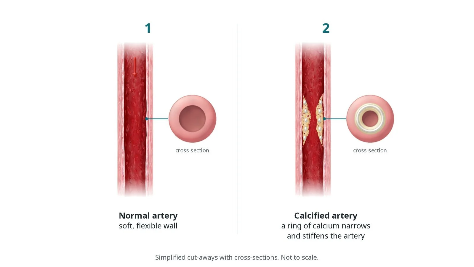
Plaque (atherosclerosis) narrows these arteries, especially in people who smoke or have diabetes, high cholesterol or kidney disease. In the thigh, plaque is often long and heavily calcified, and complete blockages (occlusions) are common. Narrowed arteries cannot increase flow when muscles work, causing calf or buttock pain on walking (claudication). When flow is very poor, the foot hurts at rest and wounds do not heal.
Treatment depends on the segment. Iliac arteries are often stented. Thigh and knee arteries are treated with balloon angioplasty, drug-coated balloons (which leave a drug in the wall to slow re-narrowing) or stents, sometimes after atherectomy (cutting or sanding plaque away) or intravascular lithotripsy (sound pressure waves that crack calcium). Below-the-knee arteries are small and are usually treated with long balloons, to get straight-line flow to a wound.
New to the acronyms? See the Cath Lab Abbreviations A–Z.
Who is it for?
It may be offered to people with:
claudication that still limits daily life after supervised exercise, stopping smoking and best medical treatment (antiplatelet, statin, blood pressure and diabetes control)
chronic limb-threatening ischaemia: rest pain, ulcers or gangrene, where restoring flow may save the leg
The choice between angioplasty, bypass surgery and medicines alone is made by a vascular team, often with surgeons and radiologists.
Who it may not suit: very long or complex blockages that are better treated with bypass surgery mild symptoms that improve with exercise and medicines
The decision is made by your heart team, who will talk through the reasons, the alternatives and the risks with you.
When is it done?
Usually planned, after a duplex ultrasound, CT or MR angiogram.
Before it, the team checks:
the ankle-brachial pressure index (ABPI) and foot pulses, often marked on the skin
kidney function and allergies, especially to contrast
blood thinners, diabetes medicines and the access plan
Threatened limbs with spreading infection or gangrene are treated urgently.
gives you a chance to ask questions and give consent
arranges blood tests, including kidney function and a blood count, and an ECG
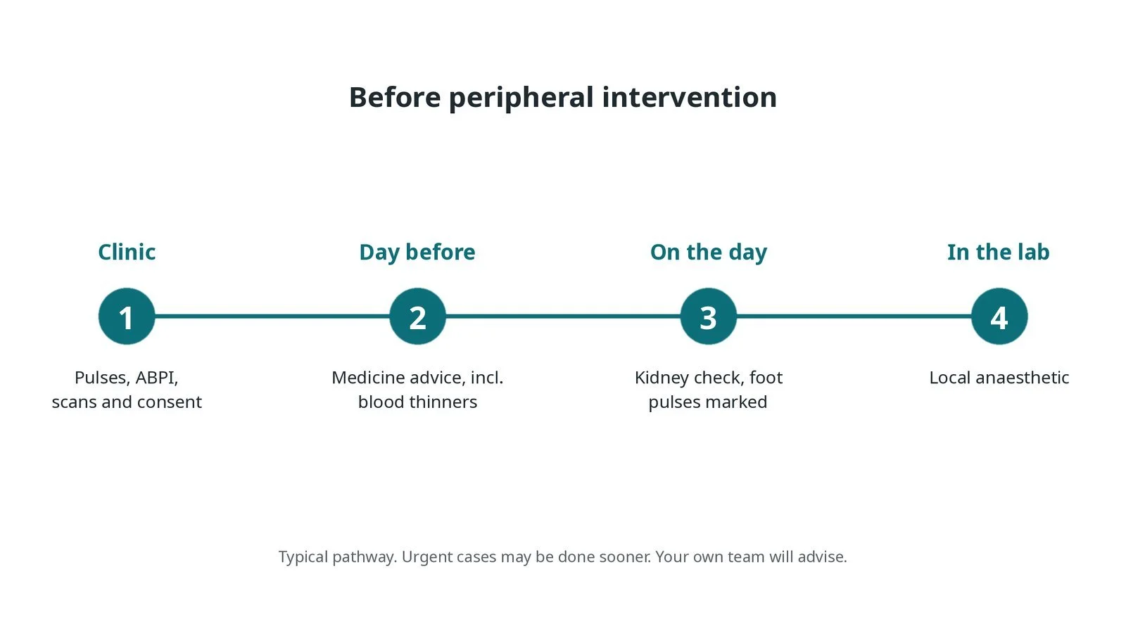
Before peripheral intervention: pulses, ABPI, scans and consent; medicine advice; kidney check and foot pulses marked; local anaesthetic in the lab.
Step by step
1. Getting ready
You lie on the X-ray table. Foot pulses are felt, or found with a Doppler probe, and marked so they can be compared at the end. Monitoring includes ECG, blood pressure and oxygen levels. Local anaesthetic is used, with light sedation if needed.
2. Access
The route depends on the target: crossover: from the opposite groin, the catheter goes up and over the aortic fork and down into the treated leg antegrade: from the groin of the same leg, pointing down the leg, for thigh and below-knee arteries pedal or tibial: a small puncture in an ankle or foot artery, when the blockage cannot be crossed from above radial or brachial: from the arm, in some centres Punctures are made under ultrasound. Heparin is given, as per centre protocol.
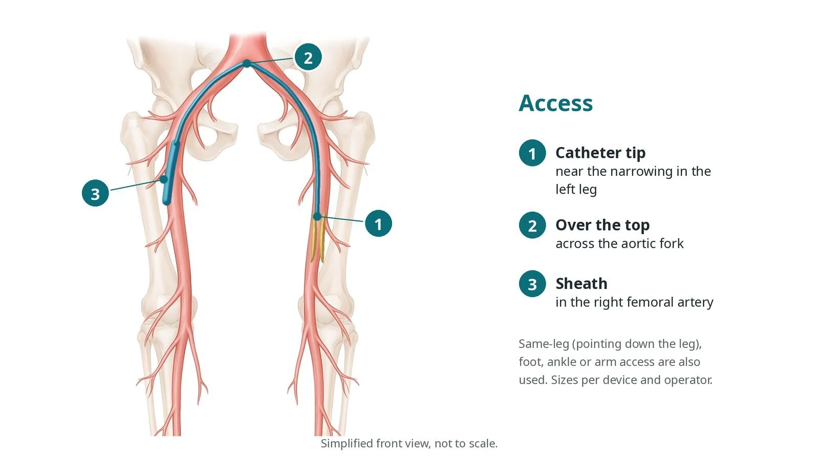
Access: (1) catheter tip near the narrowing in the left leg; (2) over the top across the aortic fork; (3) sheath in the right femoral artery. Same-leg, foot, ankle or arm access are also used.
3. Opening the artery
A. Wire across. A guidewire, with a support catheter, crosses the narrowing or blockage, sometimes through the layers of the wall (subintimal) and back into the channel beyond.
B. Prepare. A balloon stretches the narrowing. Heavily calcified plaque may first be treated with atherectomy or intravascular lithotripsy, per operator.
C. Treat. A drug-coated balloon is inflated for a set time, or a stent is placed if the artery recoils or tears (dissection). Iliac arteries are often stented first.
D. Check. An angiogram checks flow down to the foot, and the foot pulses or Doppler signals are compared with the start.
Watch: Sudden new foot pain, or vessels below the treated area no longer filling on the angiogram: tell the operator; debris or clot may have travelled down the leg (distal embolisation) and can often be removed straight away.
4. Finishing
The sheath is removed and the artery is closed with pressure or a closure device, per operator. Antegrade and pedal sites need careful, prolonged pressure.
5. Back on the ward
Checks of the access site, foot colour, warmth, feeling and pulses continue, as per centre protocol. Severe calf pain, tense swollen muscles or numbness can mean compartment syndrome, when flow returns to a leg that has been starved of blood for a long time.
Watch: A tense, swollen, very painful calf, or pain on moving the toes, after a long blockage has been opened: get urgent medical and vascular surgical review (possible compartment syndrome).
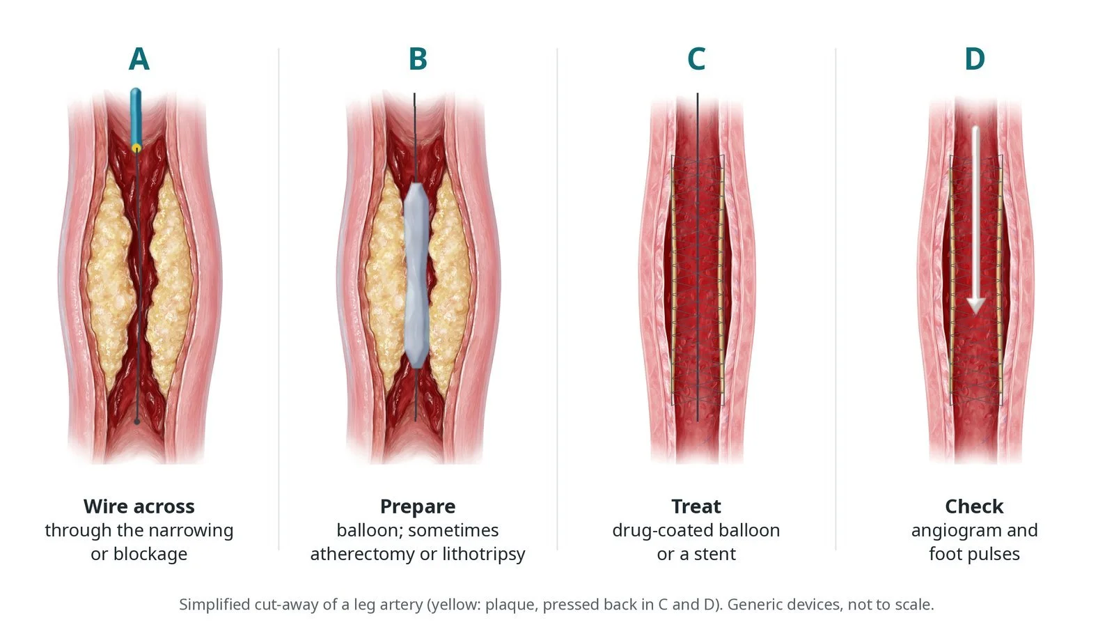
A: wire across the narrowing. B: prepare with a balloon, sometimes atherectomy or lithotripsy. C: treat with a drug-coated balloon or a stent. D: check the angiogram and foot pulses.
Recovery and aftercare
First hours: rest with checks of the access site, foot colour, warmth and pulses, and blood pressure, as per centre protocol.
Going home: often the same or next day; longer if there is a wound or infection.
Medicines: antiplatelet tablets and a statin, as prescribed; some people also take a low-dose blood thinner. Do not stop them without advice.
Activity and lifestyle: keep walking as advised; stopping smoking is the single most important thing for keeping the artery open.
Driving: ask your team; most people can drive after a few days, once the access site has healed.
Follow-up: clinic review with an ABPI or a duplex scan, and wound care with the foot team if needed.
Call 999 if you have heavy bleeding, a cold, pale, numb or very painful leg or foot, or chest pain.
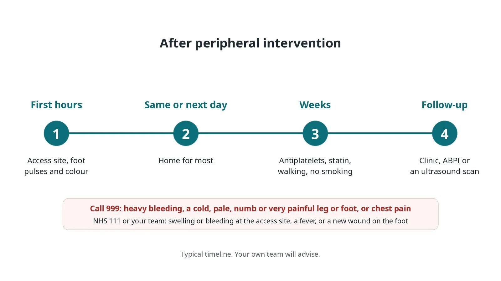
After peripheral intervention: (1) access site, foot pulses and colour checks; (2) home the same or next day for most; (3) antiplatelets, statin, walking and no smoking; (4) clinic follow-up with ABPI or a scan.
Risks and complications
Your team will explain the risks that apply to you. Possible problems include:
Bleeding, bruising or a false aneurysm at the access site
Tear (dissection) or blockage at the treated site, often fixed at the time with a balloon or stent
Debris or clot travelling down the leg (distal embolisation) (uncommon)
Artery rupture (rare)
Compartment syndrome after flow returns (rare): Swelling of the calf muscles that needs urgent surgery.
Kidney strain from the contrast dye
The artery narrowing again over time (restenosis)
Cath lab kit list (printable)
Barts example kit list
An example from one UK centre, written generically. Every lab has its own protocol, and sizes and doses vary.
arterial sheaths: crossover, long or short, size per device and operator
ultrasound for access and a micropuncture kit
guidewires of different stiffness and tip shapes, per operator
support (crossing) catheters and diagnostic catheters
angioplasty balloons of different lengths and sizes, including long below-the-knee balloons
drug-coated balloons, per operator
stents: self-expanding for the thigh, balloon-expandable or covered for the iliacs, per operator
atherectomy device or intravascular lithotripsy balloon, if needed
inflation device, contrast and heparin, as per centre protocol
hand-held Doppler probe for foot pulses
closure device or manual compression, per operator
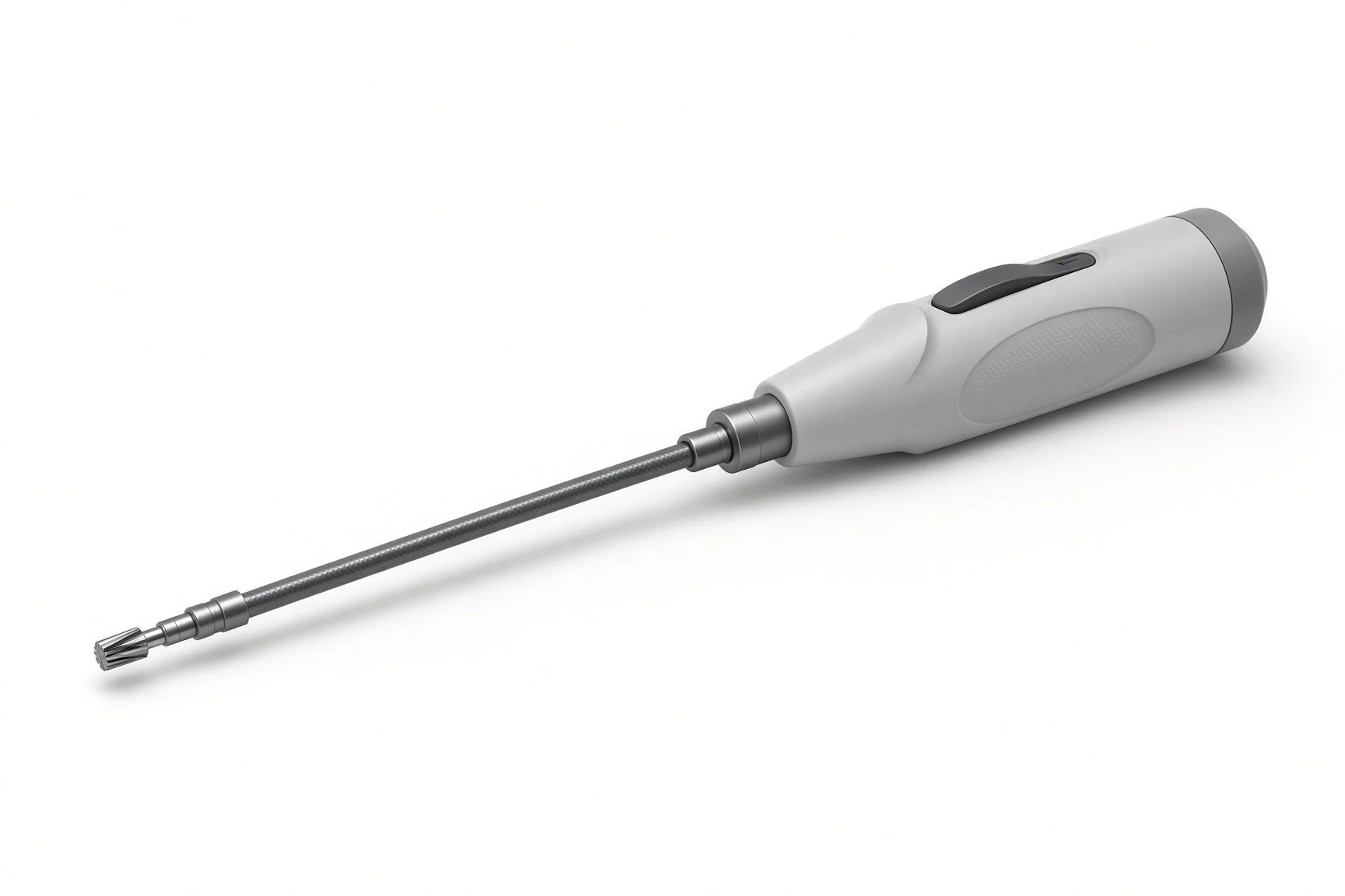
Atherectomy device.
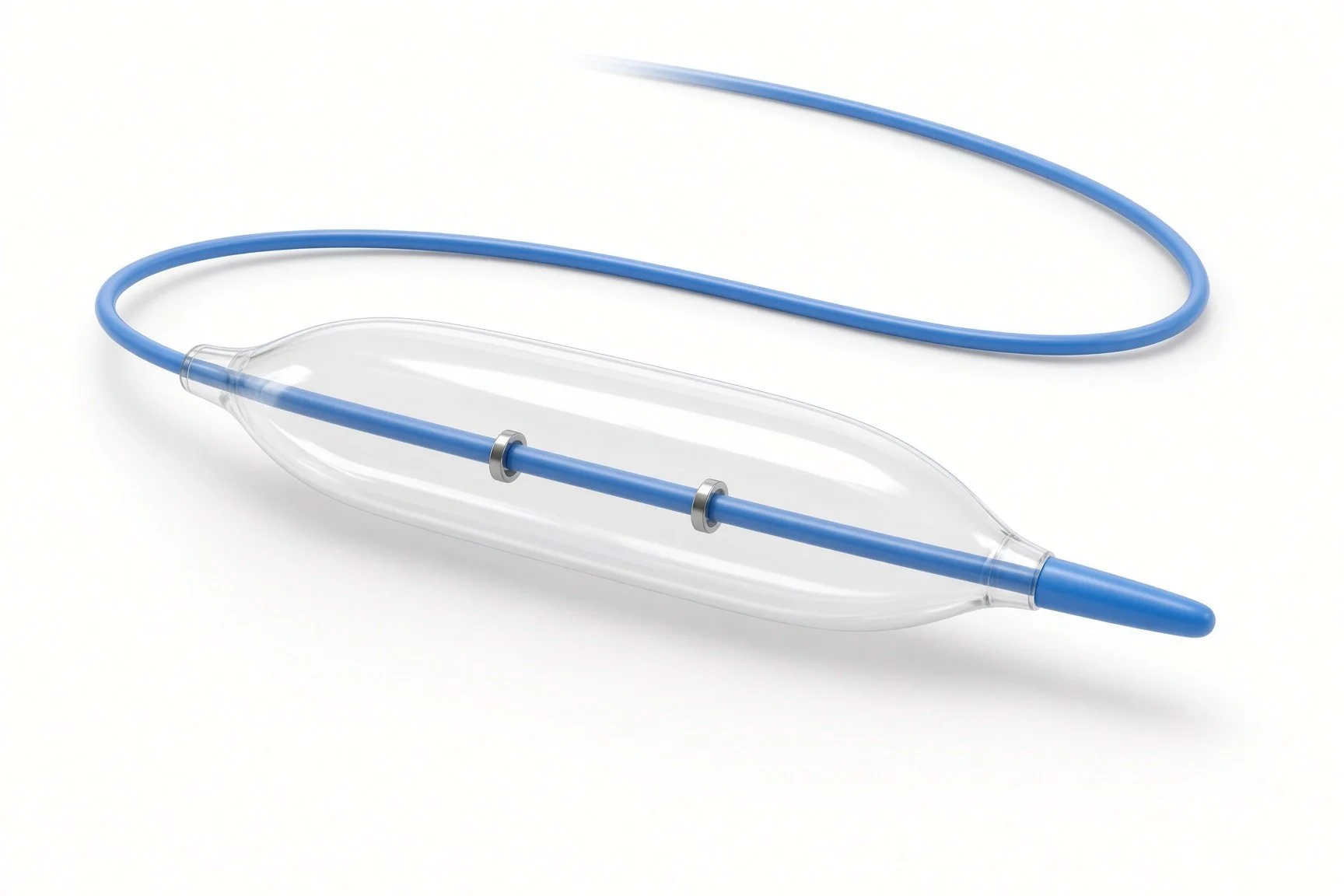
Angioplasty balloons.
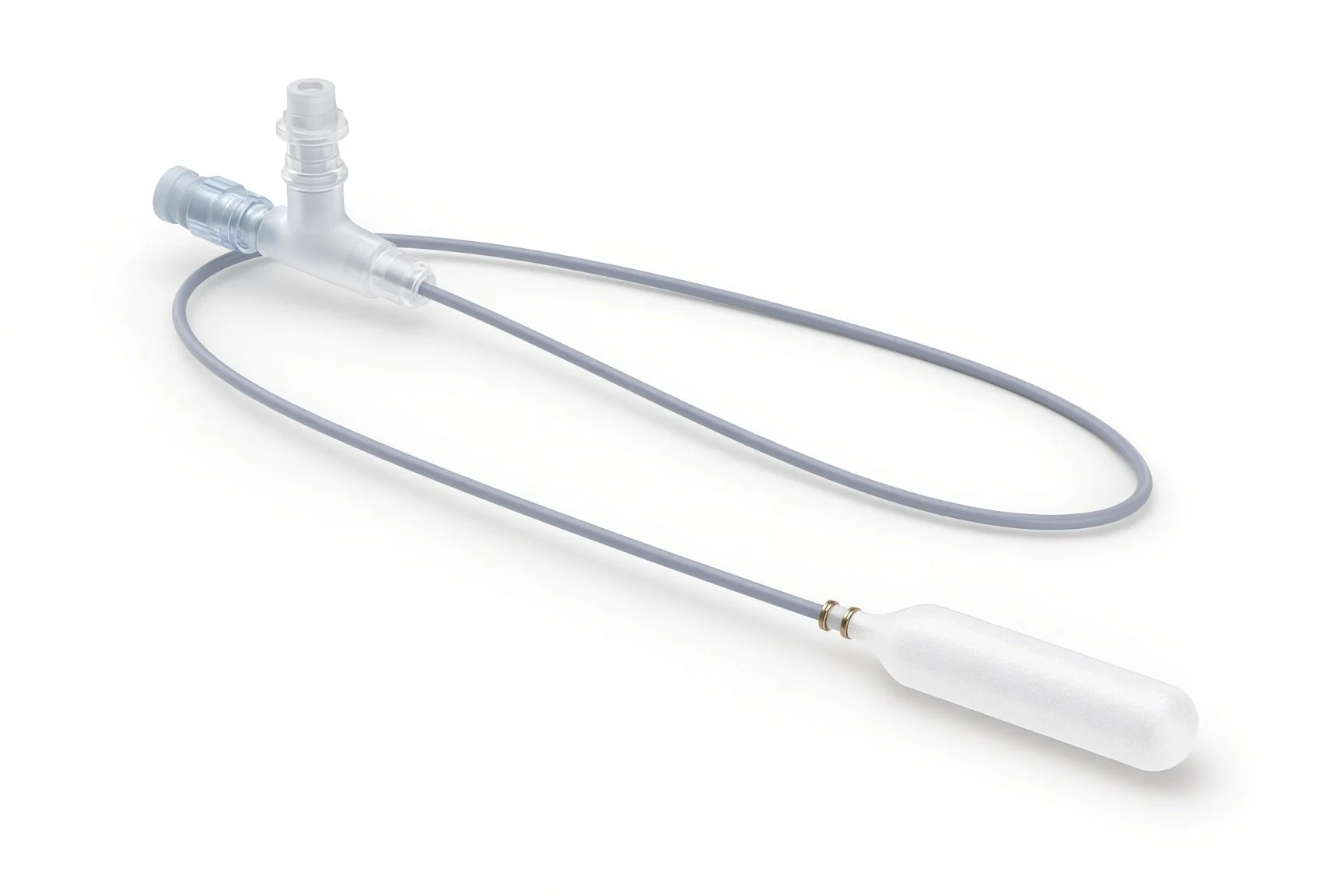
Drug-coated balloon.
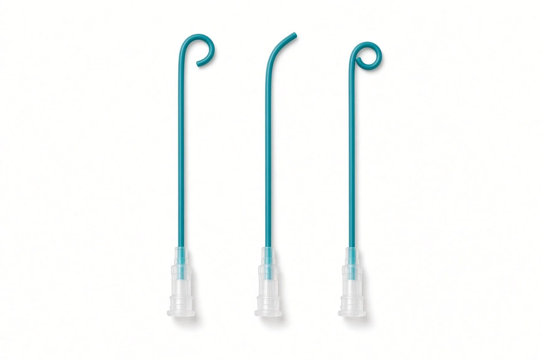
Support (crossing) catheters and diagnostic catheters.
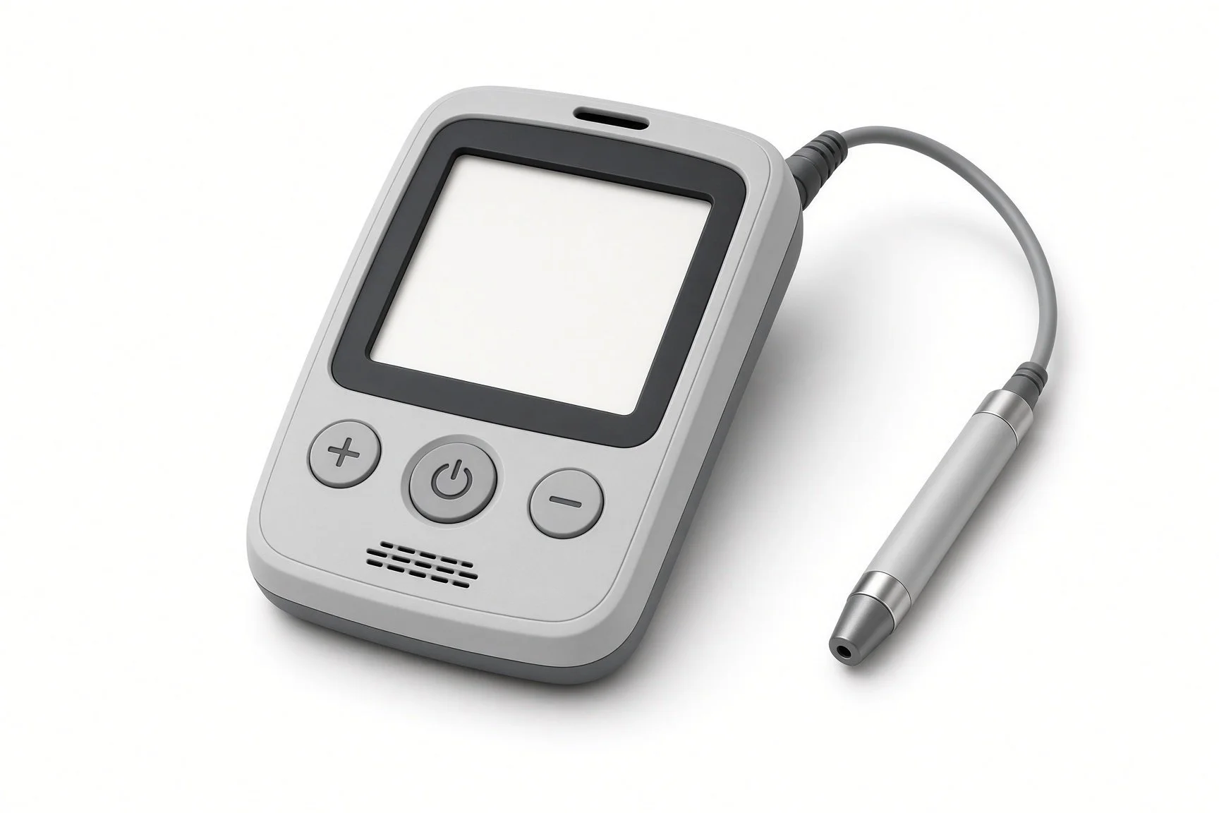
Doppler probe.
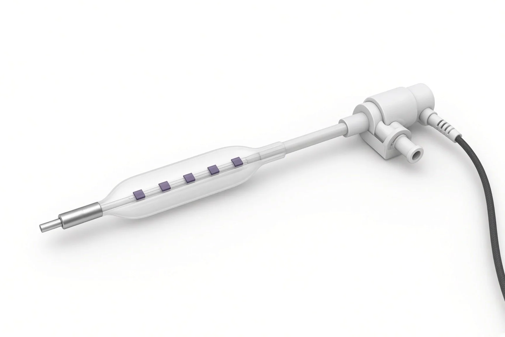
Lithotripsy balloon.
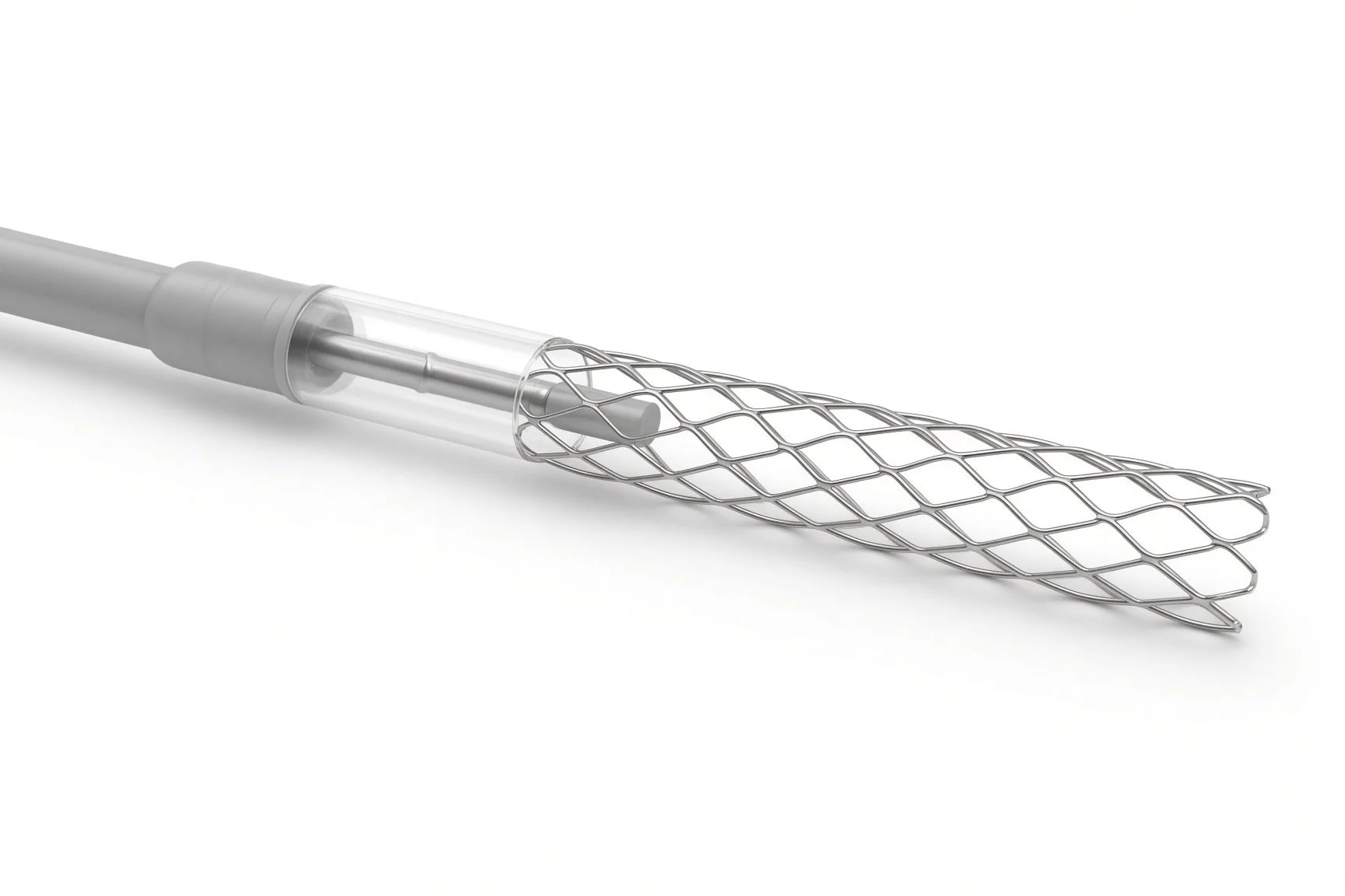
Self-expanding stent.
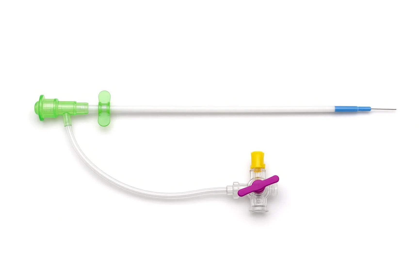
Crossover or straight sheath.
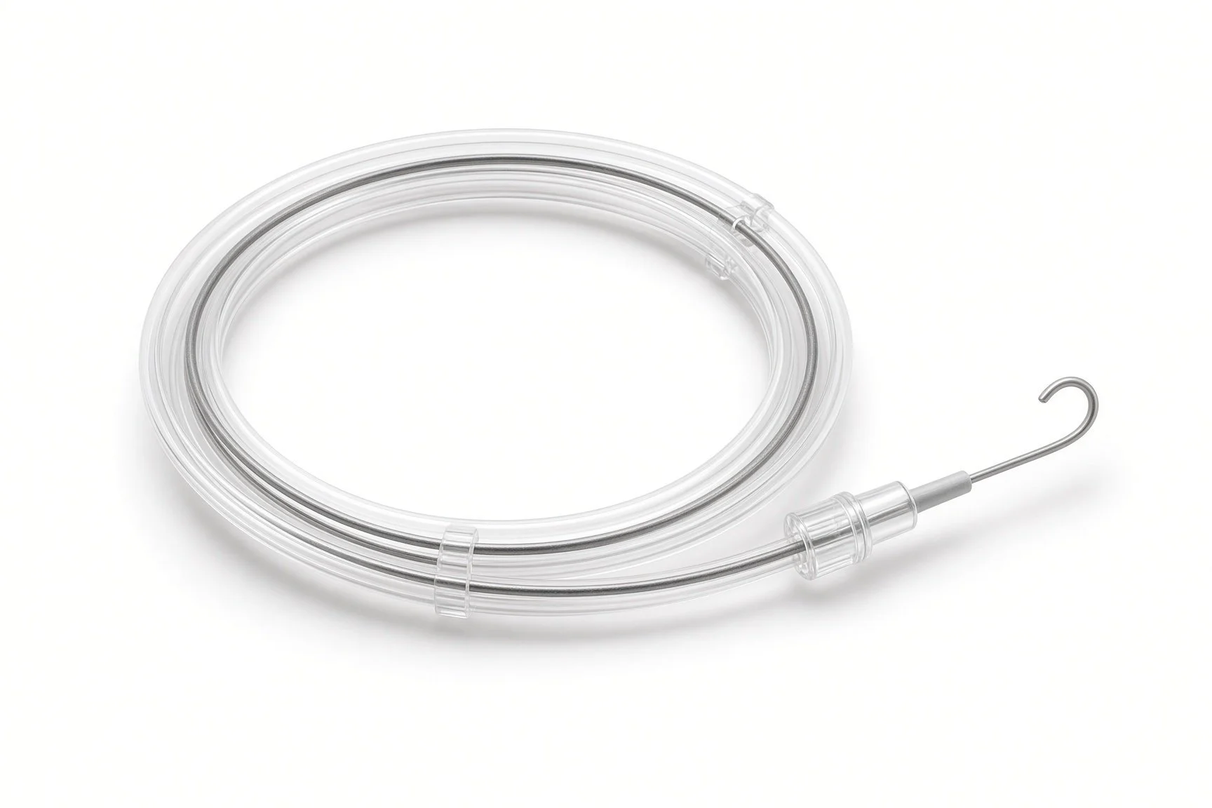
Guidewires and support catheter.
Procedural steps (Barts example)
Getting ready
Access
Opening the artery
Finishing
Back on the ward
First time in the cath lab?
Mark the pulses. Feel or Doppler the foot pulses before the case and mark them, so you can compare at the end.
Know the access plan. Crossover, antegrade and pedal access need different sheaths and positioning; check before the drapes go on.
Expect long inflations. Drug-coated balloons are inflated for a set time; watch the clock and the patient's comfort.
Watch the foot afterwards. New pain, coldness or pallor after the case needs prompt review.
Record the baseline foot. Note colour, warmth, pulses and any wounds before the case starts.
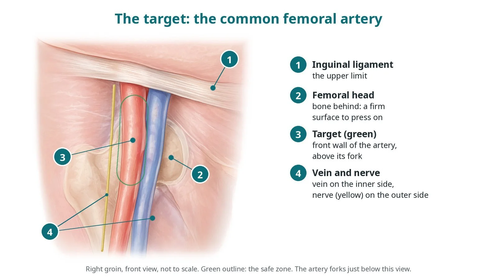
Antegrade punctures are steep and close to the groin crease. Help hold the tummy fold out of the way, and expect careful pressure at the end.
FAQ
Will I be awake?
Yes, usually with local anaesthetic and, if needed, light sedation.
Will my walking pain go away?
Many people walk further afterwards, but arteries can narrow again. Exercise, not smoking and medicines help keep the benefit.
Why a drug-coated balloon instead of a stent?
In the thigh and knee arteries, a drug-coated balloon can slow re-narrowing without leaving metal behind. The operator chooses based on the result.
What if the artery cannot be opened?
The team may try another route, such as from the foot, or discuss bypass surgery.
Related Cath Lab 101 guides
Large-bore access and closure (coming soon)
Subclavian, vertebral and mesenteric stenting (coming soon)
Sources
All checked on 12 November 2026. Each page's own date is shown where it gives one.
Mazzolai L, et al. 2024 ESC Guidelines for the management of peripheral arterial and aortic diseases. Eur Heart J 2024.
NICE. Peripheral arterial disease: diagnosis and management (CG147). nice.org.uk/guidance/cg147
Conte MS, et al. Global vascular guidelines on the management of chronic limb-threatening ischemia. Eur J Vasc Endovasc Surg 2019.
StatPearls (NCBI Bookshelf), search: peripheral arterial disease. ncbi.nlm.nih.gov/books
Disclaimer
Education only. This page is for general education for healthcare students, cath lab staff and curious readers. It isn't medical advice and doesn't replace advice from your own doctor, heart team or local unit protocols. Equipment and medicines are described generically. No product or manufacturer is recommended. If you think someone is having a heart emergency, call 999.
Disclosure: I work for a company that makes a device used in this procedure. This page is independent education, isn't sponsored or reviewed by any company, and doesn't recommend any product.
All diagrams on this page are original illustrations created for rodnieoro.com.