Intravascular lithotripsy (IVL) for calcified arteries: a Cath Lab 101 guide
Reviewed by Rodnie Oro, cath lab charge nurse · Last reviewed: October 2026 · Education only, not medical advice
Intravascular lithotripsy in one minute
What it is: some narrowed arteries are hardened with calcium, which makes them difficult to open and can stop a stent expanding properly. Intravascular lithotripsy (IVL) uses a special balloon catheter that sends sound pressure waves into the artery wall to crack the calcium, so the artery can then be opened and stented. It is part of a PCI (coronary angioplasty) procedure.
Who it is for: people having PCI whose narrowing is heavily calcified, as judged on the angiogram or intravascular imaging. It is also used in some leg (peripheral) arteries.
Main risks: the same as PCI, including bleeding at the wrist or groin, damage to the artery and heart rhythm changes. Your team will explain your own risk.
Usually: awake under local anaesthetic, as part of a PCI lasting about 1 to 2 hours, often home the same or next day.
On this page: What is intravascular lithotripsy? · Who is it for? · When is it done? · Step by step · Recovery · Risks · Kit list · First time in the lab? · FAQ
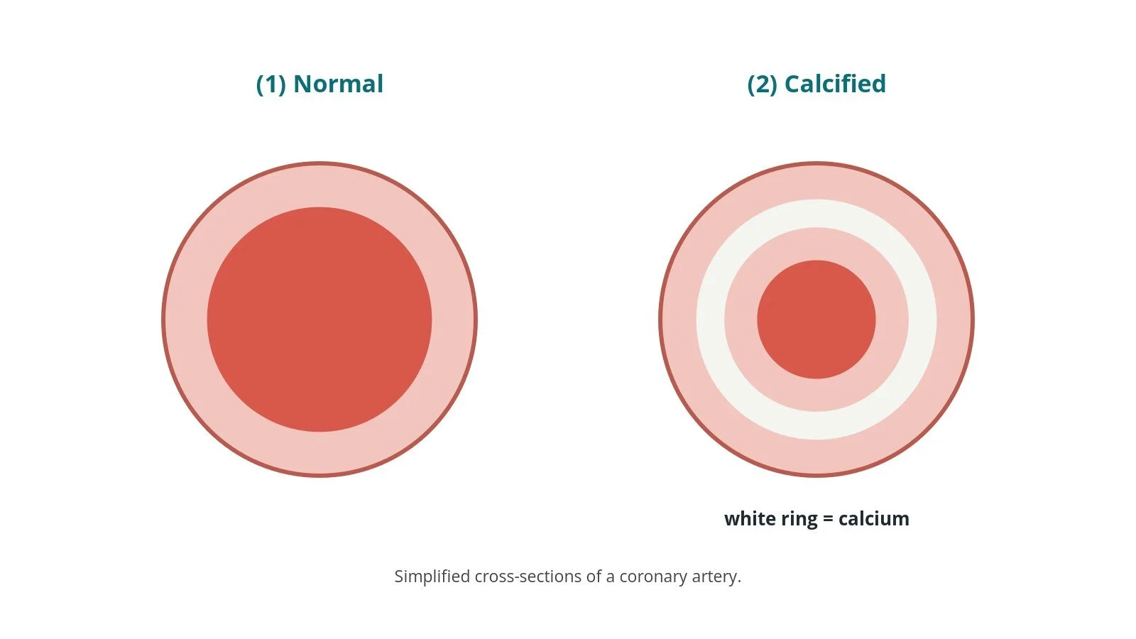
(1) Normal artery. (2) Calcified artery: a ring of calcium narrows and stiffens the artery.
What is intravascular lithotripsy?
As people age, and especially with kidney disease or diabetes, the plaque in coronary arteries can become calcified, like a ring or plate of bone in the artery wall. Calcium makes a narrowing:
hard to cross with balloons and stents
resistant to normal balloon inflation
likely to leave a stent under-expanded, which raises the chance of the stent narrowing or blocking later
Intravascular lithotripsy (IVL) borrows the idea of kidney-stone lithotripsy. The IVL catheter looks like an angioplasty balloon but contains small emitters. The balloon is inflated gently to touch the artery wall, and electrical pulses create tiny bubbles in the fluid inside the balloon that expand and collapse, sending sound pressure waves outwards. These pass through soft tissue but crack the calcium, both near the surface and deeper in the wall. The artery can then be expanded with less force, and a stent can open fully.
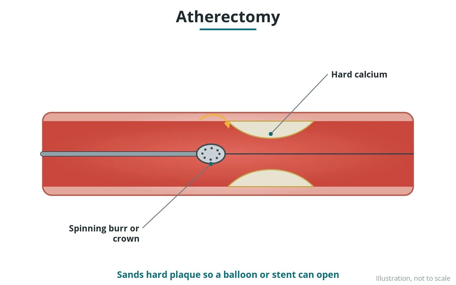
Other ways to treat calcium include cutting or scoring balloons, high-pressure balloons, and atherectomy (tiny drills or lasers that shave the calcium). The operator chooses based on the type and depth of calcium.
New to the acronyms? See the Cath Lab Abbreviations A–Z.
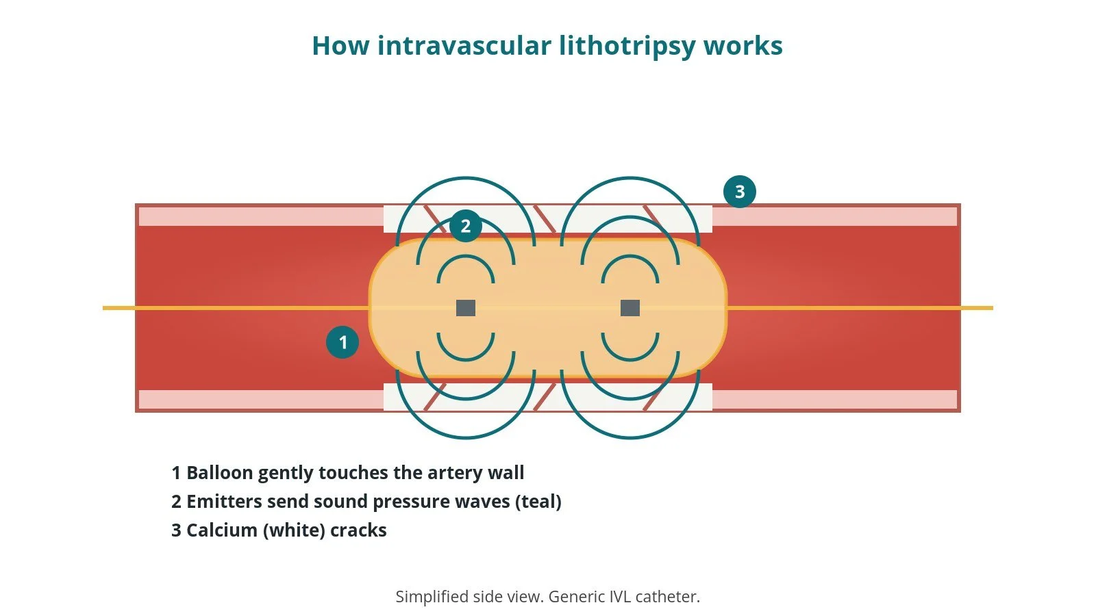
How IVL works: (1) balloon touches the wall at low pressure; (2) emitters send sound pressure waves; (3) calcium cracks.
Who is it for?
IVL is considered during PCI when a narrowing is heavily calcified, for example when:
calcium is seen on the angiogram, or on intravascular imaging (ultrasound or optical images taken from inside the artery)
a standard balloon will not expand fully ("undilatable" lesion)
the calcium is deep or forms a ring round the artery
NICE HealthTech guidance (HTG740, February 2025) says IVL can be used as an option to treat calcified coronary arteries during PCI, with standard arrangements for clinical governance, consent and audit, and that cases should be entered on the national (NICOR) audit database.
The decision to have PCI itself is made with your cardiology team. See the PCI guide.
When is it done?
Usually as part of a planned PCI, sometimes during an urgent one. Before it, the team usually:
checks kidney function, allergies and blood thinners, as per centre protocol
starts or continues antiplatelet medicines, as per centre protocol
reviews previous angiograms to plan the strategy and equipment
Step by step
1. Getting ready and access
PCI with IVL is usually done awake under local anaesthetic through the wrist (radial artery) or the groin (femoral artery). A guide catheter is placed at the opening of the coronary artery. Sheath and guide sizes are per device and operator. Heparin is given, as per centre protocol.
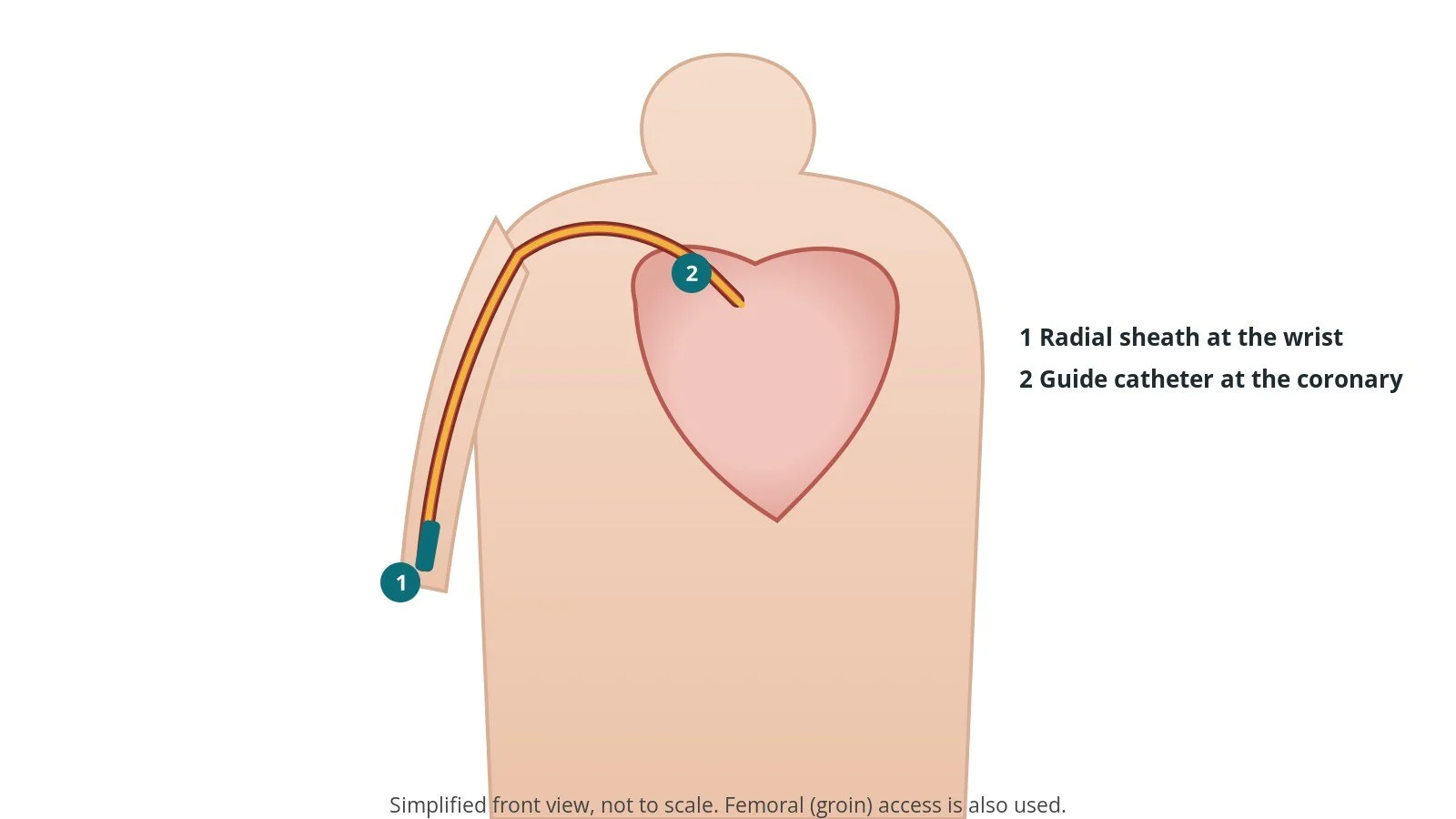
Access for PCI: (1) radial sheath at the wrist; (2) guide catheter at the coronary artery.
2. Preparing the narrowing with IVL
A. Wire. A coronary guidewire is passed across the narrowing. Intravascular imaging may be used to measure the artery and see how much calcium there is.
B. Position the IVL balloon. The IVL catheter is sized to the healthy artery either side of the narrowing, prepared with a saline and contrast mix (no air), and positioned across the calcium using its X-ray markers.
Pulses. The balloon is inflated at low pressure just to touch the wall, then a cycle of pulses is delivered, followed by a slightly higher-pressure inflation. Cycles are repeated as needed. Inflation pressures, the number of pulses per cycle and the maximum number of cycles are per device instructions and operator.
C. Check. The balloon is deflated to let blood flow and the result is checked with an angiogram or imaging.
3. Stenting
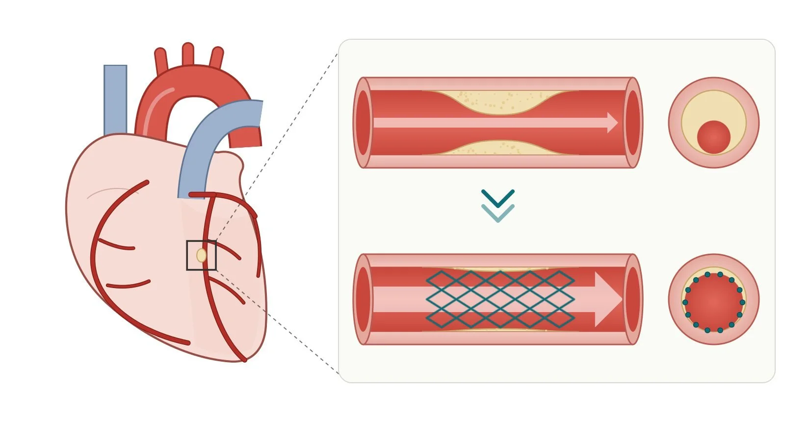
D. Stent. A drug-eluting stent is placed and expanded, often followed by a non-compliant (high-pressure) balloon to make sure it is fully open. Imaging may be used to confirm expansion.
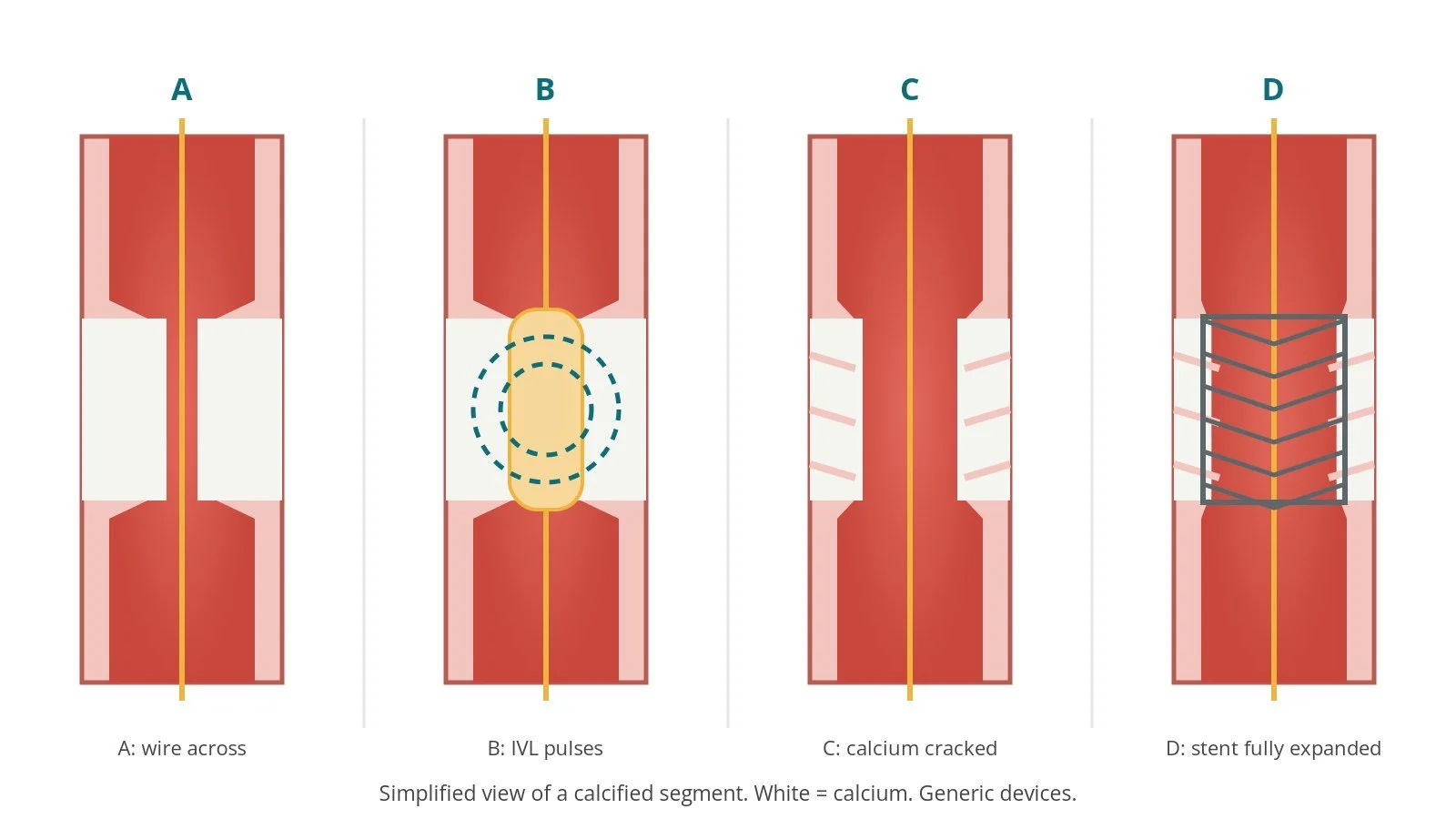
A: wire across. B: IVL pulses. C: calcium cracked; artery opens. D: stent fully expanded.
4. Finishing
The guide and sheath are removed. A compression band goes on the wrist, or the groin is closed with pressure or a closure device, per operator.
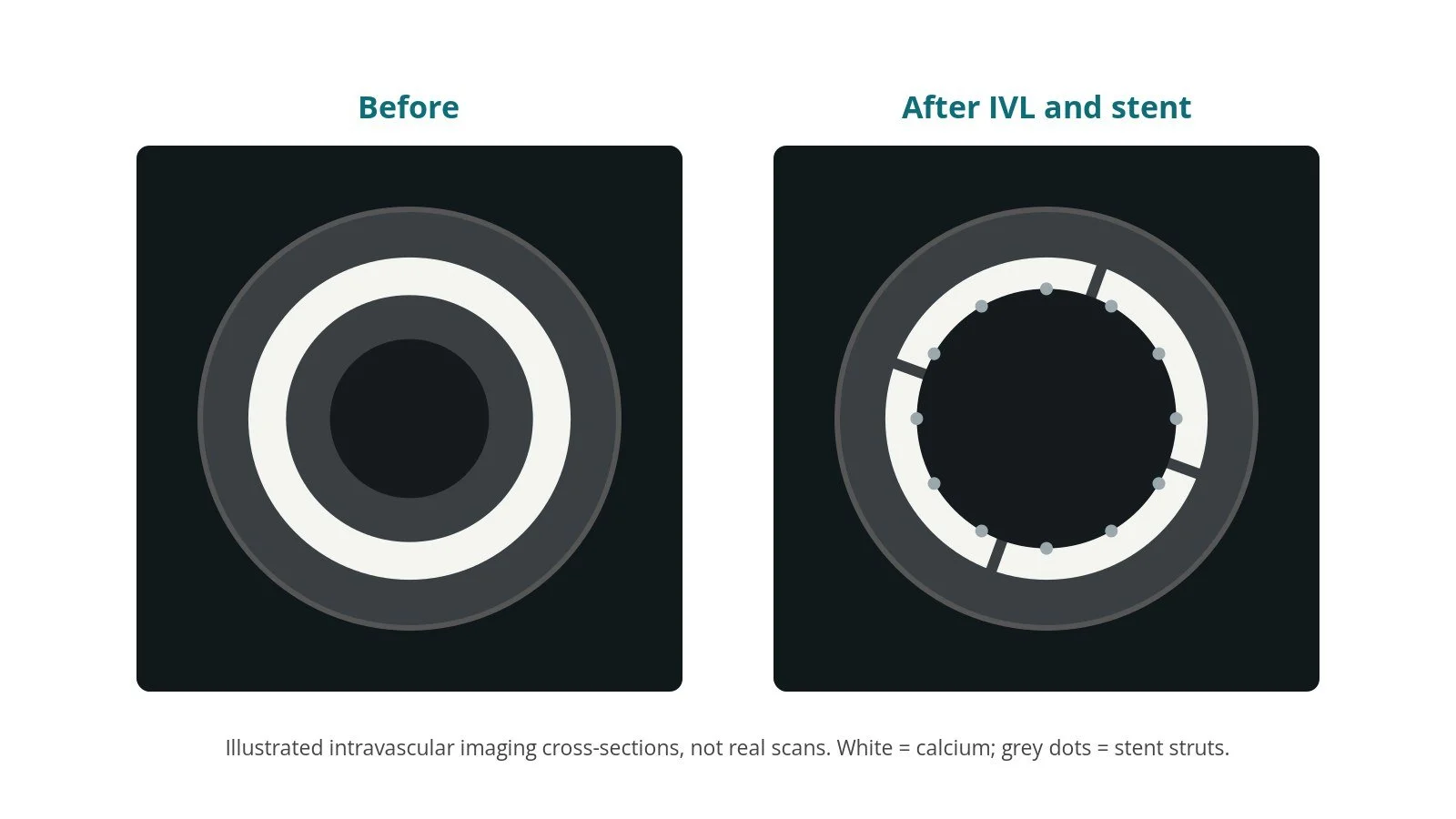
Illustrated imaging, not a real scan. Before: intact calcium ring. After: cracked calcium and a fully expanded stent.
Recovery and aftercare
Recovery is the same as after PCI:
First hours: checks of the wrist or groin, pulse, blood pressure and heart rhythm, as per centre protocol.
Going home: often the same day or the next day.
Medicines: usually dual antiplatelet therapy for a period set by your team, plus your other heart medicines. Do not stop antiplatelets without speaking to your cardiology team.
Activity: avoid heavy lifting with the arm or leg used for a few days, or as advised.
Driving: for a car licence (Group 1), DVLA guidance after a planned PCI is not to drive for at least a week; different rules apply after a heart attack and for bus and lorry licences. Check the DVLA guidance and ask your team.
Call 999 if you have chest pain that does not settle, or bleeding from the wrist or groin that does not stop with firm pressure.
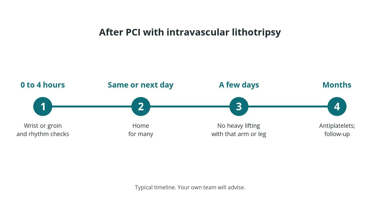
After PCI with IVL: (1) site checks; (2) home the same or next day for many; (3) no heavy lifting for a few days; (4) antiplatelets and follow-up.
Risks and complications
Your team will discuss your own risks. Possible problems include:
bleeding or bruising at the wrist or groin
a tear (dissection) or, rarely, a hole (perforation) in the artery
extra heartbeats during the pulses, usually harmless and short-lived
slow flow, heart attack, stroke or the need for emergency surgery (uncommon)
kidney strain from the contrast dye
Cath lab kit list (printable)
Example kit list
Written generically. Every lab has its own protocol, and sizes and doses vary.
standard PCI set-up: angiography pack, sheath, guide catheter, coronary guidewire, inflation device, haemostasis valve, sizes per device and operator
IVL balloon catheter, sized to the reference artery diameter, per device and operator
IVL generator and connector cable (keep the cable sterile-sleeved as per device instructions); check the generator is charged before the case
contrast–saline mix for balloon preparation, as per device instructions and centre protocol
intravascular imaging catheter, if used
drug-eluting stents and non-compliant balloons, sized to the vessel
heparin and other medicines, as per centre protocol
Procedural steps (example)
Access and guide catheter engagement
Wire across the lesion; imaging if used
IVL balloon prepared (no air) and positioned with its markers
Low-pressure inflation to touch the wall; pulse cycle; higher-pressure inflation; deflate
Repeat cycles as needed within device limits; check result
Stent deployment and post-dilatation; final imaging
Sheath removed; haemostasis
First time in the cath lab?
💡 First-time tip
No air in the balloon. Prepare the IVL balloon carefully with the correct mix; air reduces the effect and is a safety risk. Count the cycles. Each catheter has a maximum number of pulse cycles; help the team keep track. Charge the generator. Check it before the case, not when the balloon is already in the artery. Watch the ECG. Pulses can trigger extra beats; tell the operator about anything sustained. Know the backup. Know where cutting balloons, atherectomy kit and covered stents are kept.
FAQ
Does IVL hurt?
You should not feel the pulses. Some people feel brief chest discomfort when any balloon is inflated in a coronary artery.
Is it the same as kidney-stone lithotripsy?
The idea is similar, but the energy is delivered from inside the artery through a balloon, not from outside the body.
Is IVL used outside the heart?
Yes. Similar catheters are used in calcified leg arteries and sometimes to help large equipment pass through narrowed arteries, for example before TAVI.
Will I know if I had IVL?
Your discharge letter and the operator's report will say which techniques were used.
Related Cath Lab 101 guides
Sources
All checked on 10 October 2026. Each page's own date is shown where it gives one.
NICE. Intravascular lithotripsy to treat calcified coronary arteries during percutaneous coronary intervention (HTG740). nice.org.uk/guidance/htg740 (published 5 Feb 2025; replaced IPG673)
NHS. Coronary angioplasty and stent insertion. nhs.uk/conditions/coronary-angioplasty
British Heart Foundation. Coronary angioplasty and stents (PCI). bhf.org.uk
Knuuti J, et al. 2019 ESC Guidelines for the diagnosis and management of chronic coronary syndromes. Eur Heart J 2020;41(3):407–477. PubMed 31504439
DVLA / GOV.UK. Cardiovascular disorders: assessing fitness to drive. gov.uk
Disclaimer
Education only. This page is for general education for healthcare students, cath lab staff and curious readers. It isn't medical advice and doesn't replace advice from your own doctor, heart team or local unit protocols. Equipment and medicines are described generically. No product or manufacturer is recommended. If you think someone is having a heart emergency, call 999.
Disclosure: I work for a company that makes a device used in this procedure. This page is independent education, isn't sponsored or reviewed by any company, and doesn't recommend any product.
All diagrams on this page are original illustrations created for rodnieoro.com.