Large-bore access and closure: a Cath Lab 101 guide
Reviewed by Rodnie Oro, cath lab charge nurse · Last reviewed: November 2026 · Education only, not medical advice
Large-bore access and closure in one minute
What it is: Some devices, such as TAVI valves, heart pumps, ECMO cannulae and aortic stent grafts (EVAR), need a sheath many times wider than a standard catheter. Large-bore access means puncturing the common femoral artery in exactly the right place under ultrasound, then closing the hole at the end, usually with suture or plug devices.
Who it is for: Patients having a large-bore procedure through the groin. A planning CT checks that the iliac and femoral arteries are wide enough and not too calcified or twisted.
Main risks: Bleeding, including hidden bleeding behind the abdomen (retroperitoneal), a narrowed, blocked or torn artery, a false aneurysm, and poor blood flow to the leg. Your team will explain your own risk.
Usually: Part of the main procedure; access and closure add about 10 to 30 minutes. Bed rest for a few hours afterwards, as per centre protocol.
On this page: What is large-bore access and closure? · Who is it for? · When is it done? · Step by step · Recovery · Risks · Kit list · First time in the lab? · FAQ
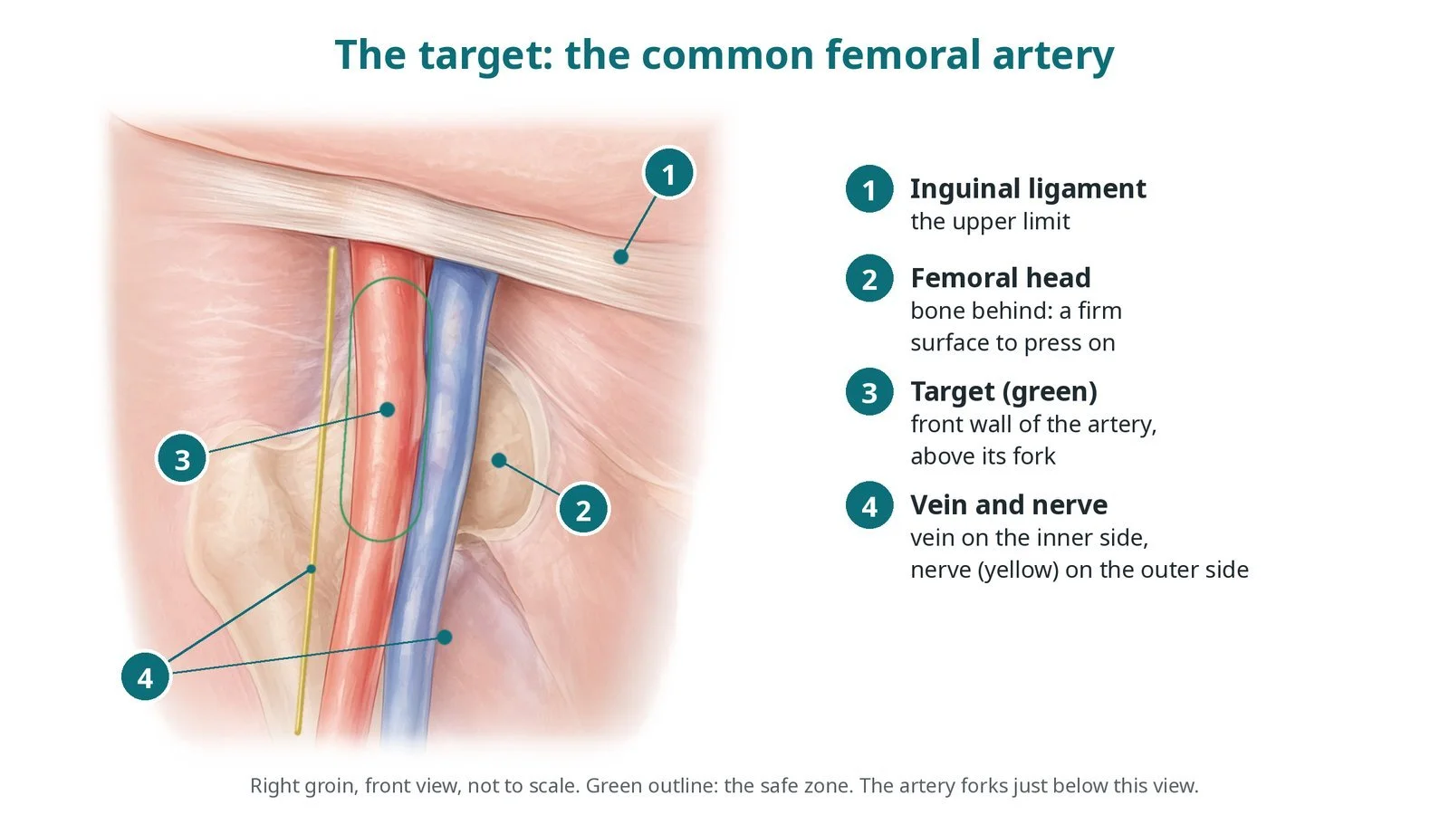
The target: (1) the inguinal ligament is the upper limit; (2) the femoral head is the firm surface behind; (3) the front wall of the common femoral artery above its fork (green); (4) vein on the inner side, nerve on the outer side.
What is large-bore access and closure?
The common femoral artery runs from under the inguinal ligament (a band from the hip bone to the pubic bone) to where it divides into the superficial femoral and profunda arteries, a few centimetres lower. It lies over the femoral head, the ball of the hip joint, which gives a firm surface to press on. The femoral vein lies on its inner side and the femoral nerve on its outer side.
The ideal puncture is in the front wall of the common femoral artery, over the femoral head, below the inguinal ligament and above the bifurcation. Too high, and bleeding can track behind the abdomen into the retroperitoneum, where it cannot be pressed on and is hard to see. Too low, and the artery is smaller, so the sheath may block it or a false aneurysm may form. Calcium in the front wall can stop closure devices working.
Large sheaths stretch the artery, so the hole cannot be closed by pressing alone. Two main methods are used: pre-closure, where one or two suture-based devices are placed before the big sheath goes in and their knots are tied at the end; and plug-based closure, where a device places a plug (often collagen) against the artery wall at the end. If bleeding continues, a balloon passed from the other groin can block the artery from inside while a covered stent or surgery repairs the hole.
New to the acronyms? See the Cath Lab Abbreviations A–Z.
Who is it for?
Patients having procedures that need much wider sheaths than usual, such as:
transcatheter aortic valve implantation (TAVI)
microaxial heart pumps and some ECMO set-ups
endovascular aneurysm repair (EVAR) and thoracic stent grafts
some other structural heart procedures
Who it may not suit: arteries too narrow, calcified or twisted for the sheath: another route, such as the axillary (arm) artery, the carotid artery or through the vena cava, may be used
The decision is made by your heart team, who will talk through the reasons, the alternatives and the risks with you.
When is it done?
It is planned on a CT angiogram, which shows:
the narrowest diameter of the iliac and femoral arteries compared with the sheath
calcium, especially in the front wall at the puncture site
bends (tortuosity) and the height of the femoral bifurcation
On the day, both groins are scanned with ultrasound, and the access, closure and bail-out plans are agreed before starting.
gives you a chance to ask questions and give consent
arranges blood tests, including kidney function and a blood count, and an ECG
Step by step
1. Planning and set-up
The team reviews the CT and agrees which groin takes the large sheath and which takes the smaller sheath for a pigtail or crossover catheter. Both groins are prepared and draped, and an ultrasound machine with a sterile probe cover is set up.
2. Puncture
Under ultrasound, the operator punctures the front wall of the common femoral artery over the femoral head, often with a micropuncture needle. Fluoroscopy or a small contrast injection confirms the height. Many teams also place a sheath in the other groin for a pigtail catheter and a safety (crossover) wire.
3. Pre-closure, sheath and closure
A. Puncture. Front-wall puncture under ultrasound, checked against the femoral head on X-ray.
B. Pre-close. One or two suture-based closure devices are deployed through the small hole, angled apart, and their sutures are kept outside, clipped and protected. For plug-based closure this step is skipped and the plug device is used at the end.
C. Large sheath. Over a stiff wire, the track and artery are dilated and the large sheath is advanced slowly under X-ray. Heparin is given, as per centre protocol.
D. Close. At the end, the sheath is withdrawn over a wire and the knots are pushed down, or the plug device is deployed. The wire stays in until bleeding has stopped, so a sheath can be put back if needed. An angiogram from the other side checks for bleeding or narrowing.
Watch: Ongoing bleeding at the groin after closure, or contrast leaking on the angiogram: tell the operator and press firmly over the puncture; a crossover balloon may be inflated and a covered stent or surgery may be needed.
4. Bail-out options
If closure fails, the options include an extra closure device, manual compression, a balloon inflated inside the artery from the other groin to stop flow at the hole, a covered stent to seal it, or vascular surgery. Heparin may be reversed, per operator.
5. After the procedure
Groin checks continue on the ward: the site, any lump or swelling, leg pulses, colour and warmth, blood pressure and heart rate, as per centre protocol. Hidden bleeding behind the abdomen may show only as back, flank or lower tummy pain, a falling blood pressure, a rising heart rate or a drop in haemoglobin.
Watch: Back, flank or lower tummy pain with a falling blood pressure or rising heart rate: suspect retroperitoneal bleeding, get urgent medical review, check the haemoglobin and prepare for a CT scan or a return to the lab, as per centre protocol.
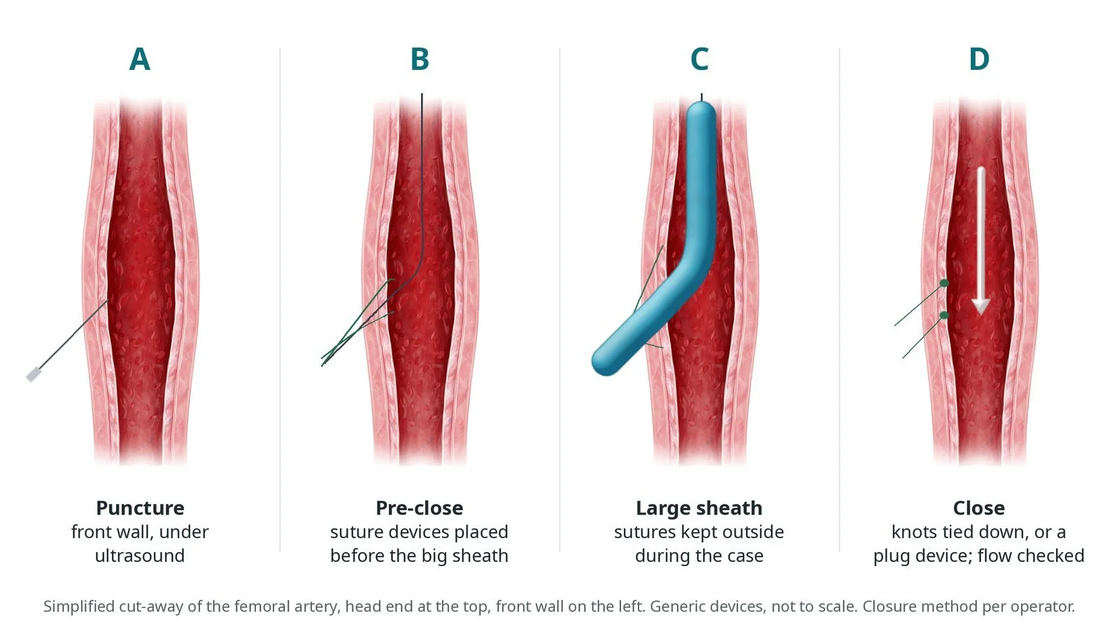
A: front-wall puncture under ultrasound. B: suture devices placed before the big sheath. C: large sheath in, sutures kept outside. D: knots tied down, or a plug device, and flow checked.
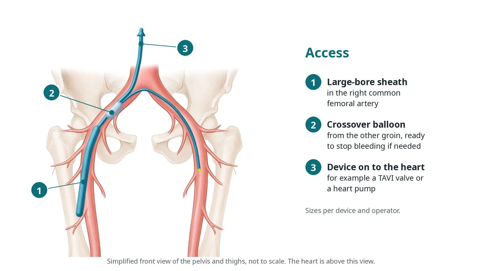
Access: (1) large-bore sheath in the right common femoral artery; (2) crossover balloon from the other groin, ready to stop bleeding; (3) device on to the heart, such as a TAVI valve or heart pump.
Recovery and aftercare
First hours: lie flat or with the head slightly raised, with checks of the groin, leg pulses, blood pressure and heart rate, as per centre protocol.
Getting up: usually later the same day or the next morning if the groin is dry; walk short distances at first.
Going home: depends on the main procedure, for example the day after TAVI for many people.
Activity: avoid heavy lifting, straining and vigorous exercise for 1 to 2 weeks, or as advised.
Driving: follow the advice for your main procedure and DVLA guidance, and wait until the groin has healed.
Follow-up: a small bruise is common; tell your team about a growing lump, pain or numbness in the leg.
Call 999 if you have heavy bleeding, a cold, pale or painful leg, chest pain, or sudden severe back or tummy pain.
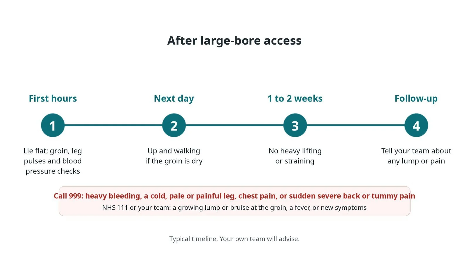
After large-bore access: (1) lie flat with groin, pulse and blood pressure checks; (2) up and walking the next day if dry; (3) no heavy lifting for 1 to 2 weeks; (4) report any lump or pain.
Risks and complications
Your team will explain the risks that apply to you. Possible problems include:
Bleeding or a haematoma at the groin
Hidden bleeding behind the abdomen (retroperitoneal) (uncommon): Often from a puncture above the inguinal ligament.
Closure device failure needing a balloon, covered stent or surgery (uncommon)
Narrowing, blockage or tear of the artery, reducing flow to the leg (uncommon)
False aneurysm or a connection between artery and vein (rare)
Infection at the groin (rare)
Numbness from irritation of the femoral nerve (rare)
Cath lab kit list (printable)
Barts example kit list
An example from one UK centre, written generically. Every lab has its own protocol, and sizes and doses vary.
ultrasound machine and sterile probe cover
micropuncture needle and wire kit
suture-based closure devices, number per operator
plug-based large-bore closure device, if used
large-bore sheath and dilators, size per device
stiff guidewires, per operator
crossover sheath, catheter and wire for the other groin
occlusion balloon for bail-out, size per artery
covered stents (stent grafts) for bail-out, per centre stock
heparin and its reversal, as per centre protocol
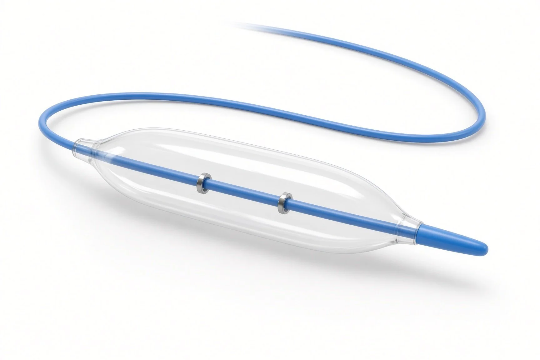
Crossover catheter and balloon.
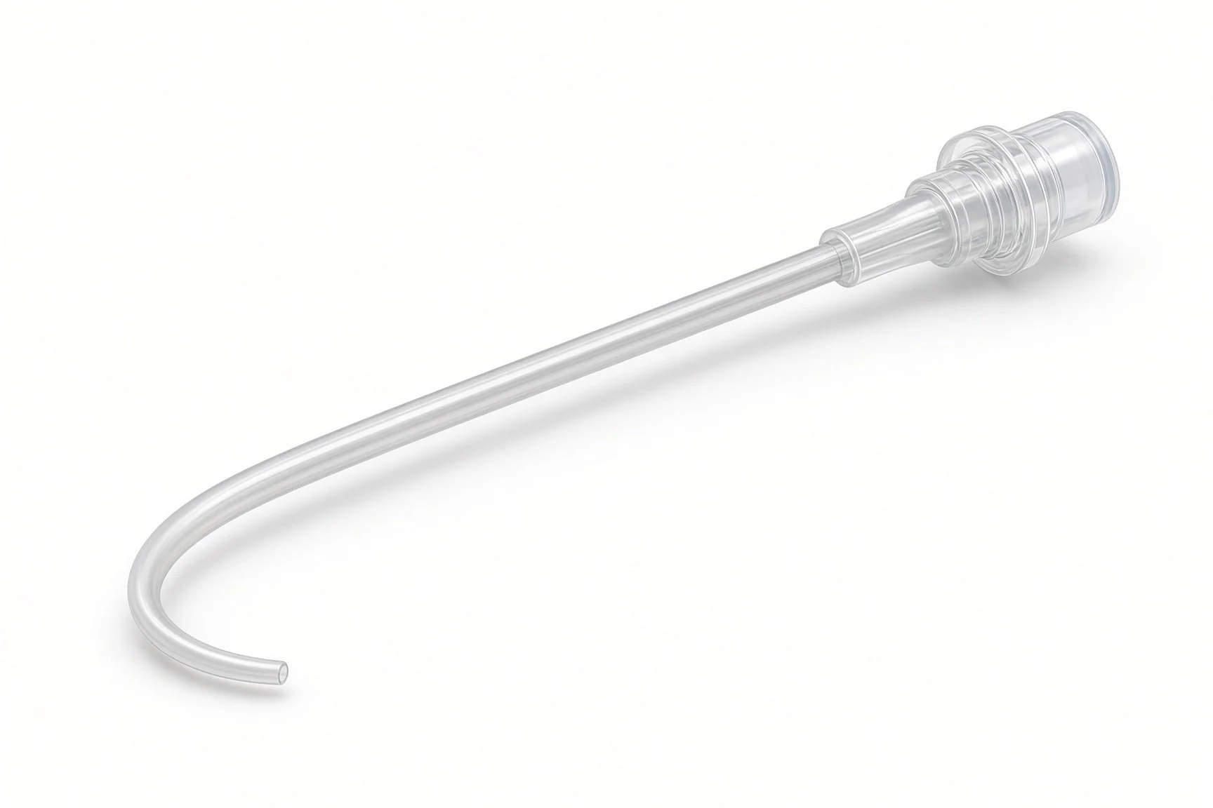
Crossover catheter and balloon.
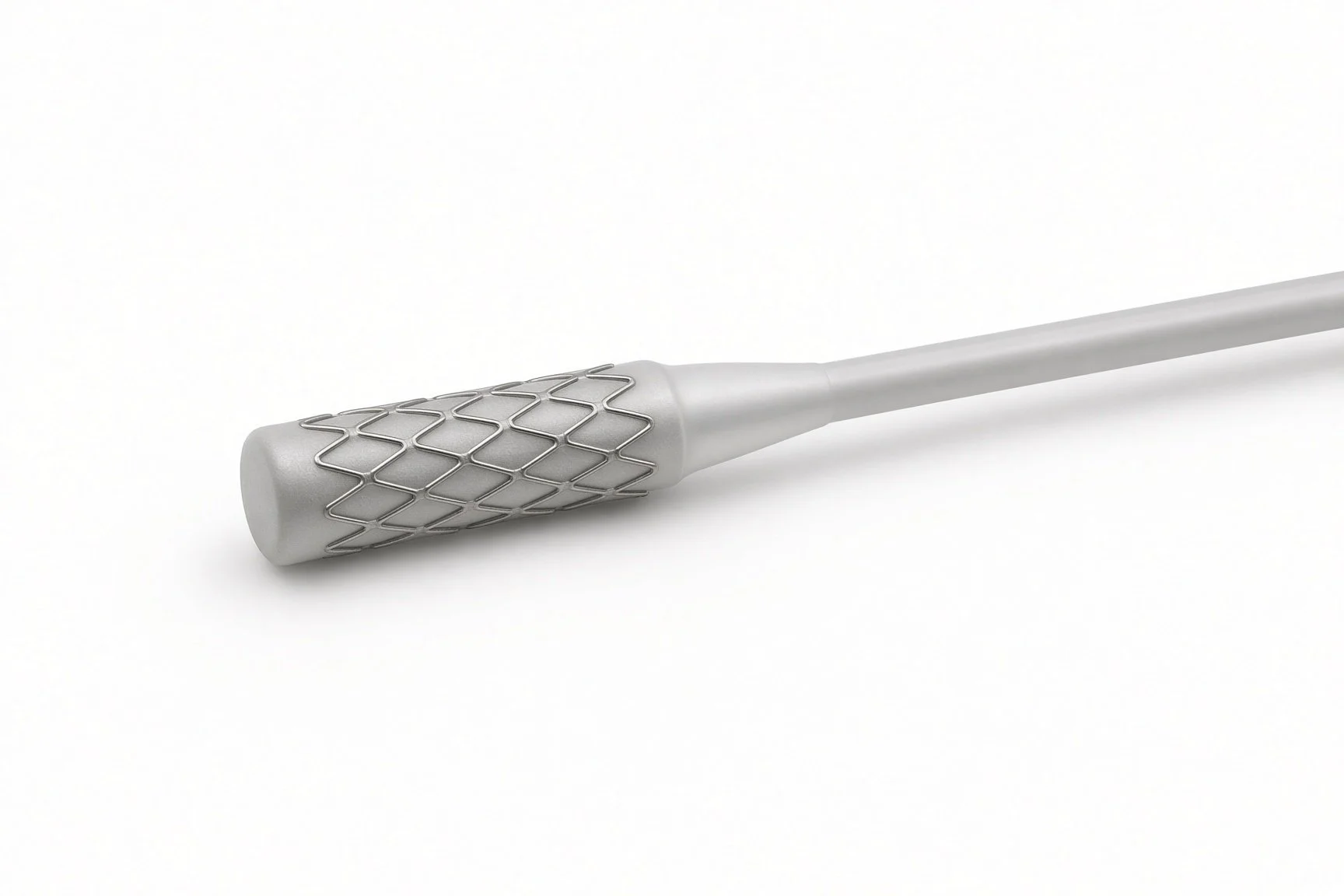
Covered stent for bail-out.
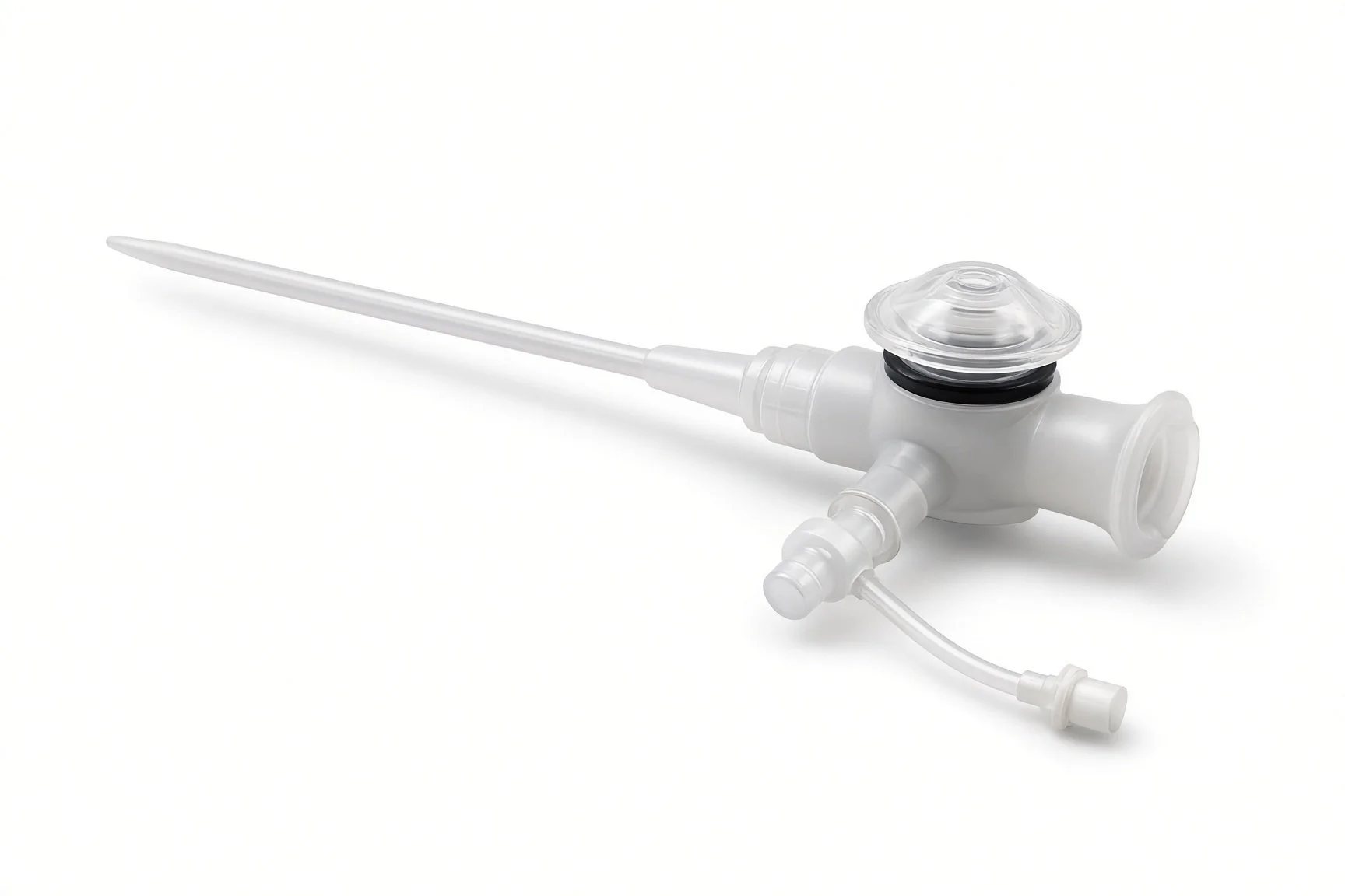
Large-bore sheath.
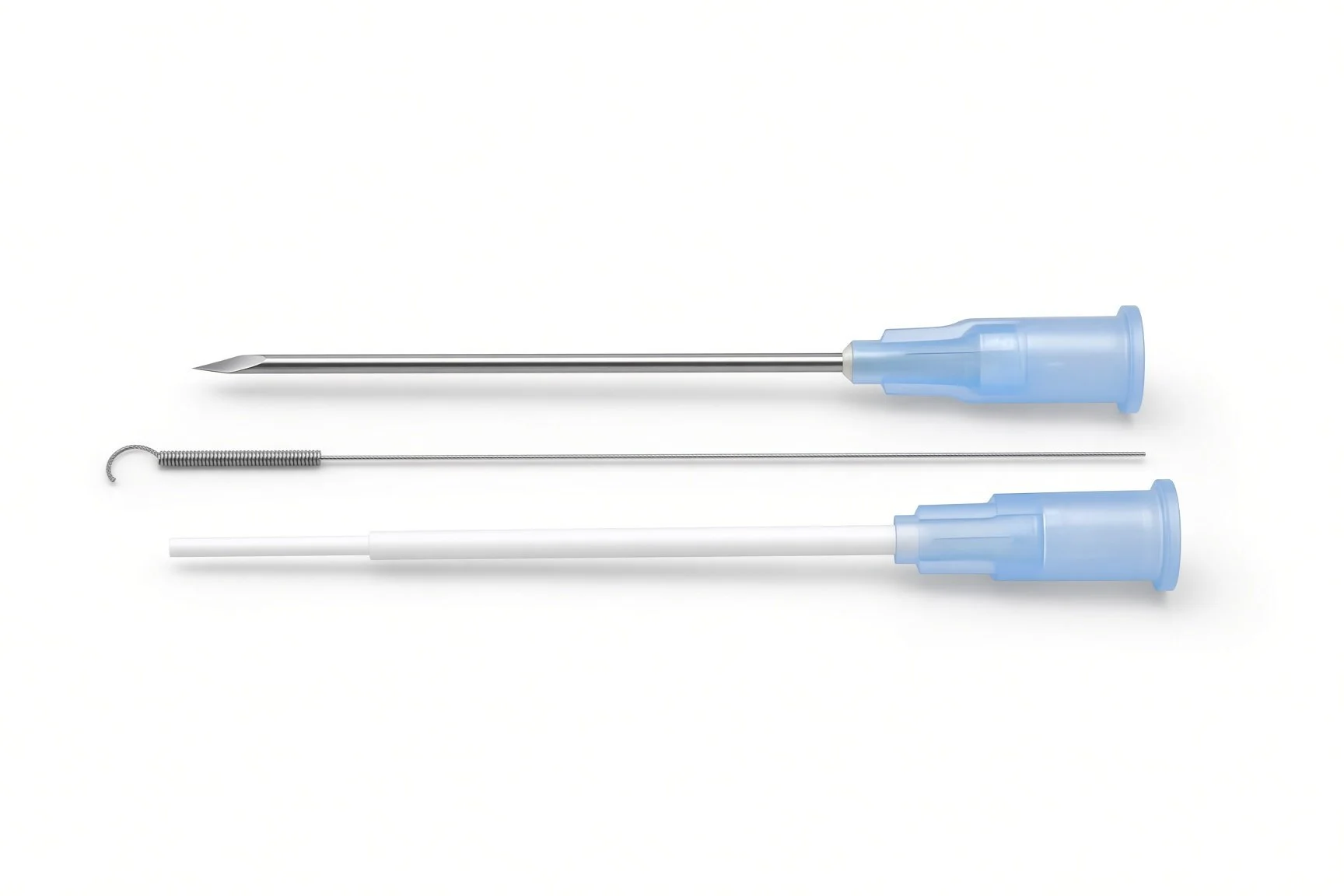
Micropuncture needle kit.
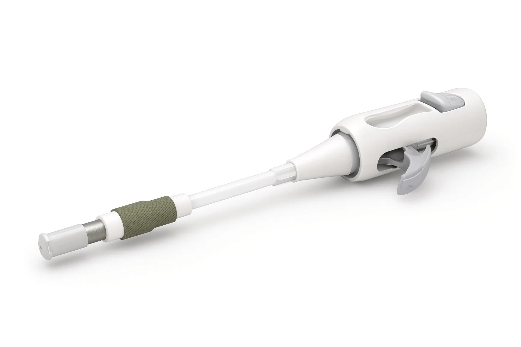
Plug-based closure device.
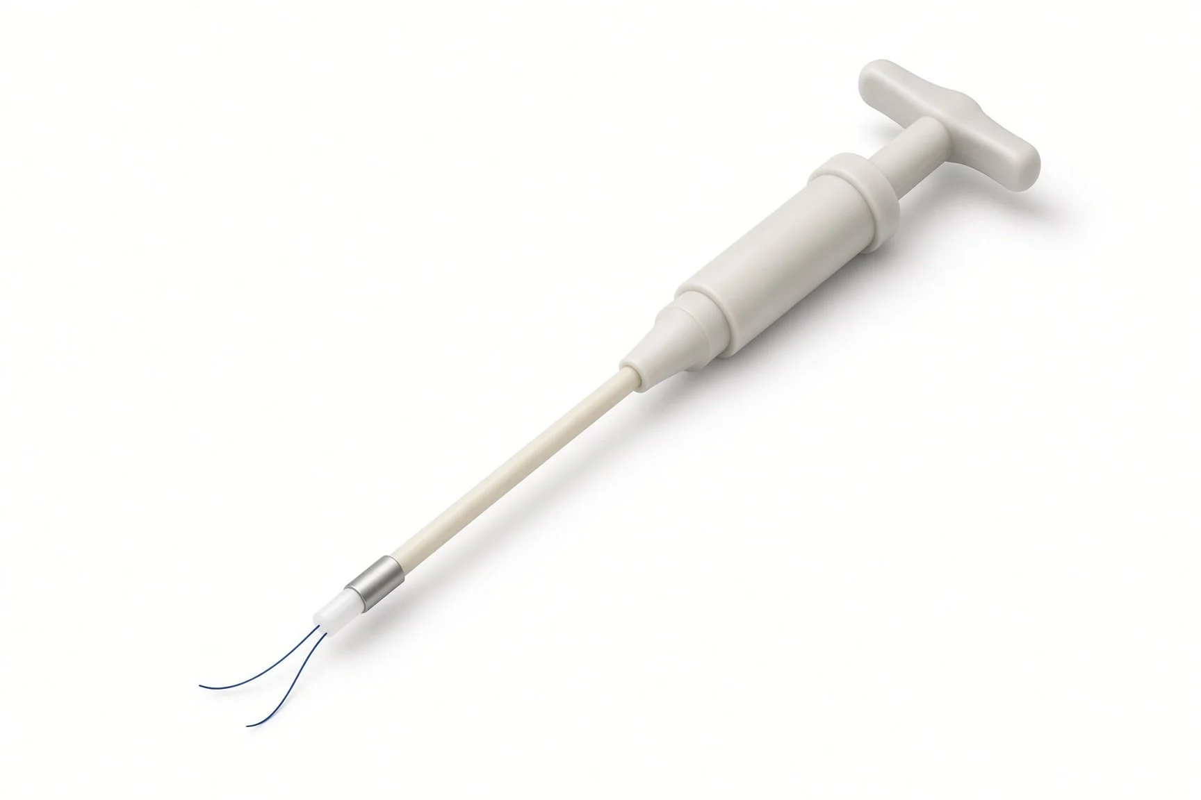
Suture-based closure devices.
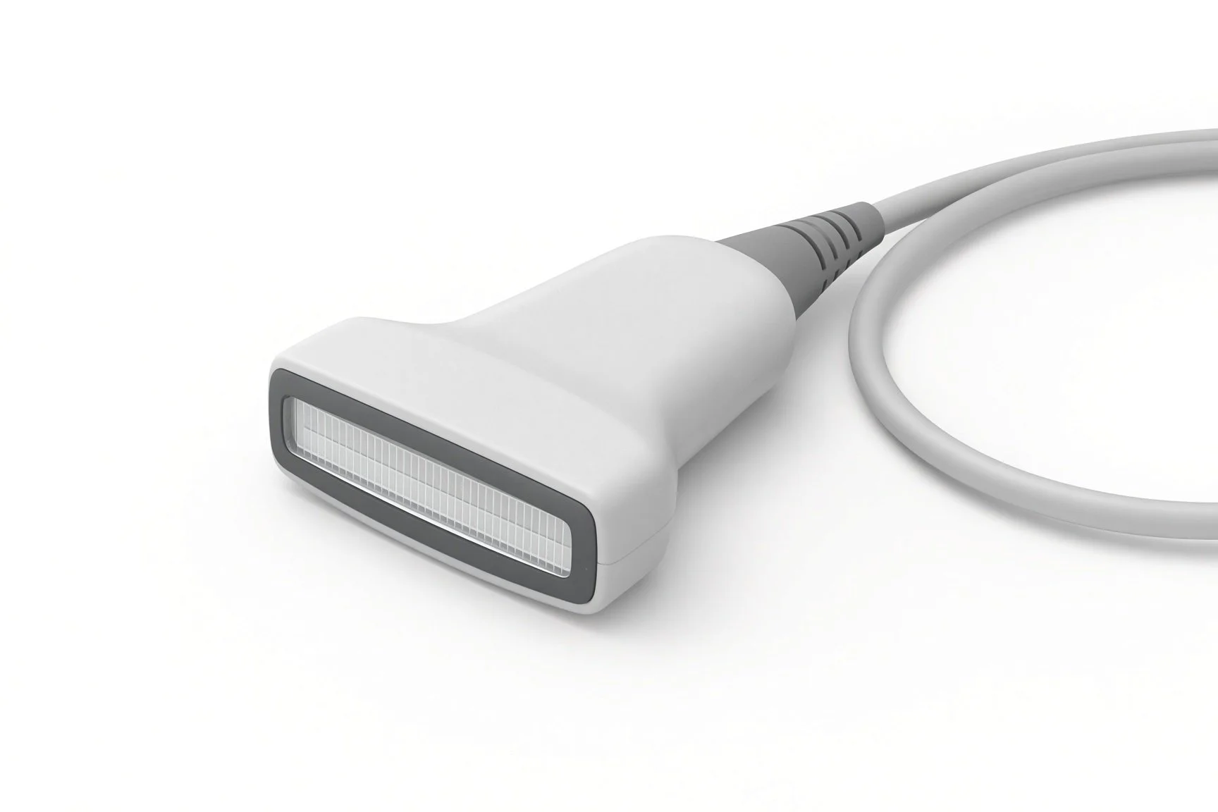
Ultrasound probe.
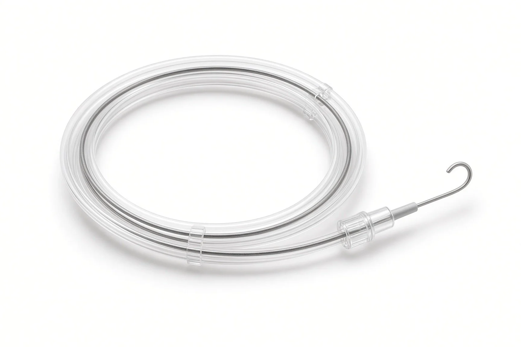
Stiff guidewire.
Procedural steps (Barts example)
Planning and set-up
Puncture
Pre-closure, sheath and closure
Bail-out options
After the procedure
First time in the cath lab?
Learn the femoral head landmark. The puncture should sit over the femoral head on X-ray; this is checked before the big sheath goes in.
Protect the sutures. Pre-closure sutures are the closure; keep them clipped, labelled and away from scissors.
Know the bail-out sequence. Ask who inflates the crossover balloon and where the covered stents are kept.
Look beyond the groin. Hidden bleeding may show only as back pain, a falling blood pressure or a rising heart rate.
Have the bail-out kit ready. Know where the crossover balloon, covered stents and long sheaths are before the big sheath goes in.
Keep the sutures tidy. Label and protect pre-closure sutures so they cannot be cut or tangled during the case.
FAQ
Why is ultrasound used for the puncture?
It shows the artery, the vein and any calcium, so the needle goes into the right place first time. This reduces bleeding.
Will I have stitches in my groin?
The artery is closed from inside with sutures or a plug, usually without skin stitches. A small dressing covers the site.
What is a crossover balloon?
A balloon passed from the other groin that can be inflated in the artery to stop bleeding quickly, if needed, while the hole is repaired.
Why does back pain matter afterwards?
Bleeding behind the abdomen may not show at the groin. Back, flank or tummy pain can be the first sign.
Related Cath Lab 101 guides
Peripheral arterial intervention (leg arteries) (coming soon)
Sources
All checked on 12 November 2026. Each page's own date is shown where it gives one.
Vahanian A, et al. 2021 ESC/EACTS Guidelines for the management of valvular heart disease. Eur Heart J 2022;43(7):561–632.
NICE. Heart valve disease presenting in adults: investigation and management (NG208). nice.org.uk/guidance/ng208
StatPearls (NCBI Bookshelf), search: femoral artery access. ncbi.nlm.nih.gov/books
StatPearls (NCBI Bookshelf), search: retroperitoneal hemorrhage. ncbi.nlm.nih.gov/books
Disclaimer
Education only. This page is for general education for healthcare students, cath lab staff and curious readers. It isn't medical advice and doesn't replace advice from your own doctor, heart team or local unit protocols. Equipment and medicines are described generically. No product or manufacturer is recommended. If you think someone is having a heart emergency, call 999.
Disclosure: I work for a company that makes a device used in this procedure. This page is independent education, isn't sponsored or reviewed by any company, and doesn't recommend any product.
All diagrams on this page are original illustrations created for rodnieoro.com.