Subclavian, vertebral and mesenteric stenting: a Cath Lab 101 guide
Reviewed by Rodnie Oro, cath lab charge nurse · Last reviewed: November 2026 · Education only, not medical advice
Subclavian, vertebral and mesenteric stenting in one minute
What it is: The subclavian arteries supply the arms and give off the vertebral arteries to the back of the brain; the mesenteric arteries supply the gut. Plaque can narrow them. Stenting opens the narrowing from inside with a balloon and a metal mesh tube (stent), passed through an artery in the groin or arm.
Who it is for: People with symptoms from the narrowing: arm pain on use, dizziness from subclavian steal, a narrowing before or after heart bypass surgery using the internal mammary artery (LIMA), symptoms from a vertebral narrowing, or chronic mesenteric ischaemia (gut pain after eating and weight loss). A vascular or heart team decides.
Main risks: Stroke (with subclavian and vertebral work), damage to the artery, bleeding at the access site and, for gut arteries, bowel injury. Your team will explain your own risk.
Usually: Under local anaesthetic, usually awake, about 1 to 2 hours, often one night in hospital.
On this page: What is subclavian, vertebral and mesenteric stenting? · Who is it for? · When is it done? · Step by step · Recovery · Risks · Kit list · First time in the lab? · FAQ
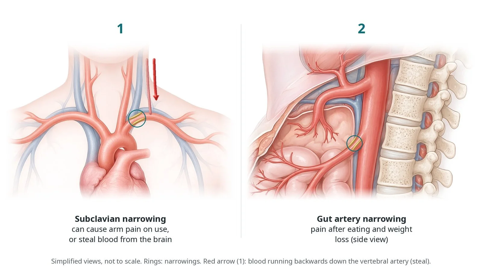
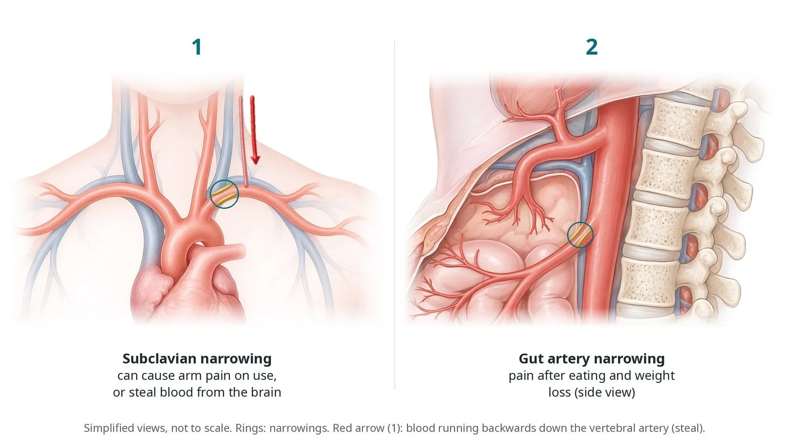
(1) A narrowed left subclavian artery can cause arm pain on use or steal blood from the brain (red arrow: flow running backwards down the vertebral artery). (2) A narrowed gut artery causes pain after eating and weight loss (side view).
What is subclavian, vertebral and mesenteric stenting?
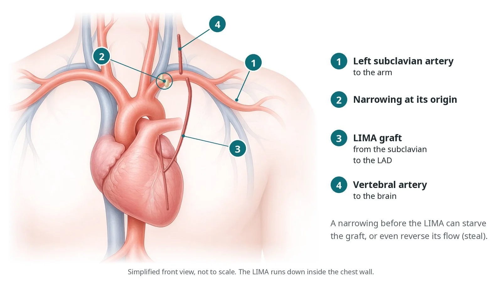
Three main branches leave the aortic arch: the brachiocephalic (innominate) artery, which divides into the right subclavian and right carotid arteries, then the left carotid and the left subclavian arteries. Each subclavian artery gives off a vertebral artery, which runs up the neck to the back of the brain, and an internal mammary (internal thoracic) artery, which runs down behind the breastbone. The left internal mammary artery (LIMA) is often used as a heart bypass graft.
A narrowing near the start of the subclavian artery, most often on the left, lowers the pressure in that arm: it may ache on use, and the blood pressure differs between the arms. If the pressure falls enough, blood can run backwards down the vertebral artery to supply the arm, 'stealing' from the brain (subclavian steal), which can cause dizziness. In someone with a LIMA bypass, the same narrowing can starve the heart of blood (coronary-subclavian steal).
The gut is supplied by the coeliac trunk and the superior mesenteric artery (SMA), which leave the front of the abdominal aorta and point downwards, and the inferior mesenteric artery lower down. Because they are linked, symptoms usually appear when two or more are narrowed. Chronic mesenteric ischaemia causes cramping tummy pain within an hour of eating, fear of eating and weight loss.
New to the acronyms? See the Cath Lab Abbreviations A–Z.
Who is it for?
Subclavian or innominate stenting may be considered for:
arm pain on use, or symptoms of subclavian steal
a narrowing before heart bypass surgery using the LIMA, or one affecting an existing LIMA graft
some people who need the arm for dialysis access
Vertebral stenting is less common, for selected people with symptoms despite best medical treatment.
For chronic mesenteric ischaemia, stenting is usually the first treatment, decided by a vascular team.
Who it may not suit: narrowings without symptoms, which usually only need medicines (unless a LIMA bypass is planned) sudden (acute) mesenteric ischaemia with bowel damage, which needs emergency surgery long blockages better treated with bypass surgery
The decision is made by your heart team, who will talk through the reasons, the alternatives and the risks with you.
When is it done?
It is planned after a CT or MR angiogram or a duplex scan. Before it, the team checks the blood pressure in both arms, kidney function, allergies and medicines, especially antiplatelets. People with mesenteric ischaemia are often underweight, so nutrition is reviewed.
gives you a chance to ask questions and give consent
arranges blood tests, including kidney function and a blood count, and an ECG
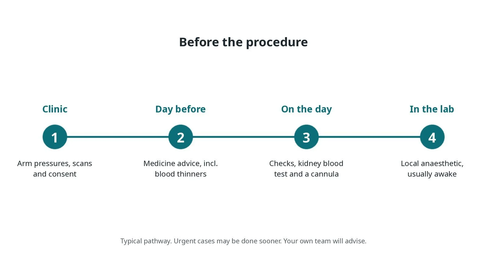
Before the procedure: arm pressures, scans and consent; medicine advice; checks and a kidney blood test; local anaesthetic, usually awake.
Step by step
1. Getting ready
You lie on the X-ray table, usually awake with local anaesthetic, so the team can check your speech and movement during subclavian or vertebral work. Monitoring includes ECG, blood pressure (on the arm that is not being treated) and oxygen levels.
2. Access
The femoral artery in the groin, or the radial or brachial artery in the arm, is used, under ultrasound. Arm access helps with the gut arteries, which point downwards; for a blocked subclavian artery, groin and arm may both be used. Heparin is given, as per centre protocol.
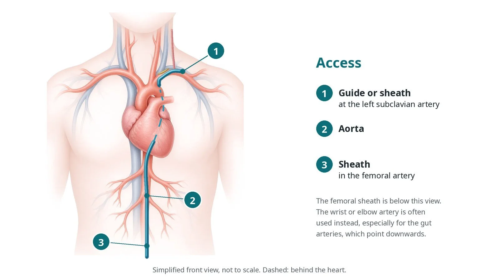
Access: (1) guide or sheath at the left subclavian artery; (2) aorta; (3) sheath in the femoral artery. The wrist or elbow artery is often used instead, especially for the gut arteries.
3. Stenting
A. Engage. A guide catheter or long sheath is placed at the opening of the artery.
B. Wire. A guidewire crosses the narrowing or blockage.
C. Stent. A balloon-expandable stent is usually placed at the opening (ostium), where it can be positioned precisely; self-expanding stents may be used further along. Vertebral narrowings usually need a small, coronary-type stent.
D. Check. An angiogram checks the result and nearby branches, such as the vertebral and internal mammary arteries, and the blood pressures in both arms may be compared.
Watch: New speech difficulty, facial droop or weakness during the case: tell the operator at once; the team will assess the patient and image the brain arteries, as per centre stroke protocol.
4. Finishing
The sheath is removed and the artery is closed with pressure, a closure device or a radial band, per operator. Pulses and neurology are checked again.
5. Afterwards
Checks continue: the access site, pulses in the treated arm, neurology and blood pressure. After mesenteric stenting, tummy pain, a tense tummy or blood in the stools are reported straight away, and eating is built up gradually as advised.
Watch: Severe tummy pain after mesenteric stenting, especially pain out of proportion to the examination: get urgent medical and surgical review, as this may be bowel ischaemia.
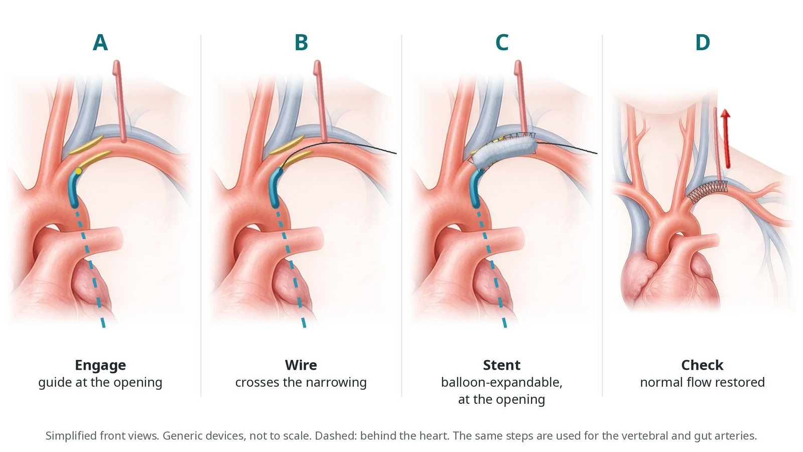
A: guide at the artery opening. B: wire across the narrowing. C: balloon-expandable stent at the opening. D: normal flow restored.
Recovery and aftercare
First hours: checks of the access site, arm pulses, neurology and blood pressure, as per centre protocol.
Hospital stay: often one night; longer after gut artery stenting if eating needs building up.
Medicines: antiplatelet tablets (often two for a time) and a statin, as prescribed.
Eating: after gut artery stenting, meals can usually be increased gradually; weight gain is a good sign.
Driving: ask your team, and follow DVLA guidance if you had stroke or TIA symptoms.
Follow-up: clinic review and a duplex ultrasound or CT scan to check the stent.
Call 999 if you have signs of a stroke, severe tummy pain, chest pain, heavy bleeding, or a cold, painful arm.
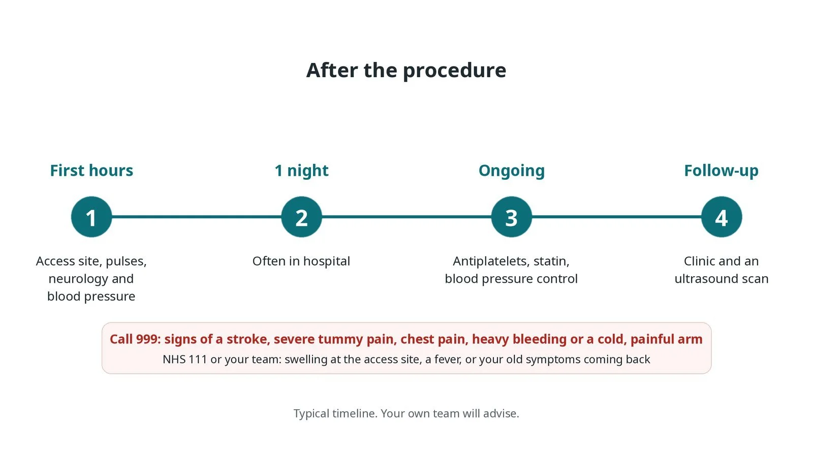
After the procedure: (1) access site, pulse, neurology and blood pressure checks; (2) often one night in hospital; (3) antiplatelets, statin and blood pressure control; (4) clinic and ultrasound follow-up.
Risks and complications
Your team will explain the risks that apply to you. Possible problems include:
Bleeding or bruising at the access site
Stroke or TIA (uncommon): Mainly with subclavian, innominate and vertebral work.
Tear (dissection) or blockage of the artery (uncommon)
Stent moving or not fully covering the opening (rare)
Bowel injury from a blocked or torn gut artery branch (rare)
Kidney strain from the contrast dye
The artery narrowing again later (restenosis): Report symptoms that come back.
Cath lab kit list (printable)
Barts example kit list
An example from one UK centre, written generically. Every lab has its own protocol, and sizes and doses vary.
arterial sheaths for the groin or arm, size per operator
ultrasound for access
guide catheters or long sheaths with different tip shapes
guidewires, per operator
balloons for pre-dilation, per operator
balloon-expandable stents for openings, per operator
self-expanding stents, per operator
coronary-type stents for vertebral narrowings, if needed
embolic protection filter, if used, per operator
inflation device, contrast and heparin, as per centre protocol
radial band or closure device, per operator
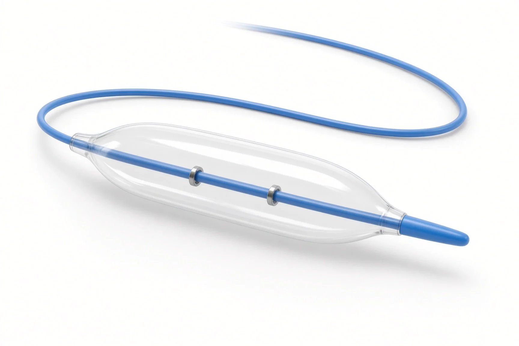
Balloon-expandable stent.
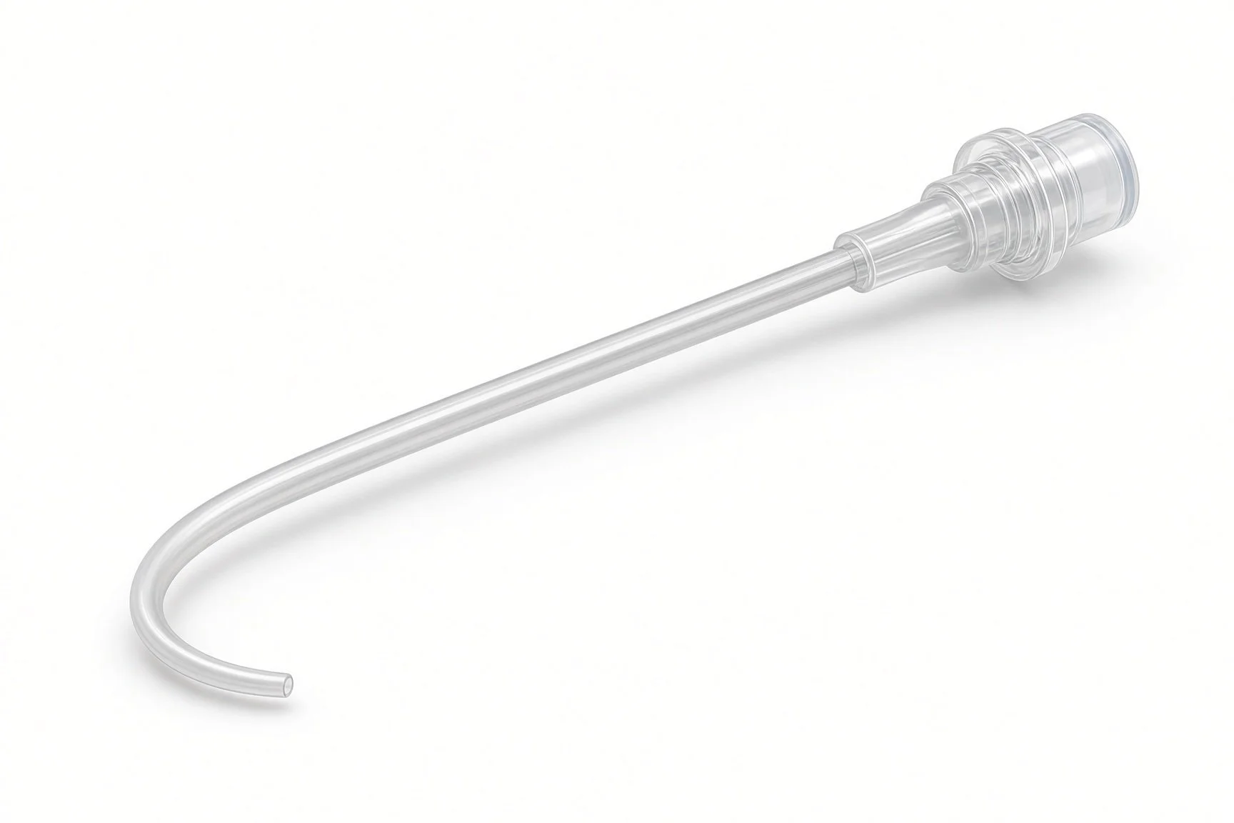
Guide catheter or long sheath.
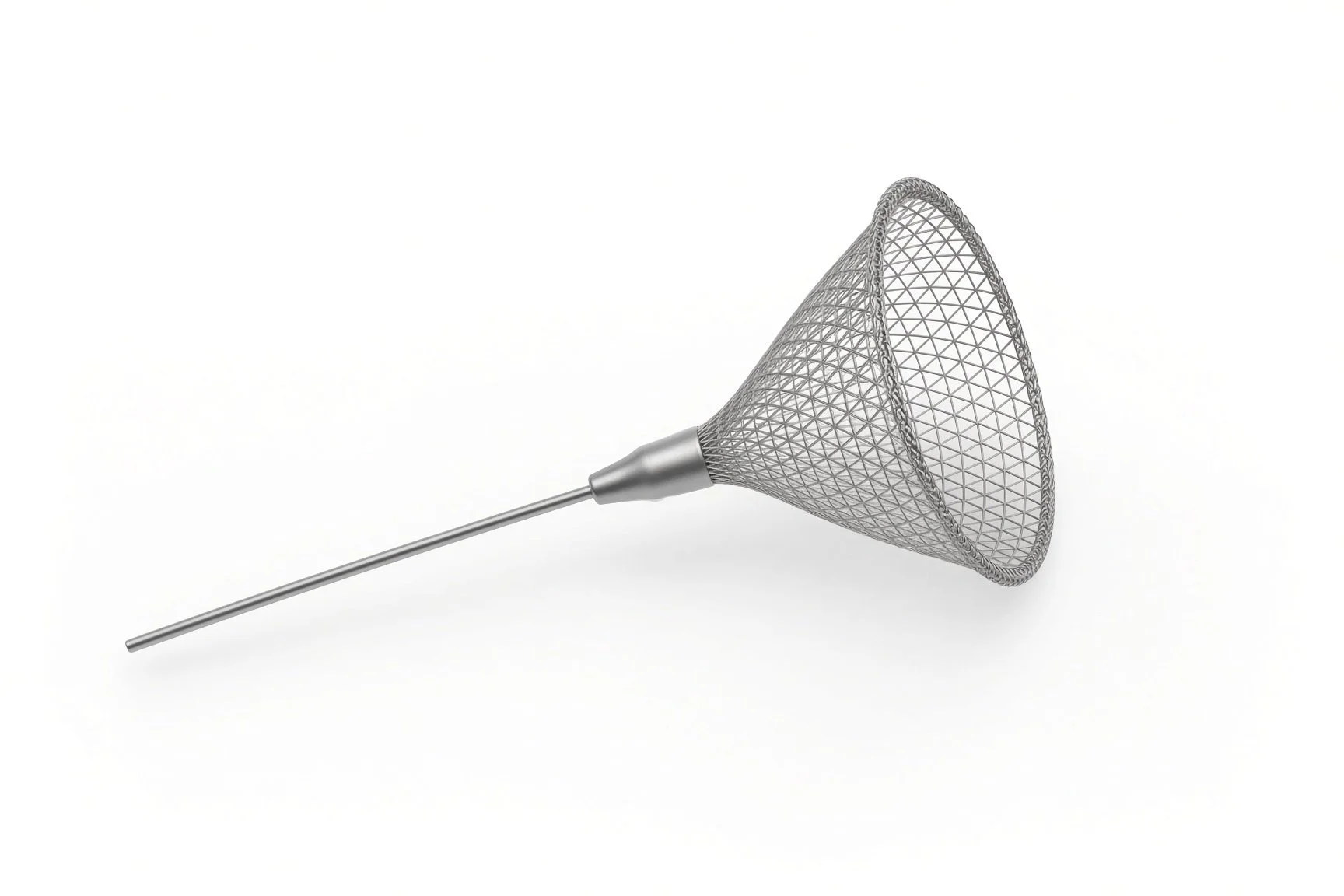
Embolic protection filter if used.
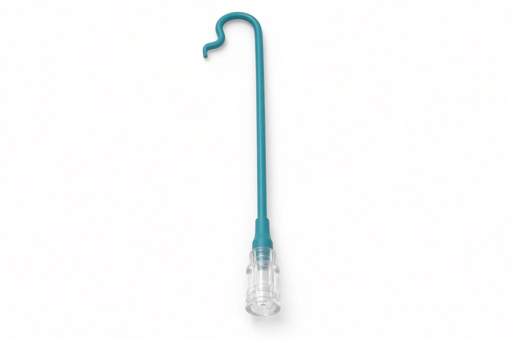
Guide catheter or long sheath.
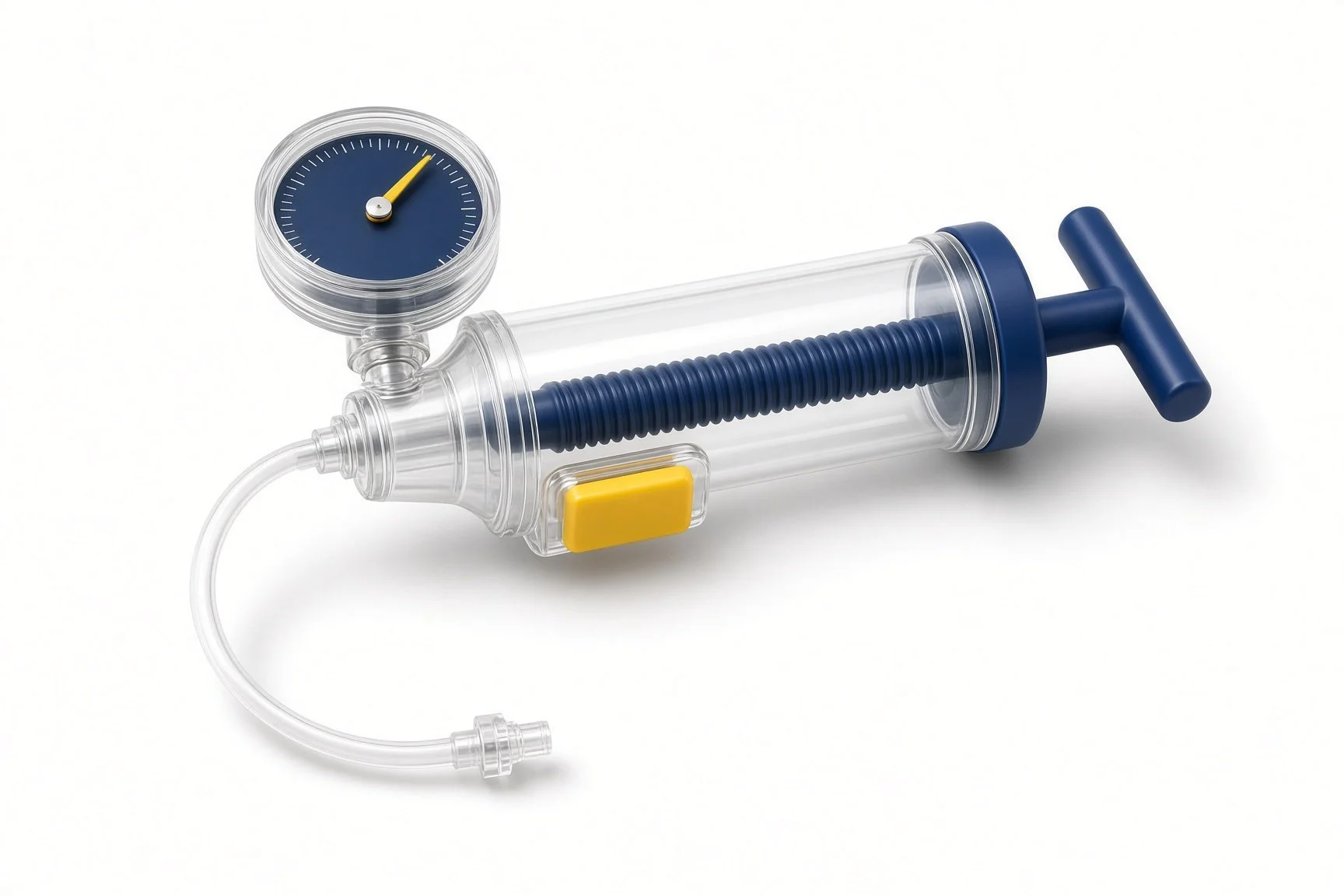
Inflation device.
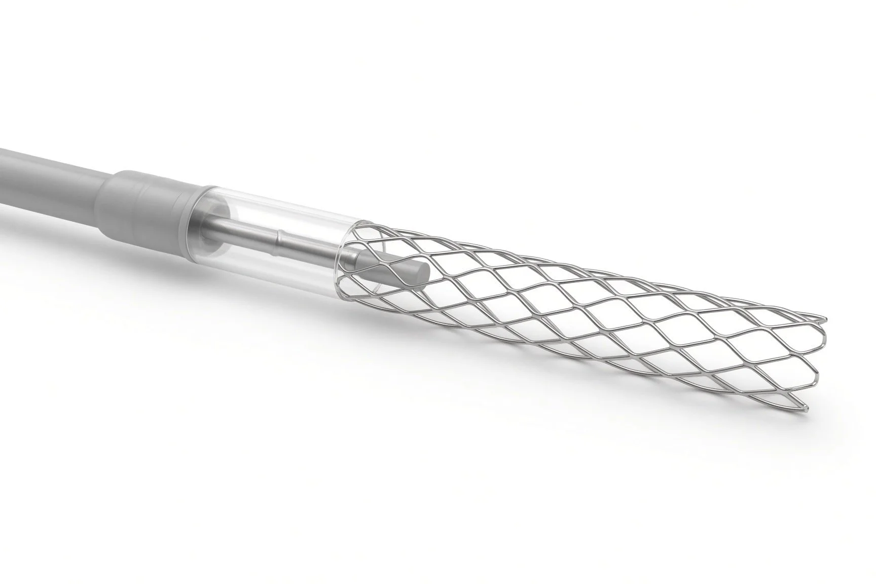
Self-expanding stent.
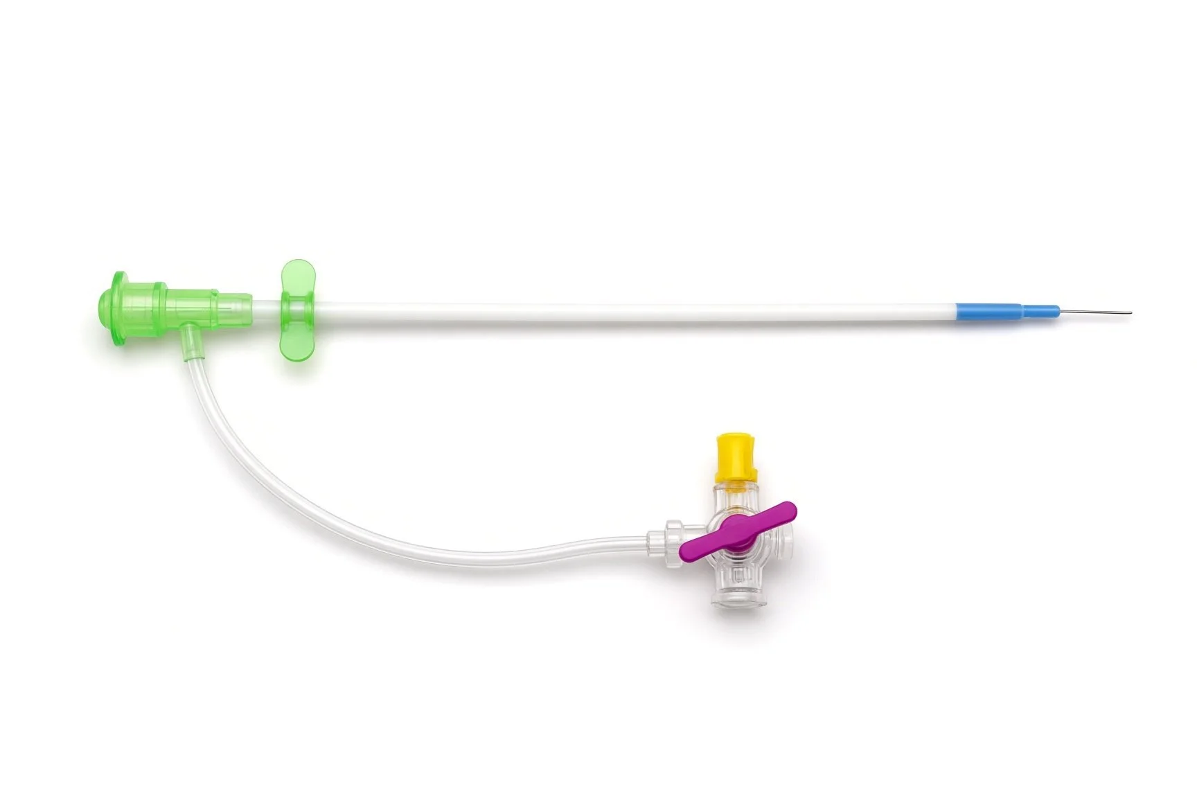
Arterial sheath.
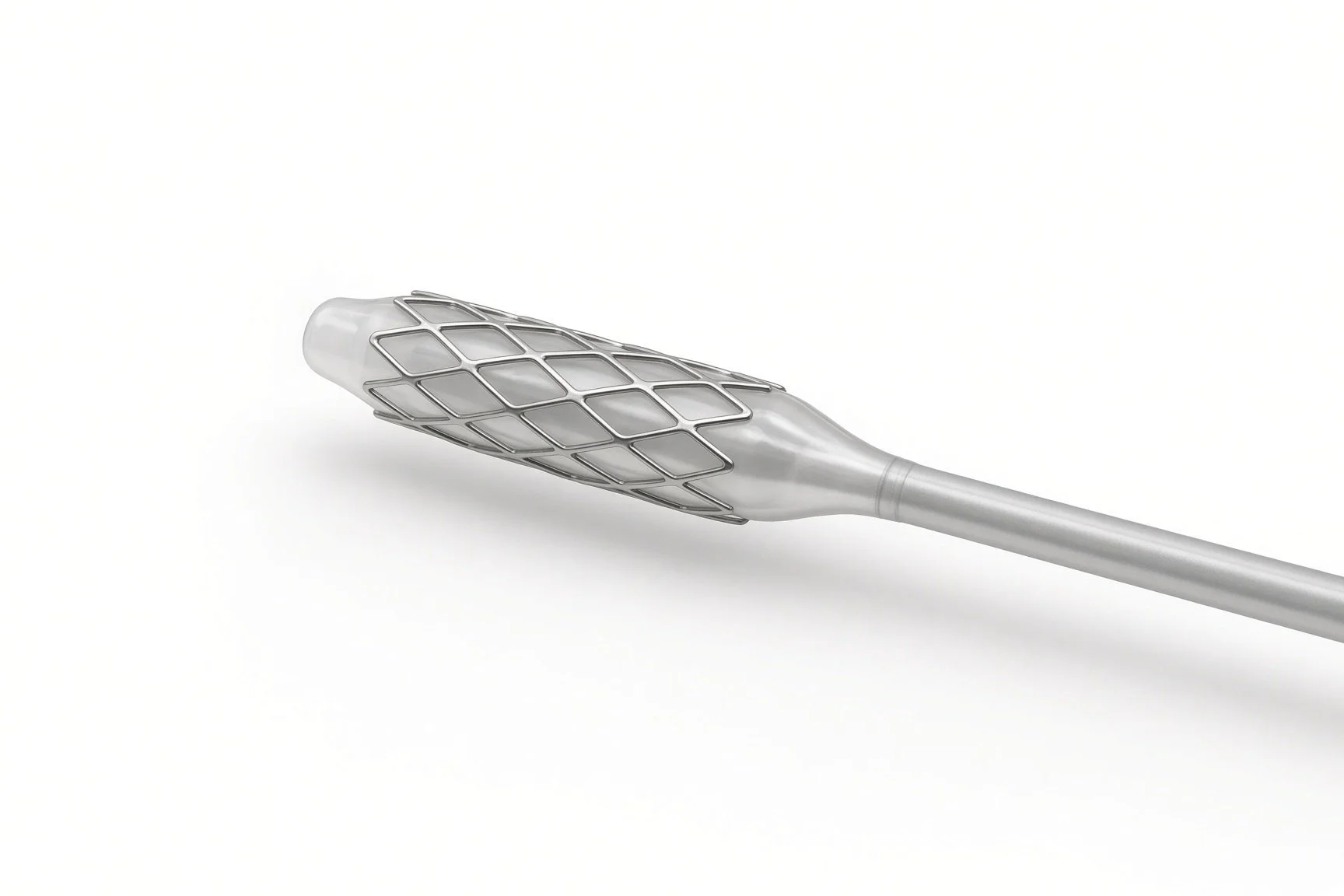
Balloon-expandable stent.
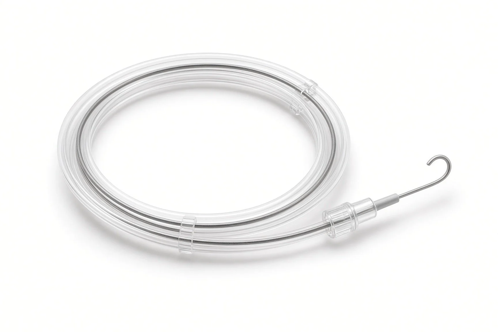
Guidewires.
Procedural steps (Barts example)
Getting ready
Access
Stenting
Finishing
Afterwards
First time in the cath lab?
Check both arm pressures. Note the difference before and after; it is part of the result.
Keep the patient talking. In subclavian and vertebral cases, simple questions and a hand squeeze help spot a stroke early.
Plan the access. Arm, groin or both: know which sites are prepared and where the second sheath goes.
Ask about eating. People with gut artery narrowing are often thin and frail; take extra care with pressure areas and fluids.
Use the other arm for the cuff. The blood pressure in the arm beyond a subclavian narrowing reads falsely low.
With arm access, keep the access arm free and reachable, and know the plan for stopping bleeding at the end.
FAQ
Why stay awake?
So the team can check your speech and movement during the procedure.
Will my arm pain go away?
Usually it improves quickly once the narrowing is opened.
Will I be able to eat normally after gut artery stenting?
Many people eat more comfortably within days to weeks and start to gain weight. Tell your team if pain after eating comes back.
Why stent before heart bypass surgery?
If the internal mammary artery is used as a bypass graft, it needs good flow from the subclavian artery to supply the heart.
Related Cath Lab 101 guides
Internal mammary angiography and subclavian stenosis (coming soon)
Peripheral arterial intervention (leg arteries) (coming soon)
Sources
All checked on 12 November 2026. Each page's own date is shown where it gives one.
Mazzolai L, et al. 2024 ESC Guidelines for the management of peripheral arterial and aortic diseases. Eur Heart J 2024.
Björck M, et al. Management of the diseases of mesenteric arteries and veins: clinical practice guidelines of the European Society for Vascular Surgery (ESVS). Eur J Vasc Endovasc Surg 2017.
StatPearls (NCBI Bookshelf), search: subclavian steal syndrome. ncbi.nlm.nih.gov/books
StatPearls (NCBI Bookshelf), search: chronic mesenteric ischemia. ncbi.nlm.nih.gov/books
Disclaimer
Education only. This page is for general education for healthcare students, cath lab staff and curious readers. It isn't medical advice and doesn't replace advice from your own doctor, heart team or local unit protocols. Equipment and medicines are described generically. No product or manufacturer is recommended. If you think someone is having a heart emergency, call 999.
Disclosure: I work for a company that makes a device used in this procedure. This page is independent education, isn't sponsored or reviewed by any company, and doesn't recommend any product.
All diagrams on this page are original illustrations created for rodnieoro.com.