IVC filter insertion and retrieval: a Cath Lab 101 guide
Reviewed by Rodnie Oro, cath lab charge nurse · Last reviewed: November 2026 · Education only, not medical advice
IVC filter insertion and retrieval in one minute
What it is: The inferior vena cava (IVC) is the large vein carrying blood from the legs and abdomen to the heart. An IVC filter is a small, umbrella-shaped metal device placed in it through a vein in the neck or groin. It traps large clots travelling up from the legs before they reach the lungs. Most filters used now can be removed.
Who it is for: People with a recent deep vein thrombosis (DVT) or pulmonary embolism (PE) who cannot have blood thinners, for example because of bleeding or urgent surgery, and a few others selected by a specialist team. Blood thinners remain the main treatment whenever they can be given.
Main risks: Bruising at the access site, the filter tilting, moving or poking through the vein wall, clot forming at the filter, and difficulty removing it, especially if it stays in a long time. Your team will explain your own risk.
Usually: Under local anaesthetic, about 30 to 60 minutes, often in interventional radiology; often home the same or next day if otherwise well.
On this page: What is IVC filter insertion and retrieval? · Who is it for? · When is it done? · Step by step · Recovery · Risks · Kit list · First time in the lab? · FAQ
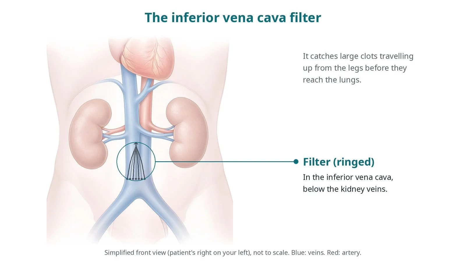
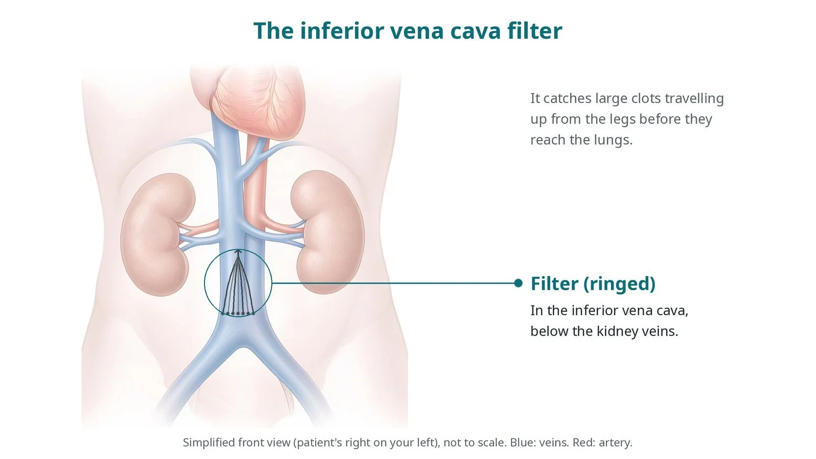
The filter (ringed) sits in the inferior vena cava below the kidney veins. It catches large clots travelling up from the legs before they reach the lungs.
What is IVC filter insertion and retrieval?
The inferior vena cava runs up the right side of the spine, next to the aorta, from where the two iliac veins join in the pelvis to the right atrium of the heart. The renal veins from the kidneys join it at about the level of the first and second lumbar vertebrae; the left renal vein crosses in front of the aorta to reach it.
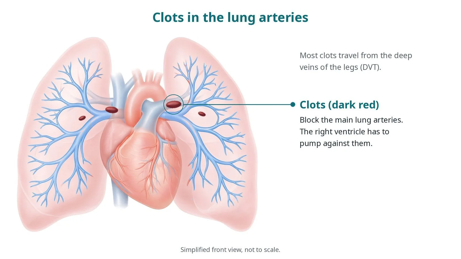
A DVT is a clot in the deep veins of the leg or pelvis. Pieces can break off, travel up the IVC and through the right heart, and lodge in the lung arteries as a pulmonary embolism. Blood thinners (anticoagulants) stop new clot forming while the body clears the old one, and are the treatment of choice.
When blood thinners cannot be used, a filter can be placed in the IVC, usually just below the renal veins (infrarenal), so it catches clots from the legs without risking the kidney veins. A typical retrievable filter is cone-shaped, with a hook at its top for removal and thin legs that hold it against the vein wall. A filter does not treat the clot or stop new clots forming in the legs, so blood thinners are started as soon as it is safe, and the filter is removed when it is no longer needed.
New to the acronyms? See the Cath Lab Abbreviations A–Z.
Who is it for?
UK guidance (NICE NG158) supports a filter mainly for people with a DVT in the thigh or pelvis, or a PE, who cannot have blood thinners, with a plan to remove it once blood thinners can be started. Examples:
bleeding, or a high risk of bleeding, at the time of a new DVT or PE
urgent surgery soon after a DVT or PE, when blood thinners must be stopped
rarely, a new PE despite well-managed blood thinners, after review by a specialist team
Who it may not suit: people who can safely take blood thinners: a filter is not a substitute for them clot extending up the IVC beyond the planned filter site, which may need a different position or plan
The decision is made by your heart team, who will talk through the reasons, the alternatives and the risks with you.
When is it done?
It can be urgent or planned. Before it, the team reviews the scan showing the clot, checks the blood count and clotting, and agrees a date or trigger for removal. Retrieval is planned as soon as blood thinners are established or the risk has passed.
gives you a chance to ask questions and give consent
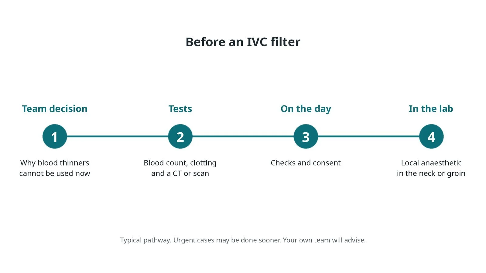
Before an IVC filter: a team decision on why blood thinners cannot be used; blood count, clotting and scans; checks and consent; local anaesthetic in the neck or groin.
Step by step
1. Getting ready
You lie on your back on the X-ray table. Monitoring includes ECG, blood pressure and oxygen levels. The neck or groin is cleaned and local anaesthetic is given; sedation is not usually needed.
2. Access
Under ultrasound, the operator places a sheath in the right internal jugular vein in the neck or the femoral vein in the groin. Sizes are per device.
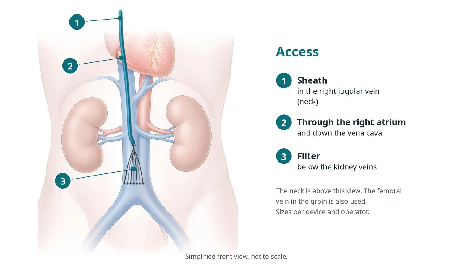
Access: (1) sheath in the right jugular vein in the neck; (2) through the right atrium and down the vena cava; (3) filter below the kidney veins. The femoral vein is also used.
3. Placing the filter
A. Cavogram. A catheter is placed in the IVC and contrast is injected to show its size, the renal veins and any clot.
B. Deploy. The filter delivery system is advanced through the sheath and positioned so the filter sits below the lowest renal vein; the sheath is pulled back to release it.
C. In place. The filter opens against the vein wall, and a final picture checks its position and tilt.
D. Retrieval later. When the filter is no longer needed, a snare or cone device passed through a sheath, usually in the neck, grabs the hook and the filter is pulled into the sheath and removed.
Watch: Breathlessness, chest pain or a sudden fall in oxygen levels during the case: tell the operator; clot may have moved to the lungs.
4. Finishing
The sheath is removed and firm pressure is applied until bleeding stops. The filter type, position and planned removal date are recorded in the notes and the filter tracking system, as per centre protocol.
5. Retrieval
Removal is usually a separate short procedure under local anaesthetic, from the jugular vein. A cavogram first checks for clot trapped in the filter; a large trapped clot may delay removal. If the filter has tilted or grown into the wall, advanced retrieval techniques may be needed, sometimes in a specialist centre.
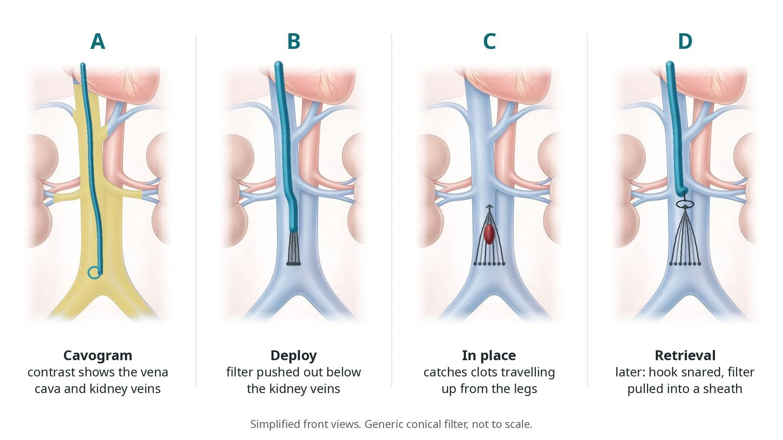
A: a cavogram shows the vena cava and kidney veins. B: the filter is pushed out below the kidney veins. C: in place, it catches clots. D: later, the hook is snared and the filter pulled into a sheath.
Recovery and aftercare
First hours: rest with checks of the neck or groin site, as per centre protocol; most people can sit up soon.
Going home: often the same or next day, depending on why the filter was needed.
Medicines: blood thinners are started or restarted as soon as your team says it is safe.
Removal: a separate short procedure is planned once you are on blood thinners or the risk has passed; keep this appointment.
Activity and driving: normal activity when you feel well; ask your team about driving.
Follow-up: carry any filter information you are given and tell other doctors you have a filter.
Call 999 if you have sudden breathlessness, chest pain, fainting, coughing up blood, or heavy bleeding.
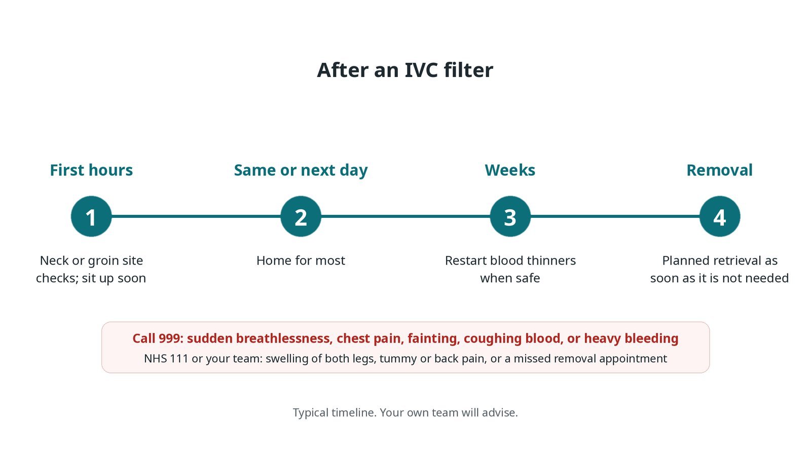
After an IVC filter: (1) neck or groin checks; (2) home the same or next day for most; (3) restart blood thinners when safe; (4) planned retrieval as soon as it is not needed.
Risks and complications
Your team will explain the risks that apply to you. Possible problems include:
Bruising or bleeding at the access site
Filter tilting or moving (uncommon)
Filter legs poking through the vein wall (uncommon)
Clot forming at or below the filter, causing leg swelling (uncommon)
Difficult or failed removal, more likely the longer it stays in (uncommon)
Part of the filter breaking off and moving (rare)
Cath lab kit list (printable)
Barts example kit list
An example from one UK centre, written generically. Every lab has its own protocol, and sizes and doses vary.
ultrasound machine and sterile probe cover
venous sheath and dilator, per device
guidewire, per operator
pigtail or marker catheter for the cavogram
IVC filter and delivery system, jugular or femoral version, per device
contrast and injector
retrieval snare or cone retrieval device with long sheath, for removal
local anaesthetic and dressing
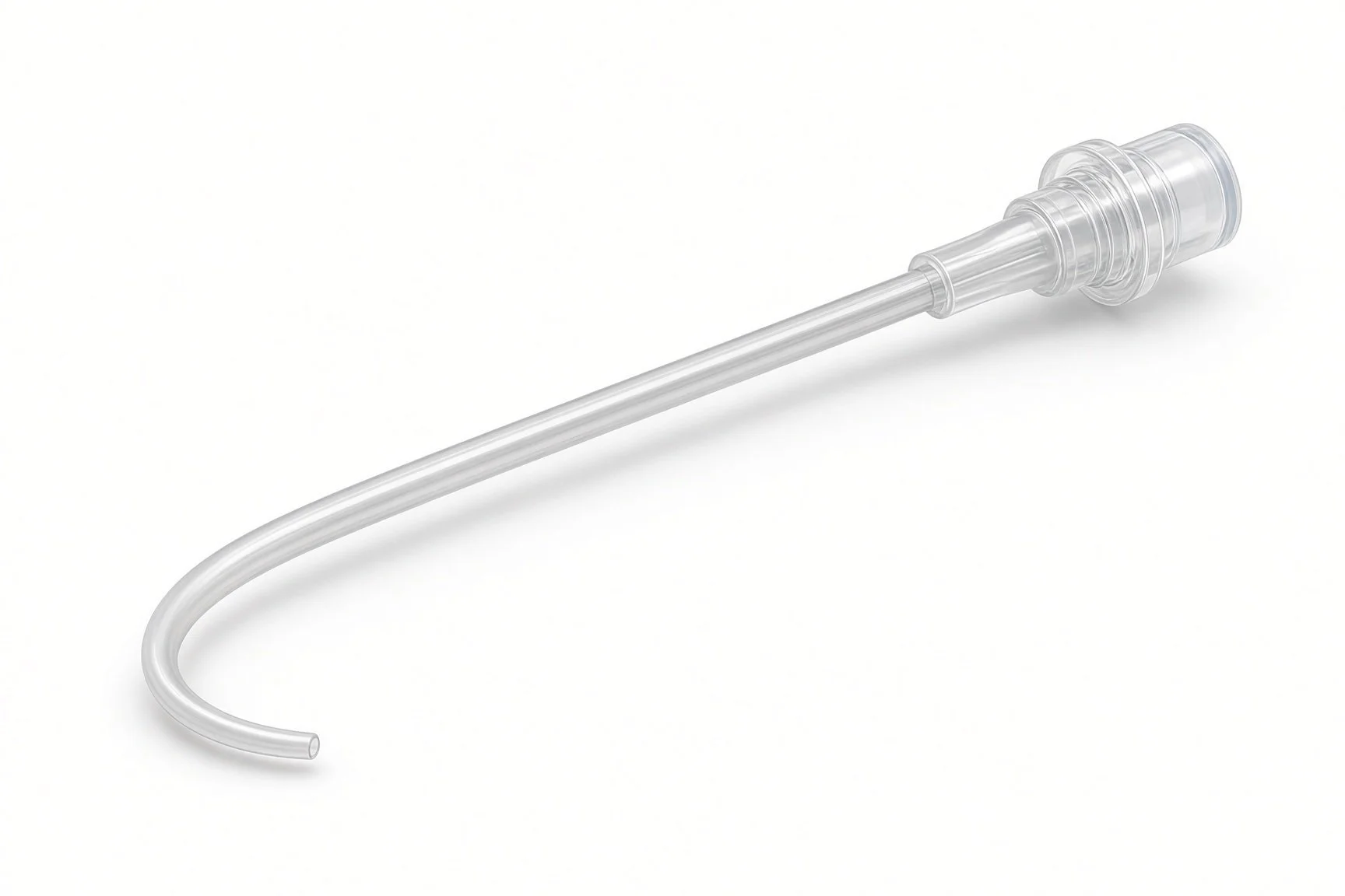
Pigtail catheter for the cavogram.
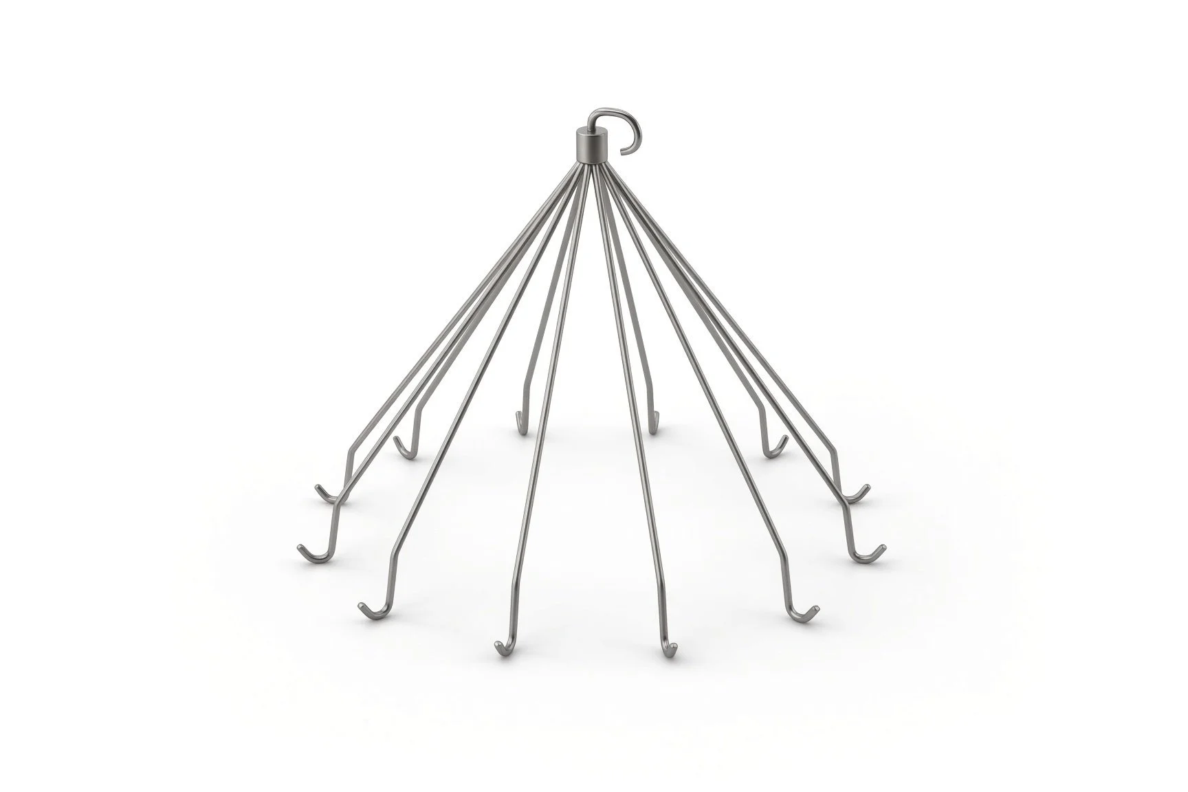
IVC filter and delivery system.
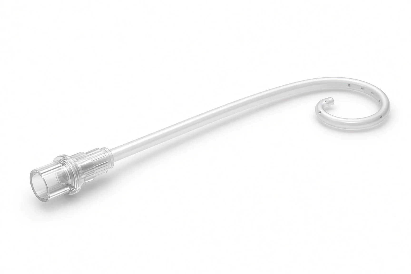
Pigtail catheter for the cavogram.
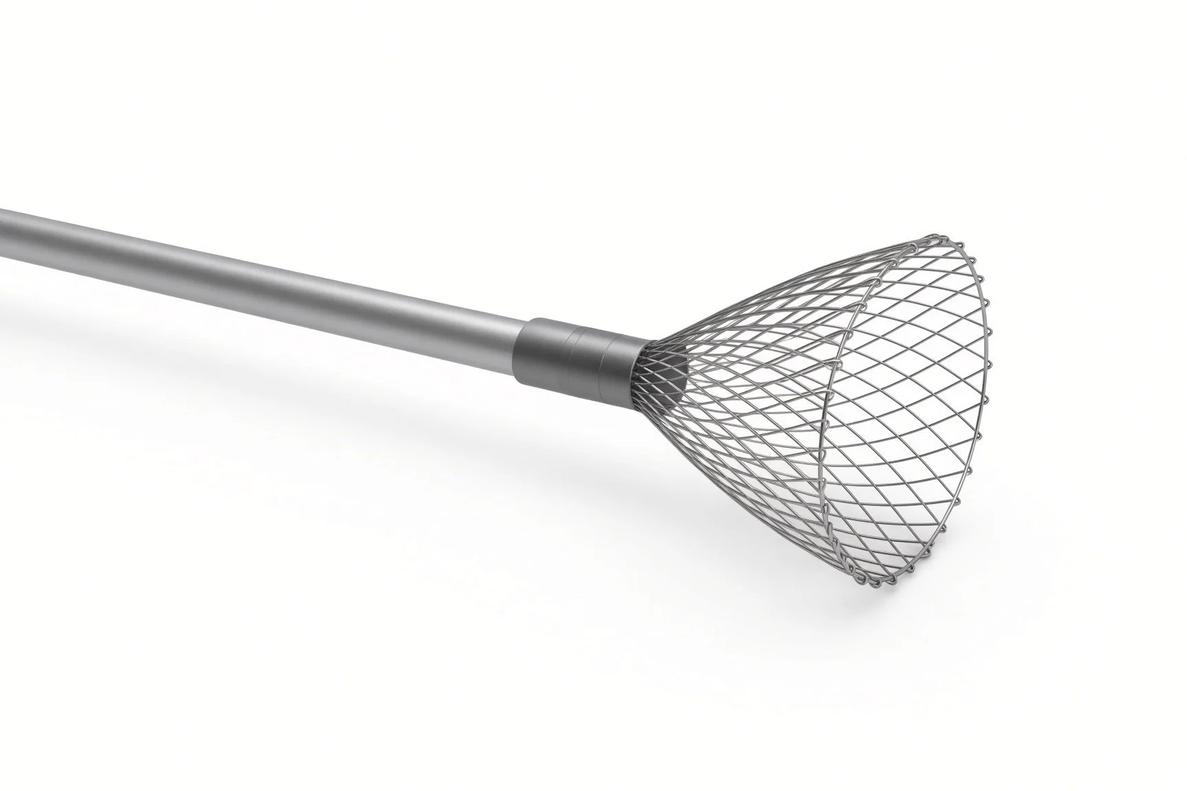
Retrieval cone device.
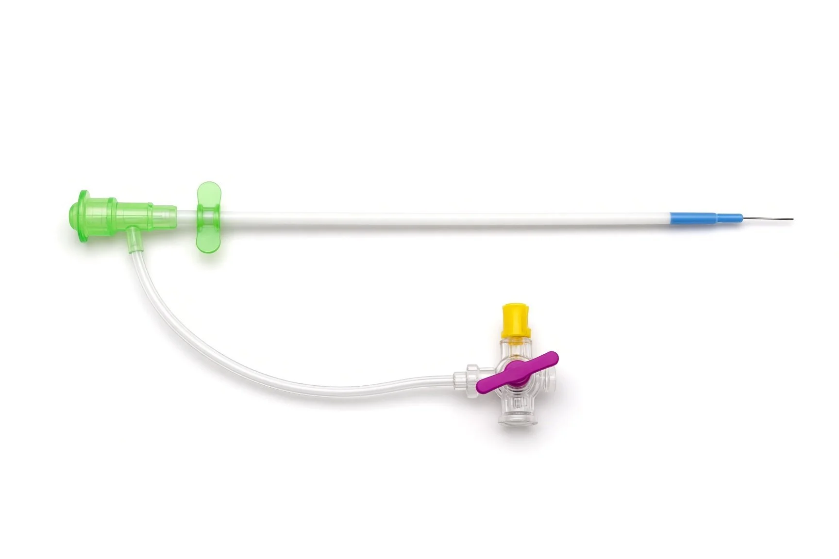
Venous sheath.
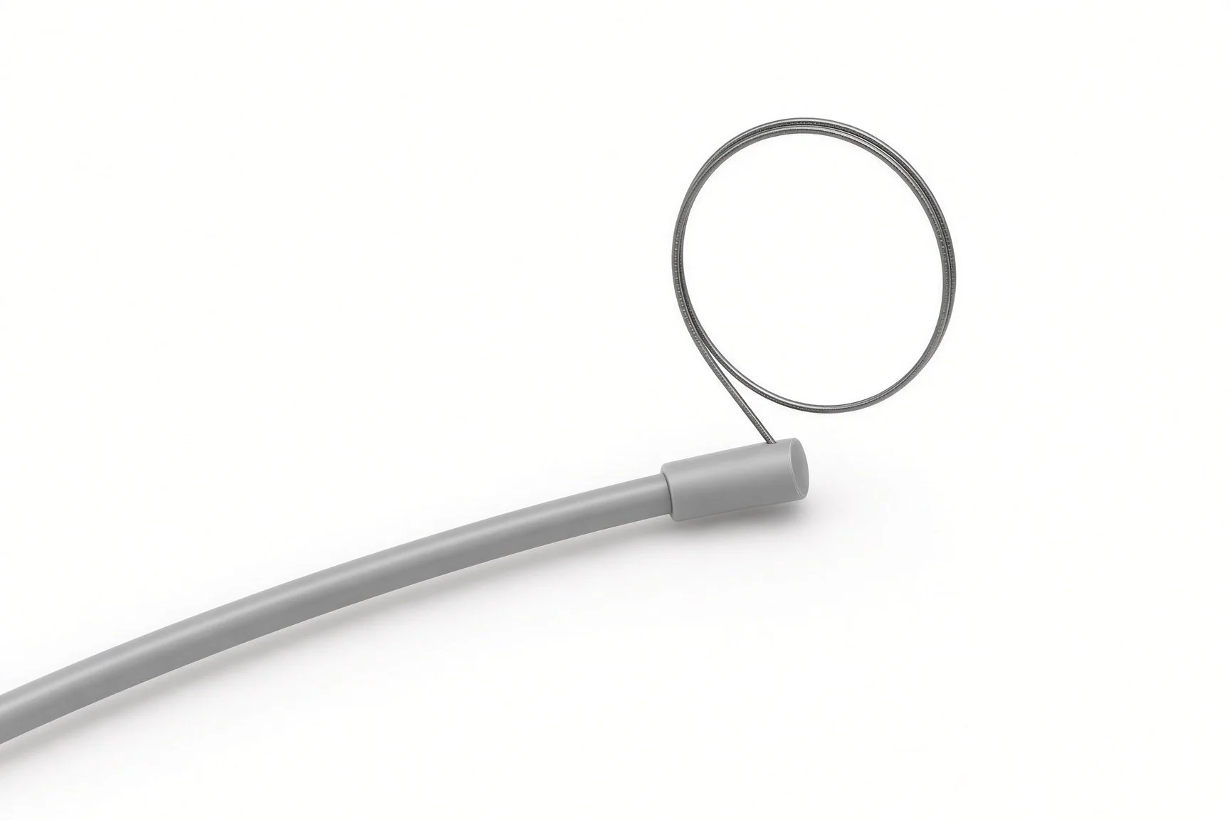
Retrieval snare.
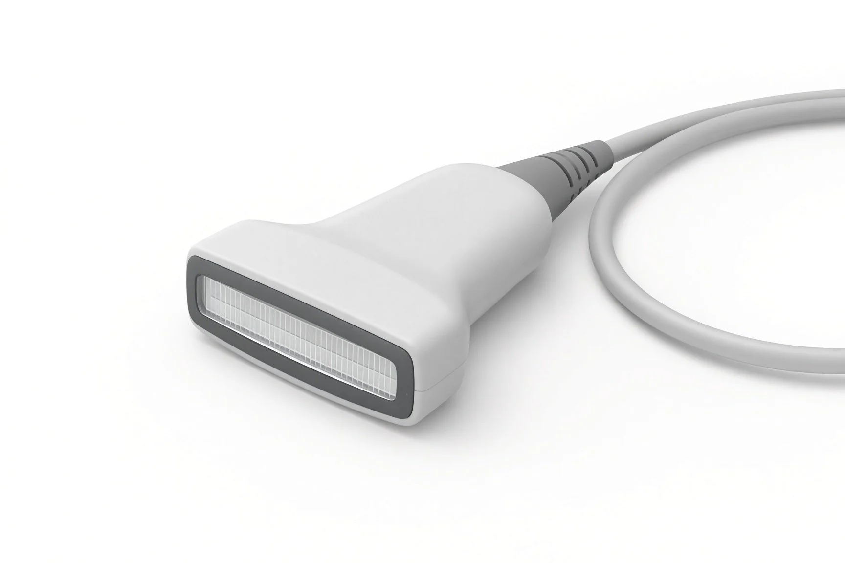
Ultrasound probe.
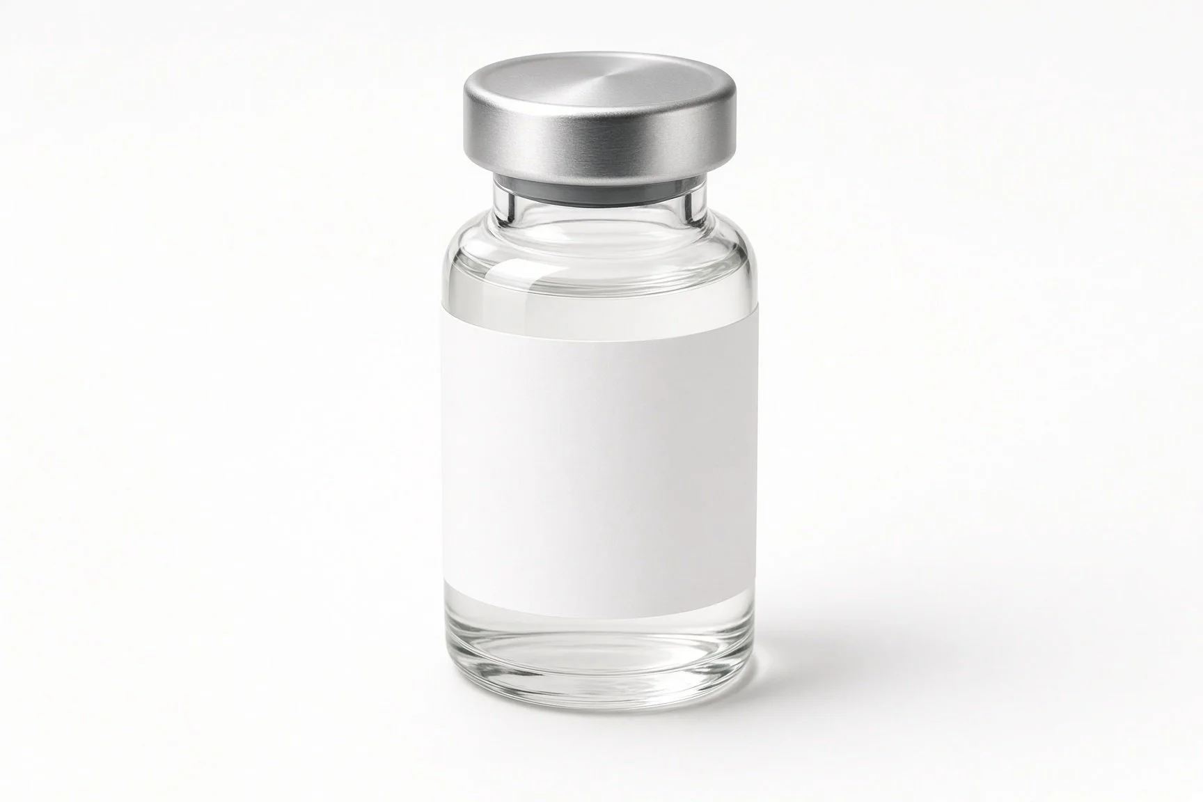
Contrast.
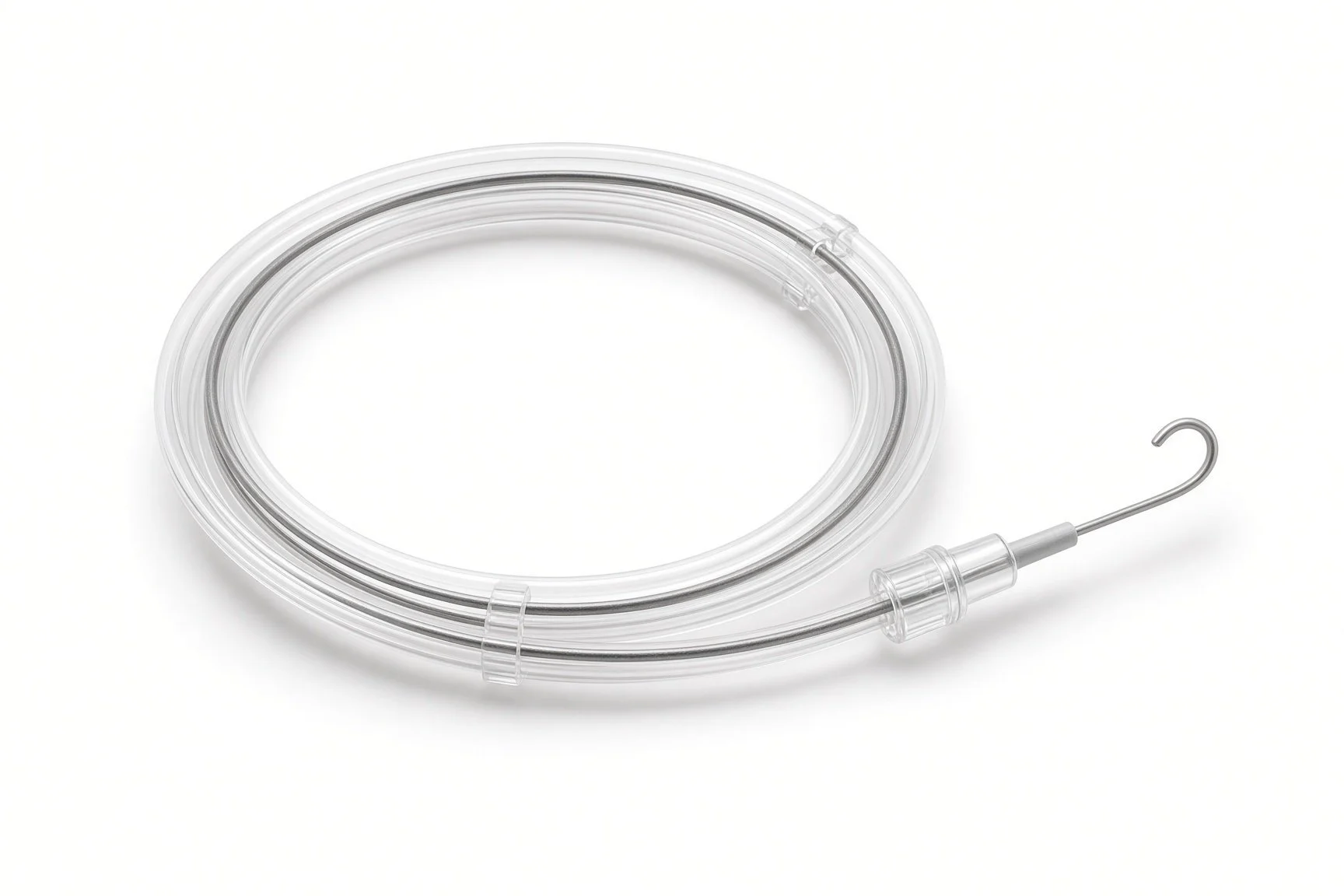
Guidewire.
Procedural steps (Barts example)
Getting ready
Access
Placing the filter
Finishing
Retrieval
First time in the cath lab?
Know the approach. Jugular and femoral filters are loaded the opposite way round; check the kit matches the access route.
Find the renal veins. The cavogram is the map; help the radiographer get a clear view before deployment.
Book the retrieval. A filter left in longer than needed causes more problems; check the removal plan is documented.
Check the side. Confirm which vein is planned and that it is free of clot on ultrasound.
Record the removal plan. Filters are often left in because no one booked the retrieval; make sure it is documented and booked.
FAQ
Does the filter treat my clot?
No. It catches clots travelling up from the legs. Blood thinners treat the clot and are started as soon as it is safe.
Can I feel the filter?
No. It sits deep inside the large vein in the abdomen.
Does it have to come out?
Most filters now are designed to be removed, and removal is advised once they are no longer needed, because problems become more likely over time.
Can I have an MRI scan with a filter?
Most modern filters are MRI-conditional. Tell the scanning team you have one so they can check the type.
Related Cath Lab 101 guides
Catheter-directed therapy for pulmonary embolism (coming soon)
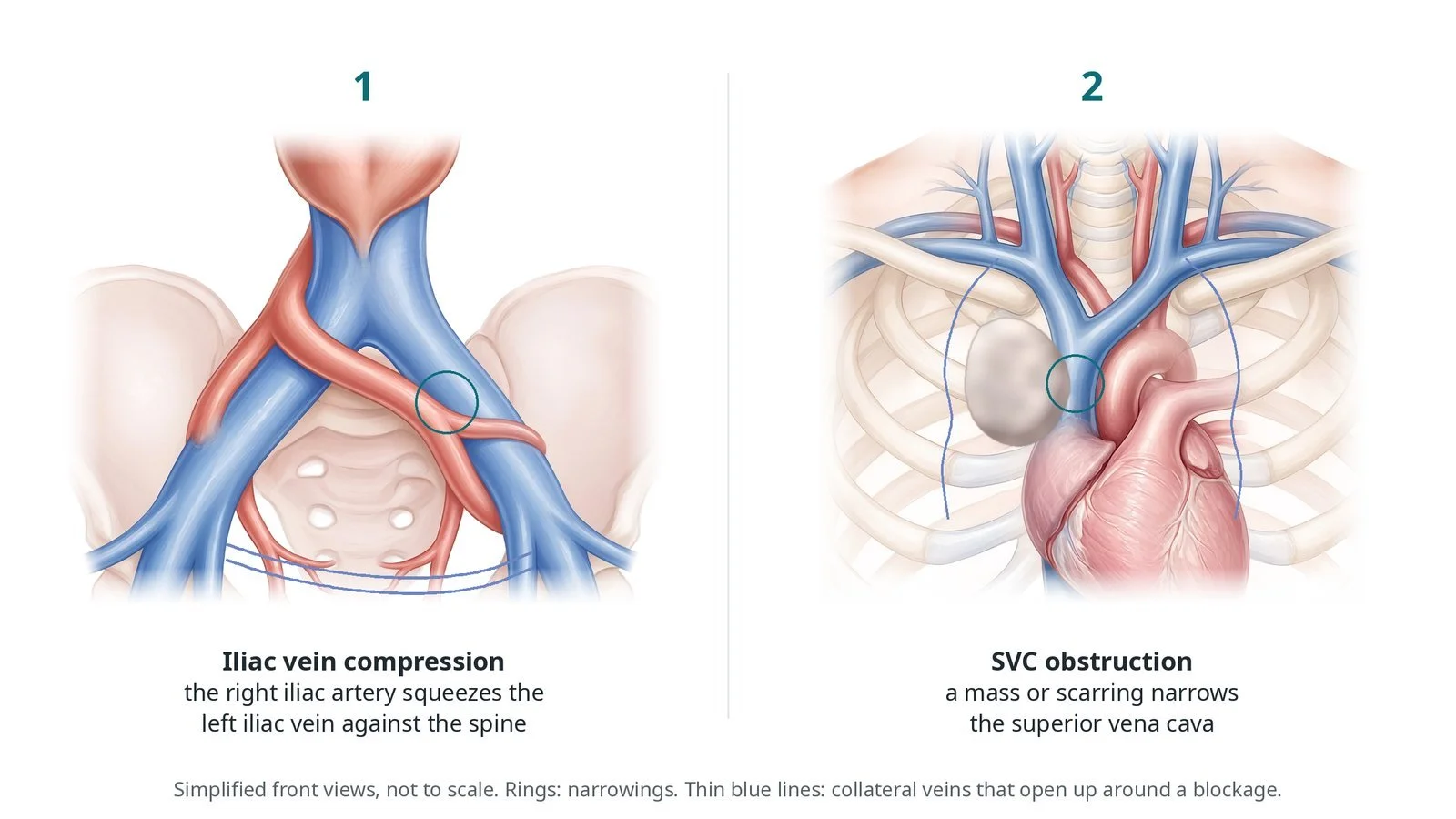
Venous stenting (iliac vein and SVC obstruction) (coming soon)
Sources
All checked on 12 November 2026. Each page's own date is shown where it gives one.
NICE. Venous thromboembolic diseases: diagnosis, management and thrombophilia testing (NG158). nice.org.uk/guidance/ng158
Konstantinides SV, et al. 2019 ESC Guidelines for the diagnosis and management of acute pulmonary embolism. Eur Heart J 2020;41(4):543–603.
British Committee for Standards in Haematology. Guidelines on use of vena cava filters. Br J Haematol 2006.
StatPearls (NCBI Bookshelf), search: inferior vena cava filter. ncbi.nlm.nih.gov/books
Disclaimer
Education only. This page is for general education for healthcare students, cath lab staff and curious readers. It isn't medical advice and doesn't replace advice from your own doctor, heart team or local unit protocols. Equipment and medicines are described generically. No product or manufacturer is recommended. If you think someone is having a heart emergency, call 999.
Disclosure: I work in the medical device industry. This page is independent education and isn't sponsored or reviewed by any company.
All diagrams on this page are original illustrations created for rodnieoro.com.