Venous stenting (iliac vein and SVC obstruction): a Cath Lab 101 guide
Reviewed by Rodnie Oro, cath lab charge nurse · Last reviewed: November 2026 · Education only, not medical advice
Venous stenting in one minute
What it is: Large veins can be squeezed, scarred or blocked, so blood backs up behind them. Venous stenting opens the vein from inside with a balloon and a strong, self-expanding stent, guided by venography and intravascular ultrasound (IVUS).
Who it is for: People with leg swelling, pain, skin changes or ulcers from a narrowed iliac vein (for example iliac vein compression, also called May-Thurner syndrome, or scarring after a DVT), and people with superior vena cava (SVC) syndrome from cancer or from pacing leads and lines. A specialist team decides.
Main risks: Bleeding or bruising, clot forming in the stent, the stent narrowing or moving, and back pain after the vein is stretched. With the SVC, a rare but serious tear of the vein. Your team will explain your own risk.
Usually: Under sedation or a general anaesthetic, about 1 to 3 hours, often home the same or next day, then blood thinners for months.
On this page: What is venous stenting? · Who is it for? · When is it done? · Step by step · Recovery · Risks · Kit list · First time in the lab? · FAQ
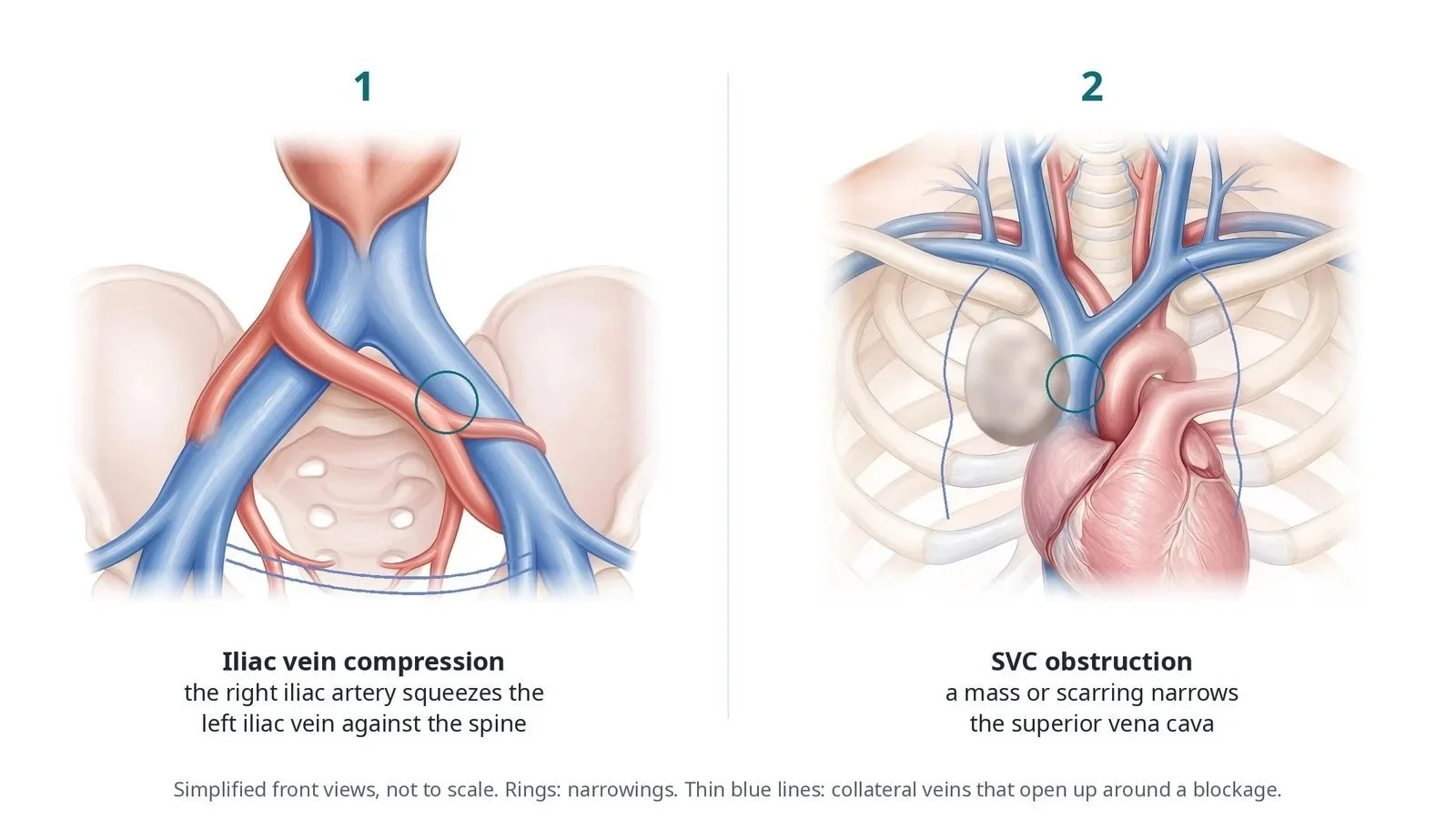
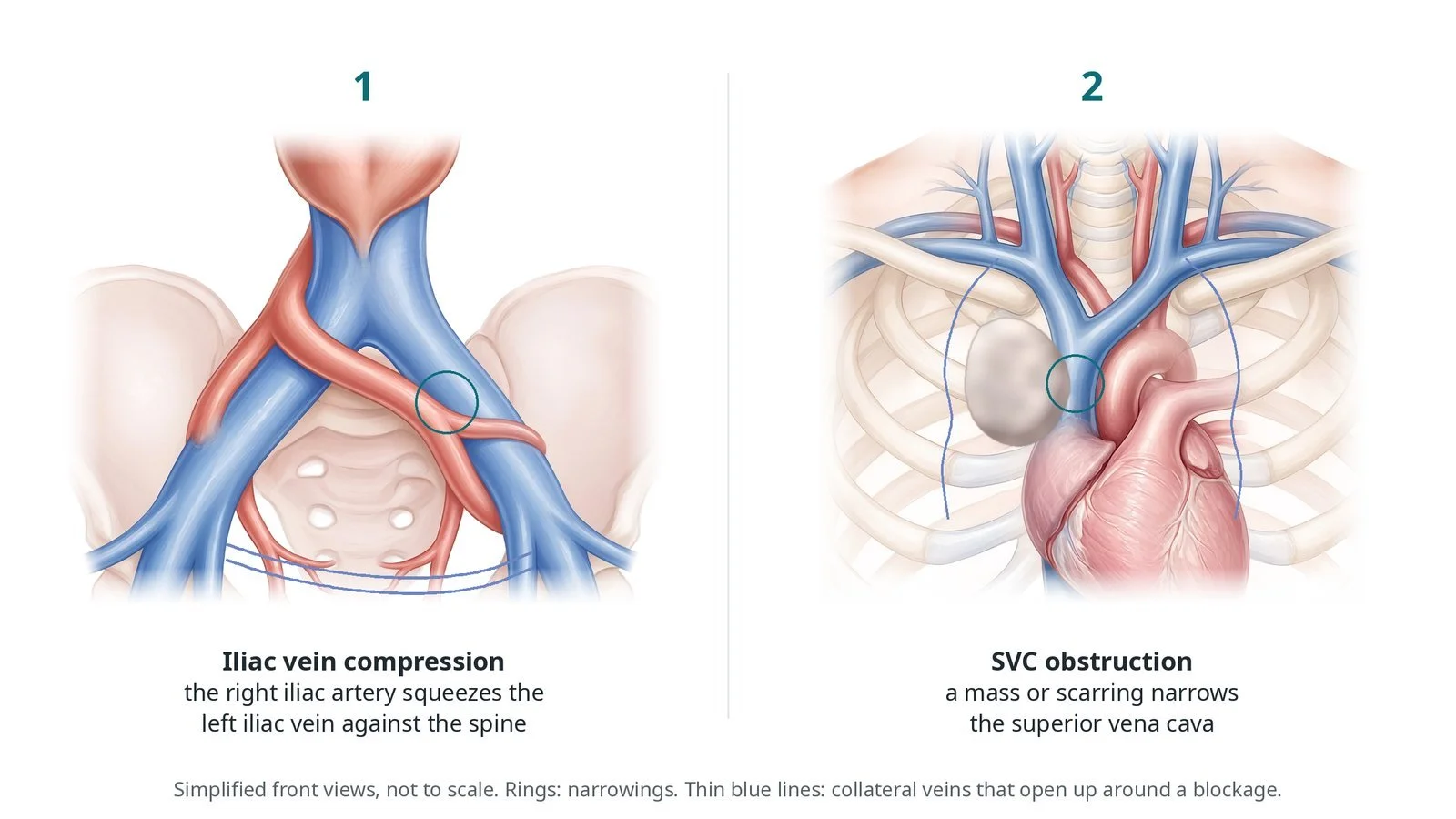
(1) Iliac vein compression: the right iliac artery squeezes the left iliac vein against the spine. (2) SVC obstruction: a mass or scarring narrows the superior vena cava. Thin blue lines are collateral veins.
What is venous stenting?
Blood from each leg drains up the femoral vein into the external and common iliac veins in the pelvis, which join to form the inferior vena cava (IVC). The left common iliac vein passes behind the right common iliac artery, just in front of the lower spine. Blood from the head, neck and arms drains through the brachiocephalic veins into the superior vena cava (SVC) and on into the right atrium.
In iliac vein compression (May-Thurner syndrome), the right iliac artery presses the left iliac vein against the spine. This can cause swelling and heaviness of the left leg and makes a DVT more likely. After a DVT, the vein can heal with scarring that narrows or blocks it (post-thrombotic obstruction), causing pain, swelling, skin changes and ulcers. Small collateral veins open up around the blockage but cannot carry enough blood.
The SVC can be squeezed by a tumour, most often lung cancer or lymphoma, or scarred from inside by pacing leads, dialysis lines or other catheters. SVC syndrome causes swelling of the face, neck and arms, headache that is worse lying down, and breathlessness. Venous stents are wide, flexible and strong enough to resist the squeeze; IVUS is used because a venogram can underestimate the narrowing.
New to the acronyms? See the Cath Lab Abbreviations A–Z.
Who is it for?
It may be considered for:
leg swelling, pain, skin changes or ulcers from iliac vein compression or post-thrombotic obstruction, after duplex ultrasound and CT or MR venography, when compression and other treatment have not helped enough
some people with a fresh DVT in the thigh and pelvis, after clot removal, when an underlying compression is found
SVC syndrome from cancer, often for quick relief while cancer treatment starts, or from scarring around pacing leads or long-term lines
The decision is made by a specialist venous or interventional radiology team.
Who it may not suit: problems limited to surface (varicose) veins narrowings that cause no symptoms veins that cannot be crossed, or where inflow from the leg is too poor to keep a stent open
The decision is made by your heart team, who will talk through the reasons, the alternatives and the risks with you.
When is it done?
Usually planned; SVC stenting may be urgent when symptoms are severe. Before it, the team checks blood tests, kidney function, blood thinners and the access plan and, for the SVC, whether pacing leads are present.
gives you a chance to ask questions and give consent
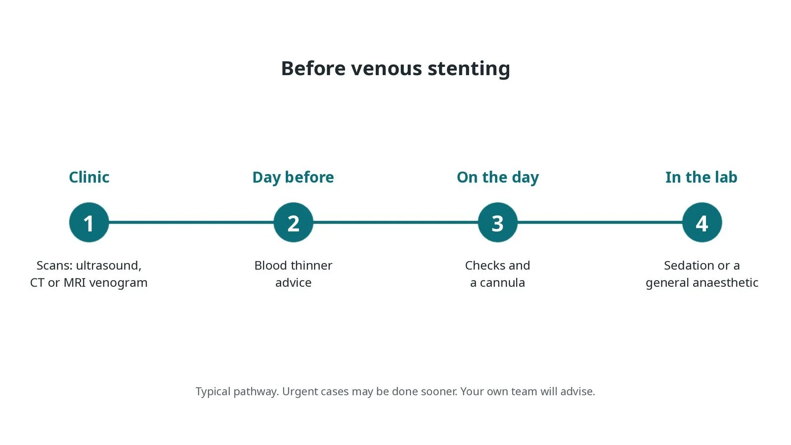
Before venous stenting: ultrasound, CT or MRI venogram; blood thinner advice; checks and a cannula; sedation or a general anaesthetic in the lab.
Step by step
1. Getting ready
You lie on the X-ray table, face down or on your back depending on the access. Many centres use a general anaesthetic or deep sedation, because stretching the vein can be painful. Monitoring includes ECG, blood pressure and oxygen levels.
2. Access
Under ultrasound, a sheath is placed in the femoral vein in the thigh, the popliteal vein behind the knee or the jugular vein in the neck. For the SVC, the jugular, arm or femoral veins are used. Heparin is given, as per centre protocol.
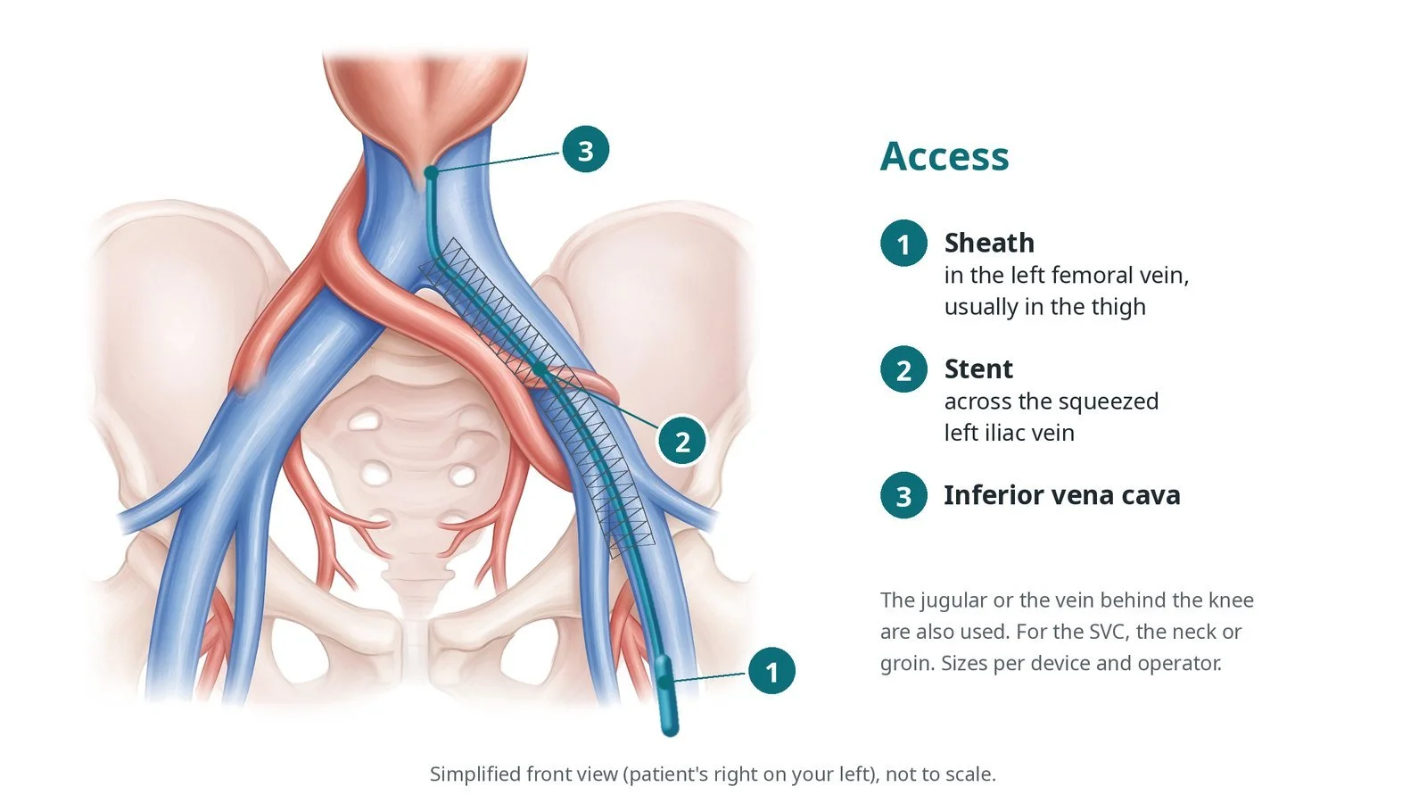
Access: (1) sheath in the left femoral vein, usually in the thigh; (2) stent across the squeezed left iliac vein; (3) inferior vena cava. The jugular or the vein behind the knee are also used.
3. Opening the vein
A. Venogram and IVUS. Contrast shows the narrowing and the collateral veins, and an IVUS catheter measures the vein size and where the narrowing starts and ends.
B. Balloon. A high-pressure balloon stretches the narrowing. This can be painful, so more pain relief may be needed.
C. Stent. A self-expanding venous stent, sized from the IVUS, is placed across the whole narrowing, often extending into the IVC or down into the femoral vein.
D. Check. The stent is ballooned to full size, and a final venogram and IVUS check good flow and that the collateral veins no longer fill.
Watch: In SVC cases, a sudden fall in blood pressure, chest pain or breathlessness: tell the operator at once, as the vein may have torn with bleeding around the heart or lung; prepare for a balloon to seal it, pericardiocentesis or surgery, as per centre protocol.
4. Finishing
The sheaths are removed and pressure is applied. Compression stockings may be started, per operator.
5. Afterwards
Walking soon afterwards is encouraged. Blood thinners are started, as per centre protocol, and usually continue for several months, sometimes with an antiplatelet. A duplex ultrasound scan checks the stent before or soon after discharge, and at follow-up.
Watch: New or worse leg swelling or pain after stenting: arrange an urgent duplex ultrasound, as the stent may have blocked with clot.
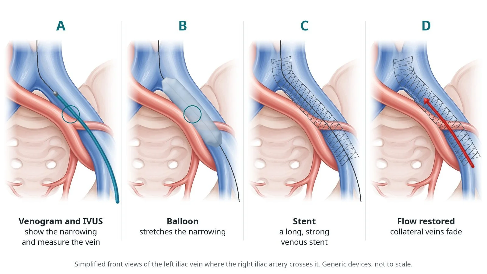
A: venogram and IVUS show and measure the narrowing. B: a balloon stretches it. C: a long, strong venous stent is placed. D: flow restored and collateral veins fade.
Recovery and aftercare
First hours: checks of the access site, leg circulation and observations, as per centre protocol; walking early is encouraged.
Going home: usually the same or next day.
Medicines: blood thinners, usually for several months, sometimes with an antiplatelet; take them exactly as prescribed.
Compression: wear compression stockings if advised, and keep walking.
Driving: ask your team; do not drive for at least 24 hours after sedation or a general anaesthetic.
Follow-up: duplex ultrasound scans of the stent at set times, and clinic review.
Call 999 if you have sudden breathlessness, chest pain, coughing up blood, heavy bleeding, or signs of a stroke.
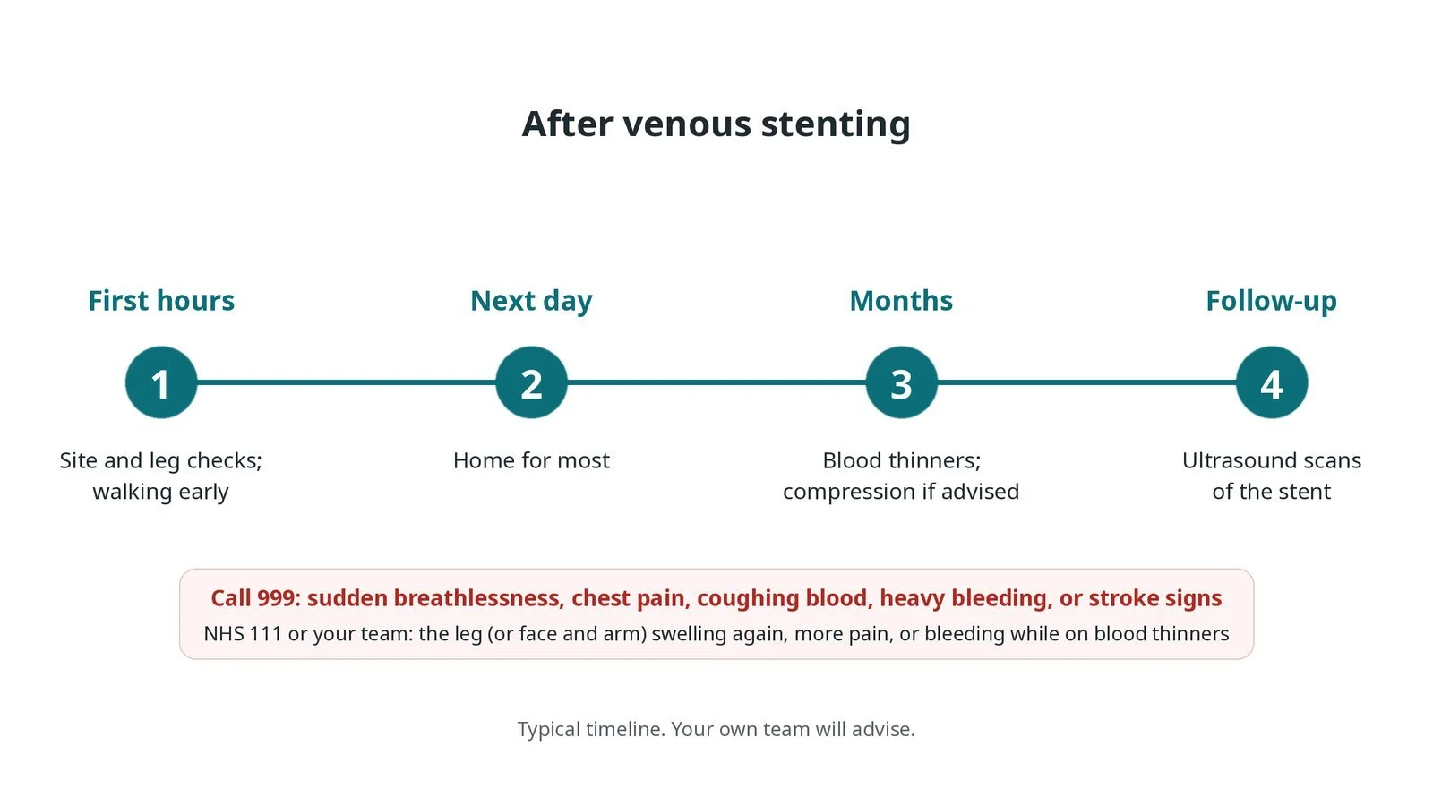
After venous stenting: (1) site and leg checks with early walking; (2) home the next day for most; (3) blood thinners and compression if advised; (4) ultrasound scans of the stent.
Risks and complications
Your team will explain the risks that apply to you. Possible problems include:
Bleeding or bruising at the access site
Back or pelvic ache for a few days after stretching the vein
Clot forming in the stent (uncommon)
Stent narrowing again or moving (uncommon)
Bleeding while on blood thinners
Tear of the vein, especially the SVC, causing serious bleeding (rare)
Cath lab kit list (printable)
Barts example kit list
An example from one UK centre, written generically. Every lab has its own protocol, and sizes and doses vary.
ultrasound machine and sterile probe cover
venous sheaths, including large sheaths for the stent, per device
guidewires, including stiff and crossing wires, per operator
support (crossing) catheters
IVUS catheter and console
high-pressure balloons, size per IVUS
self-expanding venous stents, size and length per IVUS
contrast for venography
heparin, pain relief and sedation or anaesthetic, as per centre protocol
compression stockings, per operator
IVUS catheter.
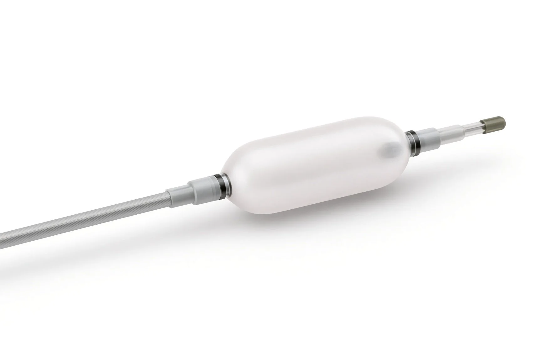
High-pressure balloons.
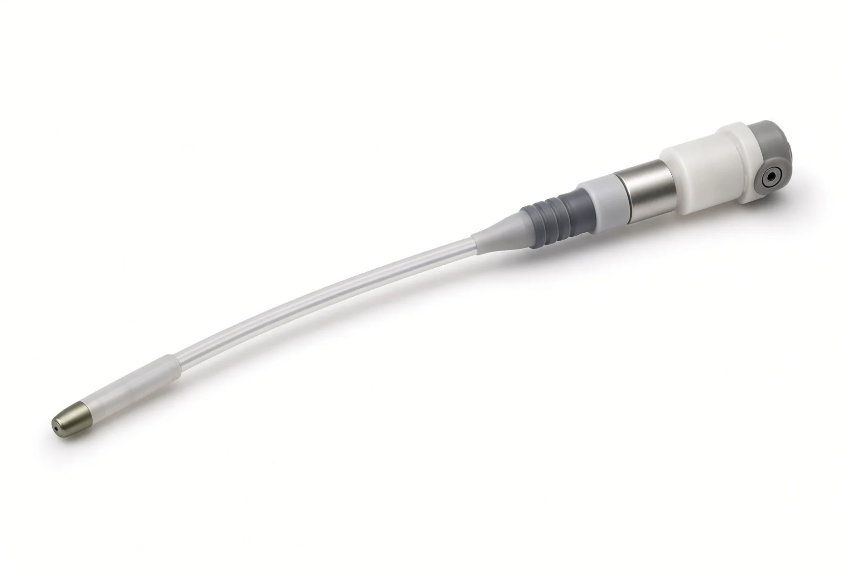
IVUS catheter.
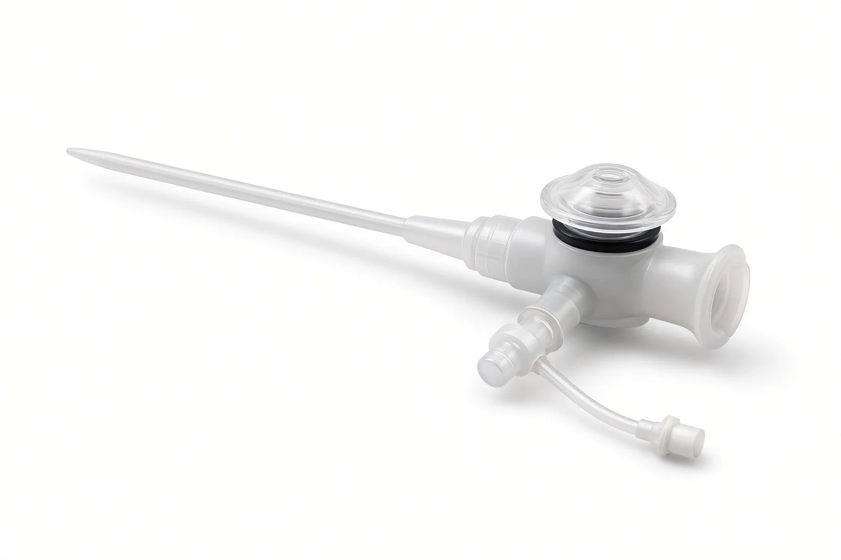
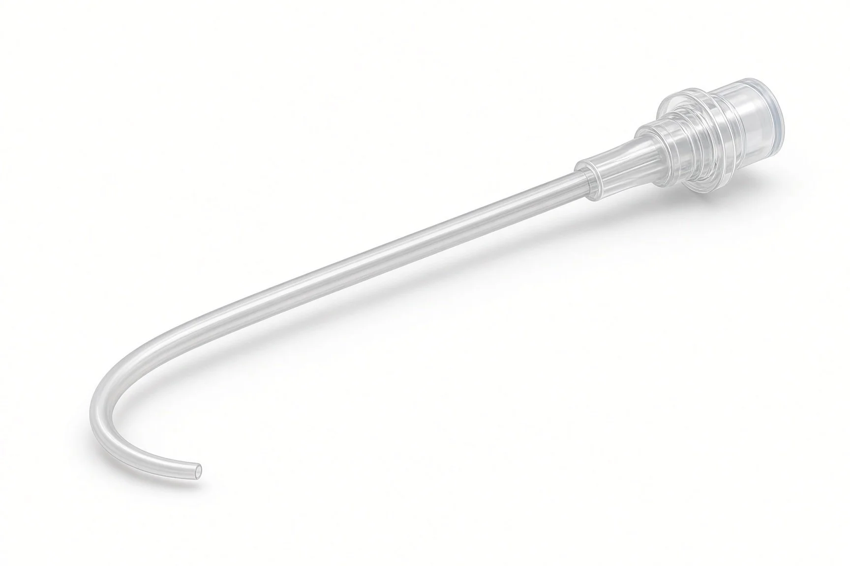
Large venous sheath.
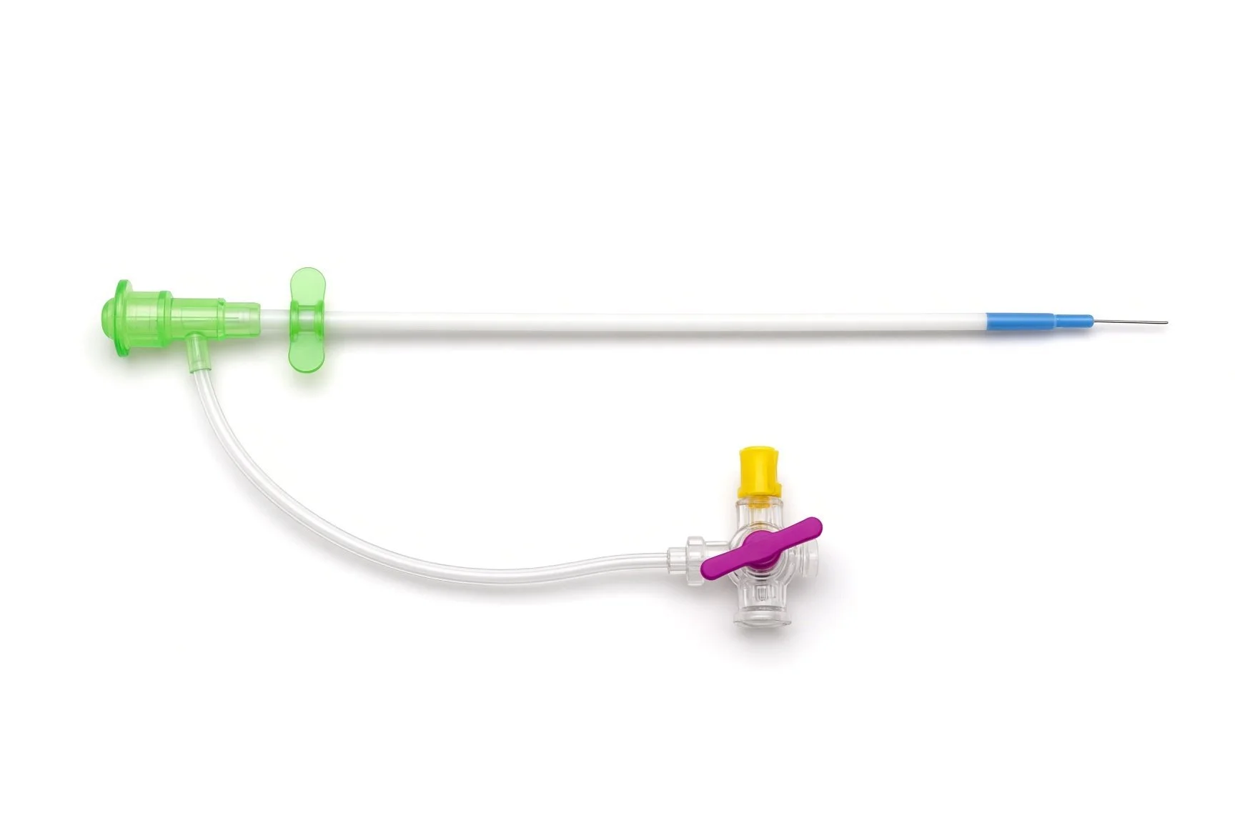
Large venous sheath.
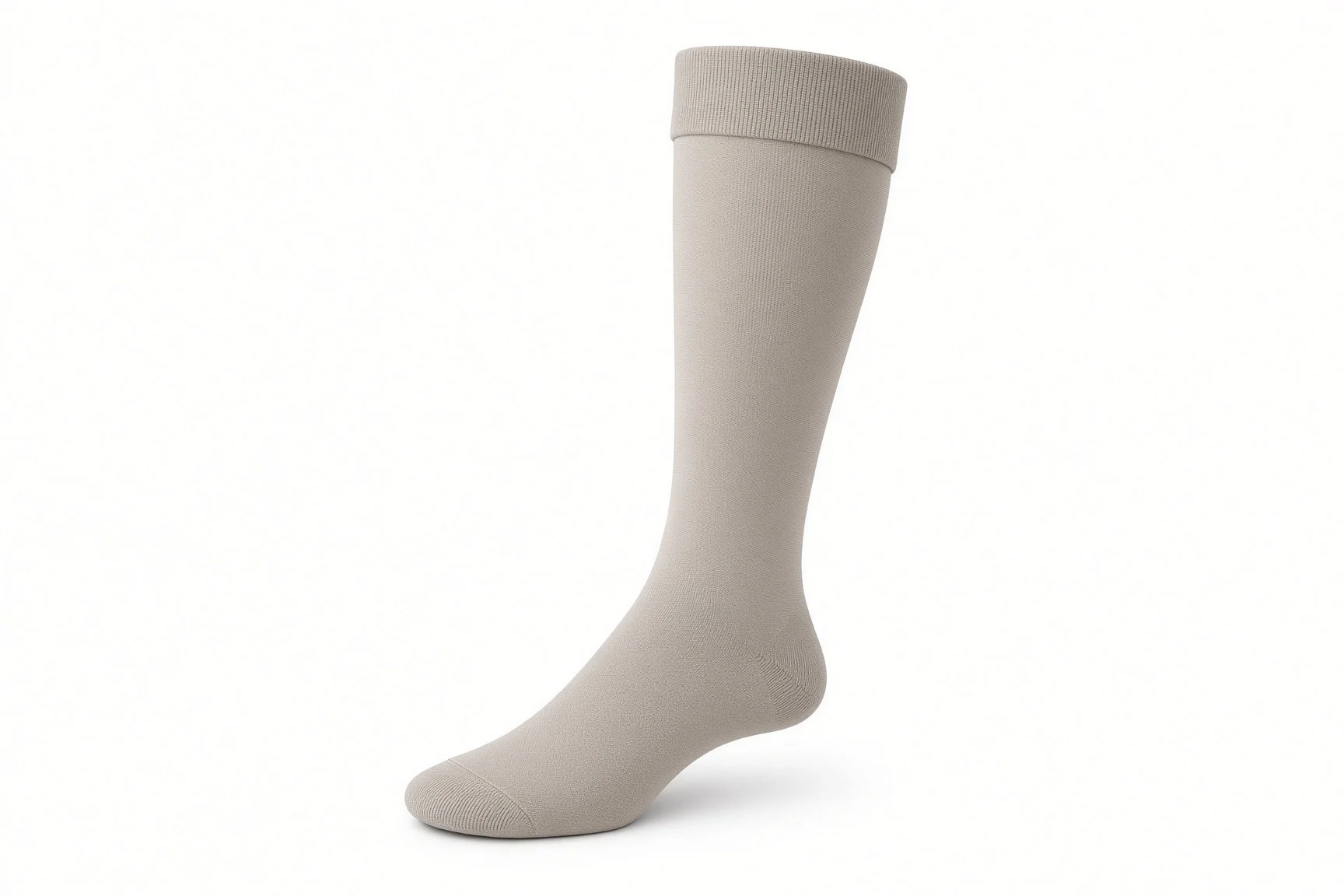
Compression stockings.
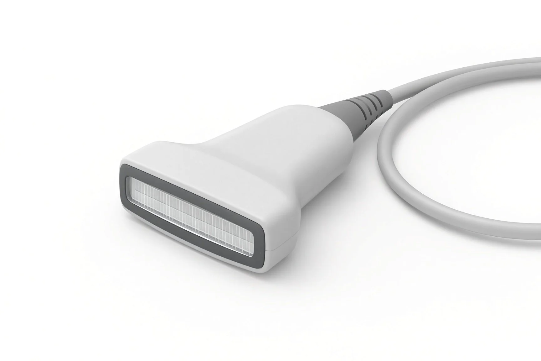
Ultrasound probe.
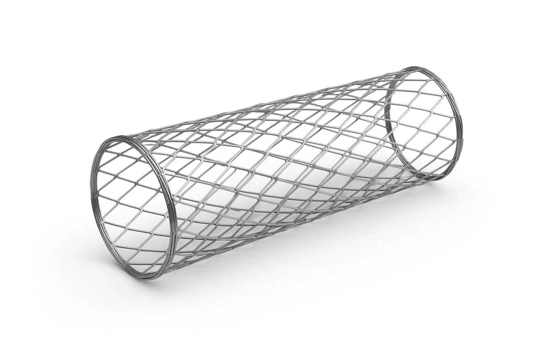
Self-expanding venous stent.
Contrast.
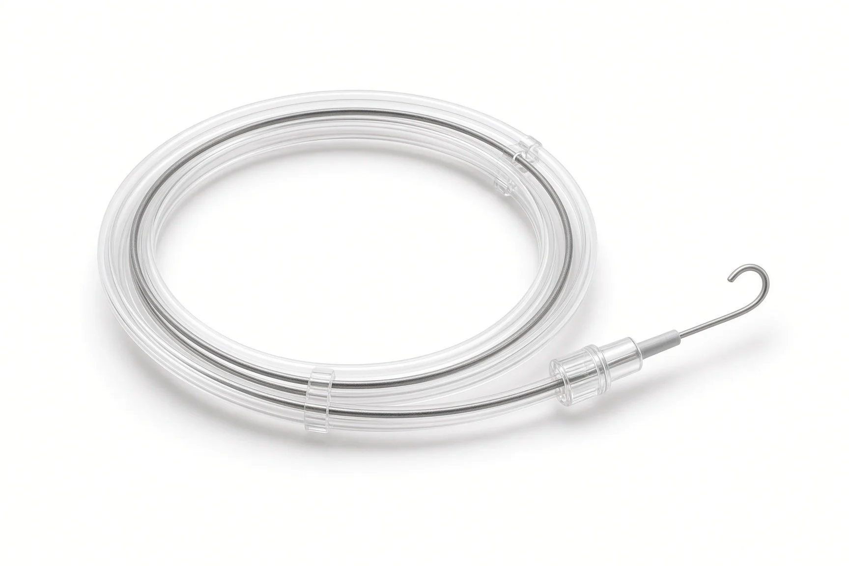
Guidewires.
Procedural steps (Barts example)
Getting ready
Access
Opening the vein
Finishing
Afterwards
First time in the cath lab?
Expect pain on ballooning. Stretching a vein hurts more than many artery cases; check pain relief or anaesthetic depth before the balloon goes up.
Help with IVUS. The vein size and landing zones decide the stent size; keep the console ready and record the numbers.
Know the SVC emergency plan. In SVC cases, check where the balloons, covered stents and pericardiocentesis kit are.
Confirm the blood thinner plan. Check with the operator when anticoagulation starts after the case.
Check the position plan. Access behind the knee needs the patient face down; plan pressure areas and the airway with the anaesthetist.
FAQ
Will my leg swelling go away?
Many people notice less swelling, heaviness and pain over weeks to months, and ulcers may heal faster. Compression and walking still help.
How long will I need blood thinners?
Usually several months after the stent, sometimes longer. Your team will advise.
Can I feel the stent?
No. Some people have a mild back or pelvic ache for a few days.
Why is IVUS used?
It measures the vein from inside and shows narrowing that a venogram can miss, so the right size of stent is used.
Related Cath Lab 101 guides
IVC filter insertion and retrieval (coming soon)
Catheter-directed therapy for pulmonary embolism (coming soon)
Sources
All checked on 12 November 2026. Each page's own date is shown where it gives one.
Kakkos SK, et al. European Society for Vascular Surgery (ESVS) 2021 clinical practice guidelines on the management of venous thrombosis. Eur J Vasc Endovasc Surg 2021.
De Maeseneer MG, et al. European Society for Vascular Surgery (ESVS) 2022 clinical practice guidelines on the management of chronic venous disease of the lower limbs. Eur J Vasc Endovasc Surg 2022.
StatPearls (NCBI Bookshelf), search: May-Thurner syndrome. ncbi.nlm.nih.gov/books
StatPearls (NCBI Bookshelf), search: superior vena cava syndrome. ncbi.nlm.nih.gov/books
Disclaimer
Education only. This page is for general education for healthcare students, cath lab staff and curious readers. It isn't medical advice and doesn't replace advice from your own doctor, heart team or local unit protocols. Equipment and medicines are described generically. No product or manufacturer is recommended. If you think someone is having a heart emergency, call 999.
Disclosure: I work in the medical device industry. This page is independent education and isn't sponsored or reviewed by any company.
All diagrams on this page are original illustrations created for rodnieoro.com.