Dialysis fistula angioplasty (fistuloplasty): a Cath Lab 101 guide
Reviewed by Rodnie Oro, cath lab charge nurse · Last reviewed: November 2026 · Education only, not medical advice
Dialysis fistula angioplasty in one minute
What it is: An arteriovenous (AV) fistula joins an artery to a vein, usually in the arm, so the vein grows big and strong enough for dialysis needles. A graft does the same with a soft synthetic tube. Narrowings often develop. Fistuloplasty passes a balloon into the fistula and stretches the narrowing from inside.
Who it is for: People on haemodialysis whose fistula or graft is failing: poor flow or high pressures on dialysis, long bleeding after the needles come out, a weak buzz (thrill), arm swelling, or a narrowing seen on ultrasound. It is mostly done in interventional radiology; some cardiac labs also do it.
Main risks: Bruising or bleeding, a tear of the vein, clot blocking the fistula and, rarely, poor blood supply to the hand (steal). Your team will explain your own risk.
Usually: Under local anaesthetic, about 30 to 60 minutes, home the same day, with dialysis continuing as planned.
On this page: What is dialysis fistula angioplasty? · Who is it for? · When is it done? · Step by step · Recovery · Risks · Kit list · First time in the lab? · FAQ
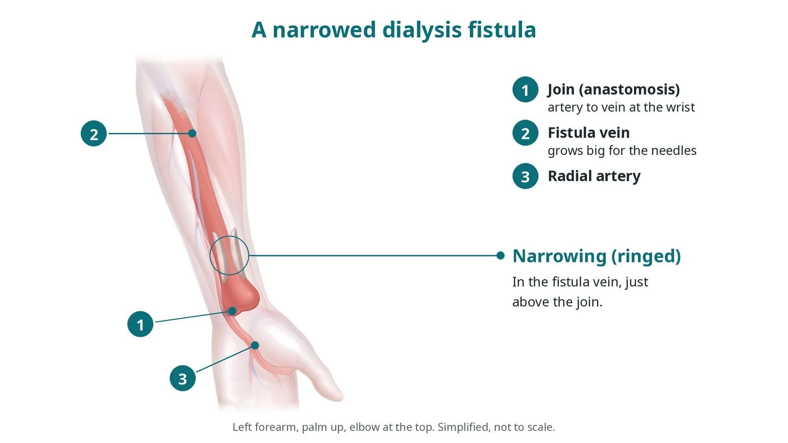
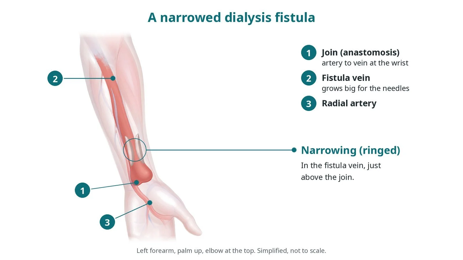
A narrowed dialysis fistula: (1) the join (anastomosis) of artery to vein at the wrist; (2) the fistula vein; (3) the radial artery. The narrowing (ringed) is in the vein just above the join.
What is dialysis fistula angioplasty?
In a radiocephalic fistula, the cephalic vein is joined to the radial artery at the wrist; in a brachiocephalic fistula, it is joined to the brachial artery at the elbow. Arterial blood flows straight into the vein, which enlarges and thickens over weeks (matures) until it can take two dialysis needles. A graft joins an artery to a vein with a synthetic tube.
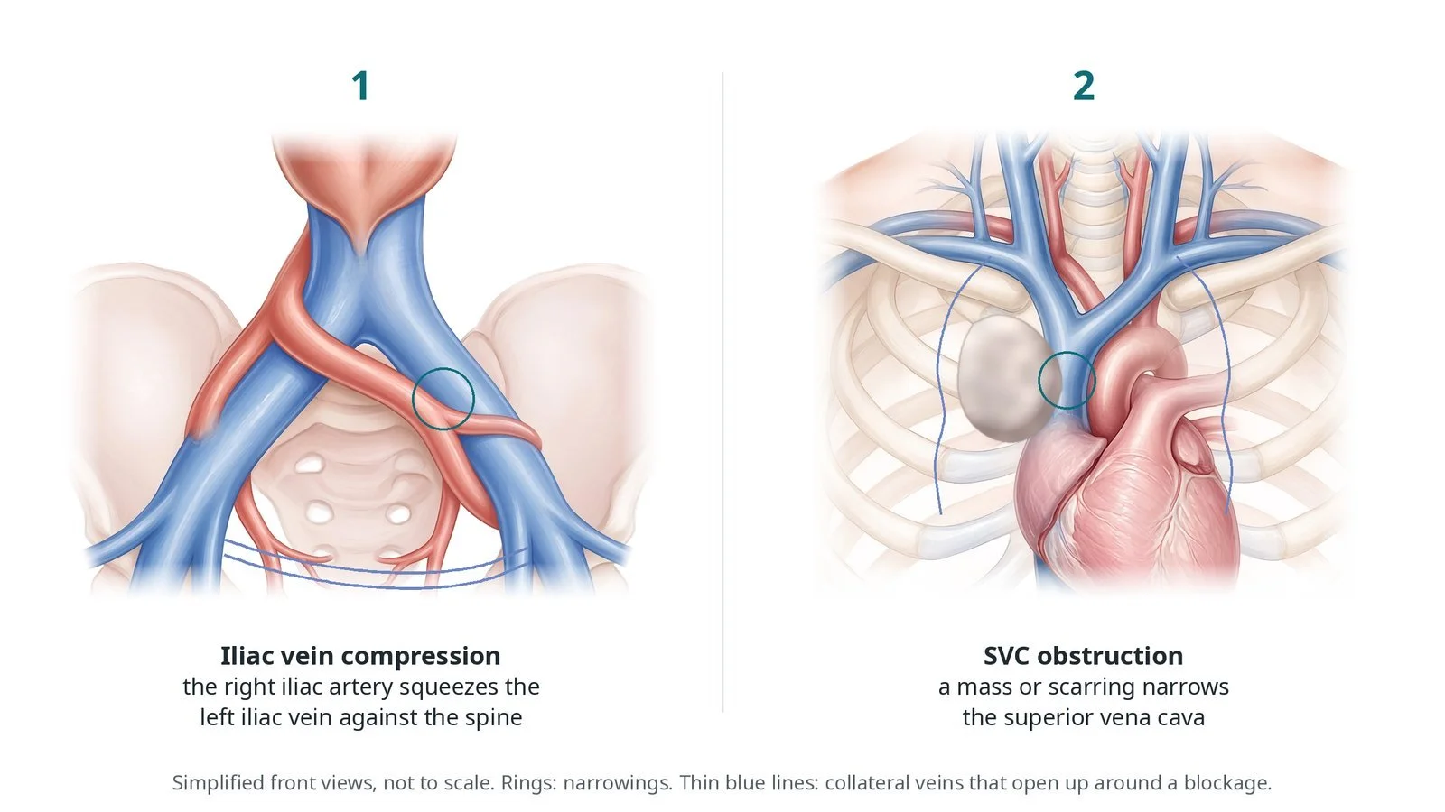
The high flow and repeated needling make the vein wall thicken, so narrowings form, most often just beyond the join (juxta-anastomotic), where veins bend or join deeper veins and, in grafts, where the graft joins the vein. Narrowing of the large central veins in the chest, often after previous lines or pacing leads, causes arm swelling.
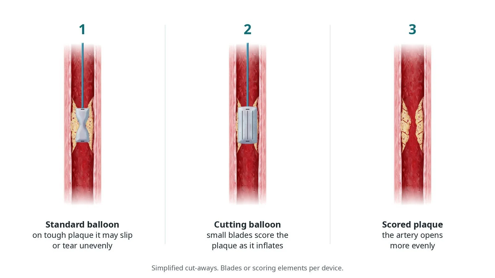
Fistuloplasty is balloon angioplasty of the fistula. Fistula narrowings are often tough and fibrous, so high-pressure balloons are used, sometimes drug-coated balloons to slow re-narrowing, or covered stents (stent grafts) for narrowings that keep coming back or for tears. A strong, continuous buzz (thrill) felt over the fistula is a sign of good flow.
New to the acronyms? See the Cath Lab Abbreviations A–Z.
Who is it for?
People on dialysis, or about to start, whose fistula or graft shows signs of a narrowing:
poor flow or high pressures on the dialysis machine, or less effective dialysis
long bleeding after the needles come out
a weak, pulsing or absent thrill
swelling of the arm, face or breast (central vein narrowing)
a narrowing seen on duplex ultrasound or a fistulogram
a fistula that is not maturing as expected
Who it may not suit: a fistula that has fully clotted may also need clot removal (thrombectomy) or surgery narrowings that keep coming back may be better treated with surgery, a stent graft or a new fistula
The decision is made by your heart team, who will talk through the reasons, the alternatives and the risks with you.
When is it done?
Usually planned between dialysis sessions. On the day, potassium and blood count are checked, because people on dialysis can have a high potassium level that may be dangerous; dialysis may be needed first. No blood pressure cuff, drip or blood test is taken from the fistula arm.
gives you a chance to ask questions and give consent
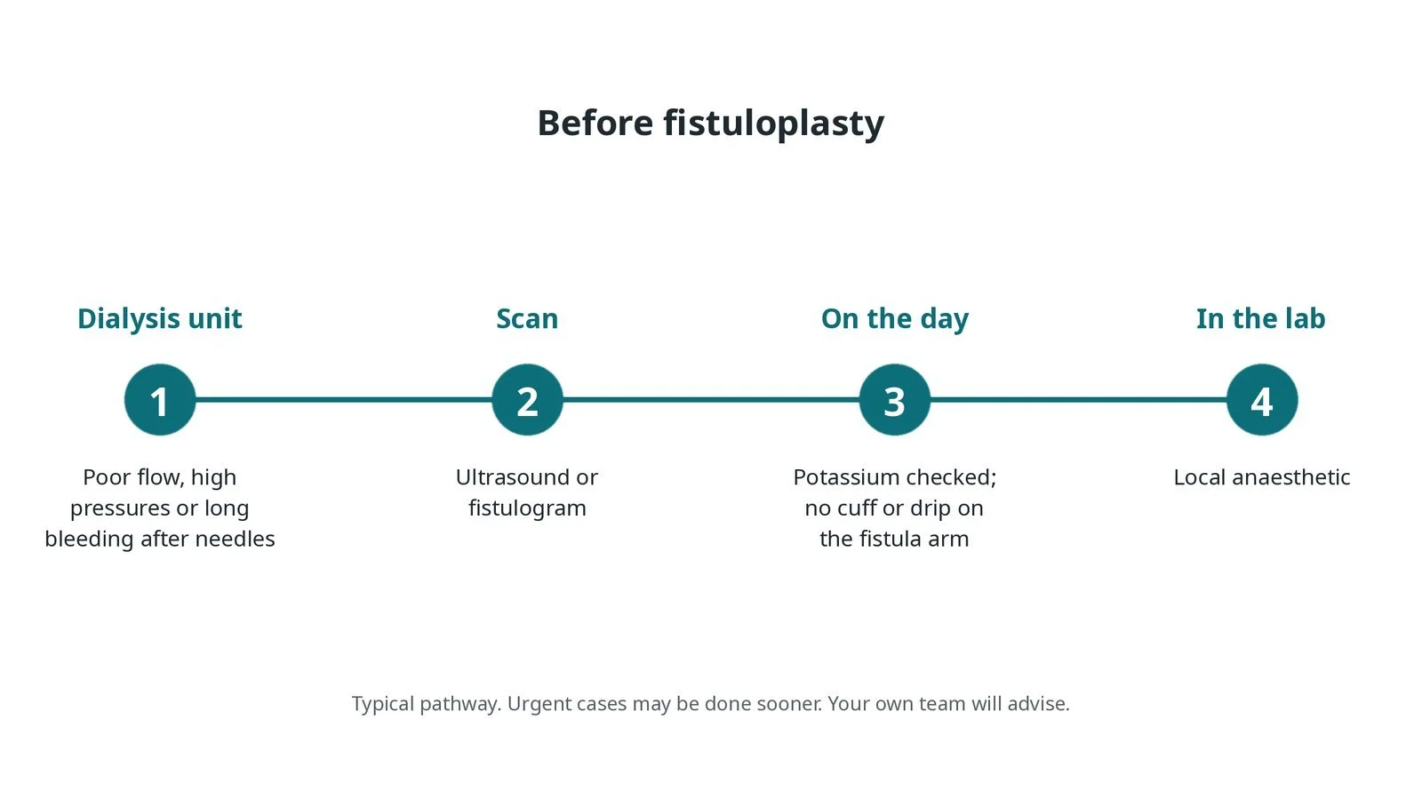
Before fistuloplasty: poor flow, high pressures or long bleeding noticed at dialysis; an ultrasound or fistulogram; potassium checked and no cuff or drip on the fistula arm; local anaesthetic.
Step by step
1. Getting ready
You lie on the table with the fistula arm out on an arm board. Monitoring includes ECG and oxygen levels, and blood pressure is taken on the other arm or a leg. The fistula is felt and listened to, and often scanned with ultrasound.
Watch: A high potassium result, or new ECG changes, before starting: tell the operator and the kidney team; dialysis may be needed first.
2. Access
Under local anaesthetic, a short sheath is placed straight into the fistula vein, usually under ultrasound. It points towards the narrowing: towards the join for narrowings near the artery, or up the arm for narrowings closer to the heart. Occasionally the brachial artery is used. Heparin may be given, as per centre protocol.
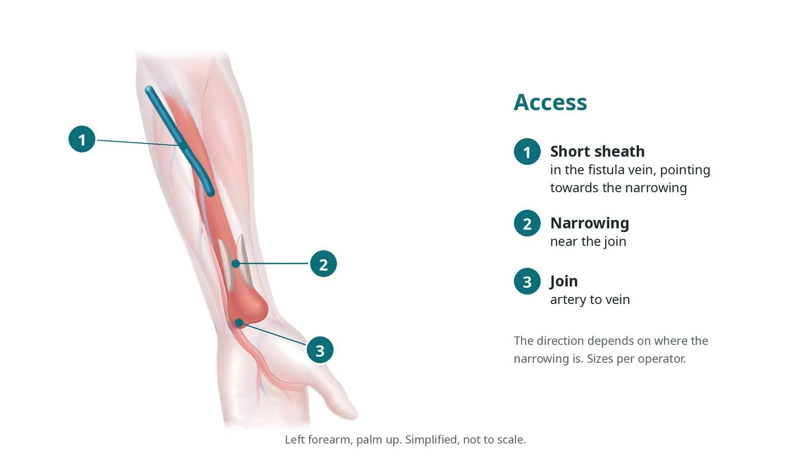
Access: (1) short sheath in the fistula vein, pointing towards the narrowing; (2) the narrowing near the join; (3) the join of artery to vein.
3. Opening the narrowing
A. Fistulogram. Contrast is injected to show the whole circuit, from the join with the artery to the central veins in the chest.
B. Wire across. A guidewire crosses the narrowing.
C. High-pressure balloon. A balloon sized to the vein is inflated, often to high pressure, until its waist disappears. This can hurt for a short time. A drug-coated balloon or a covered stent may be used, per operator.
D. Check. A repeat fistulogram checks the result, and the thrill is felt again.
Watch: Contrast leaking outside the vein after ballooning, or a fast-growing swelling: tell the operator; a balloon may be gently reinflated to seal it, or a covered stent placed.
4. Finishing
The sheath is removed and the site is held with firm but gentle pressure, or closed with a purse-string stitch, without blocking flow in the fistula. The thrill is checked before you leave the lab.
5. Afterwards
You can usually go home the same day. Dialysis continues as planned. Any stitch is removed as advised. Feel for the buzz every day and report any change straight away.
Watch: A cold, pale, numb or painful hand after the procedure: get urgent review, as this may be steal (blood bypassing the hand through the fistula) or a clot.
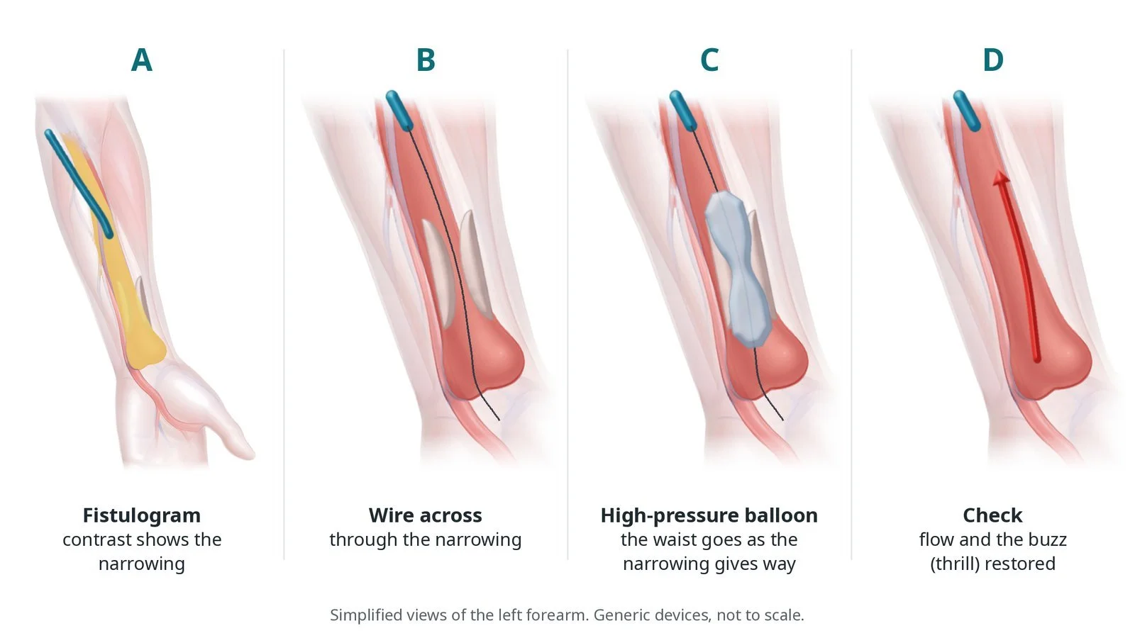
A: a fistulogram shows the narrowing. B: a wire crosses it. C: a high-pressure balloon; the waist goes as the narrowing gives way. D: flow and the buzz (thrill) restored.
Recovery and aftercare
First hours: the site is checked for bleeding and the thrill is felt before you go, as per centre protocol.
Going home: usually the same day.
Dialysis: continues as planned; tell the dialysis team about the fistuloplasty so they can avoid the puncture site if advised.
Protecting the fistula: no blood pressure cuffs, tight sleeves or watches, and no sleeping on the arm; feel for the buzz every day.
Driving: ask your team; do not drive if you have had sedation.
Follow-up: flow and pressure checks at dialysis and ultrasound scans, as per your unit's surveillance plan.
Call 999 if you have bleeding from the fistula that does not stop with firm pressure, chest pain or breathlessness.
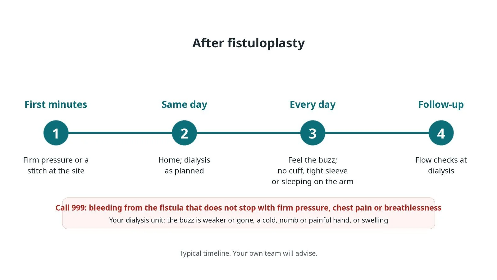
After fistuloplasty: (1) firm pressure or a stitch at the site; (2) home the same day, dialysis as planned; (3) feel the buzz every day and protect the arm; (4) flow checks at dialysis.
Risks and complications
Your team will explain the risks that apply to you. Possible problems include:
Bruising or bleeding at the puncture site
Pain during balloon inflation (common, brief)
Tear of the vein (uncommon): Usually managed with a balloon or covered stent.
Clot blocking the fistula (uncommon)
Poor blood supply to the hand (steal) (rare)
The narrowing coming back, needing repeat treatment: Common over months; regular checks find it early.
Cath lab kit list (printable)
Barts example kit list
An example from one UK centre, written generically. Every lab has its own protocol, and sizes and doses vary.
ultrasound machine and sterile probe cover
short sheaths, size per balloon and operator
guidewires, per operator
angled catheter for crossing, if needed
high-pressure balloons, size per vein
drug-coated balloons, if used, per operator
covered stents (stent grafts), if needed
inflation device with pressure gauge
contrast, local anaesthetic and heparin, as per centre protocol
purse-string suture or dressing, per operator
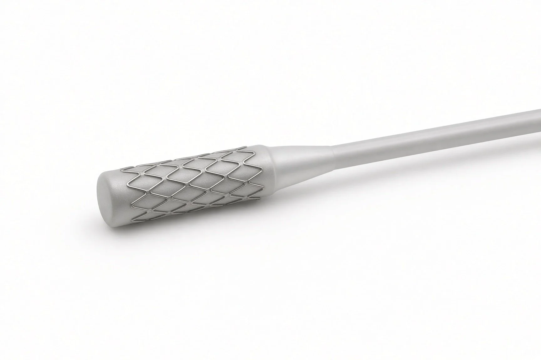
Covered stent if needed.
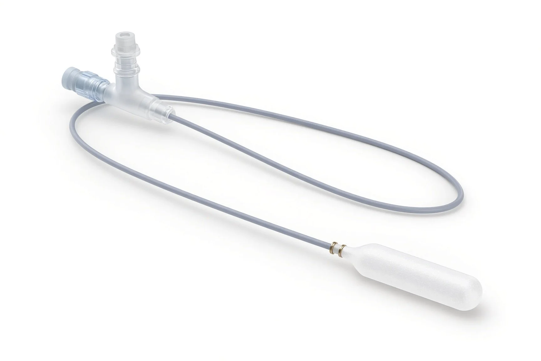
Drug-coated balloon if used.
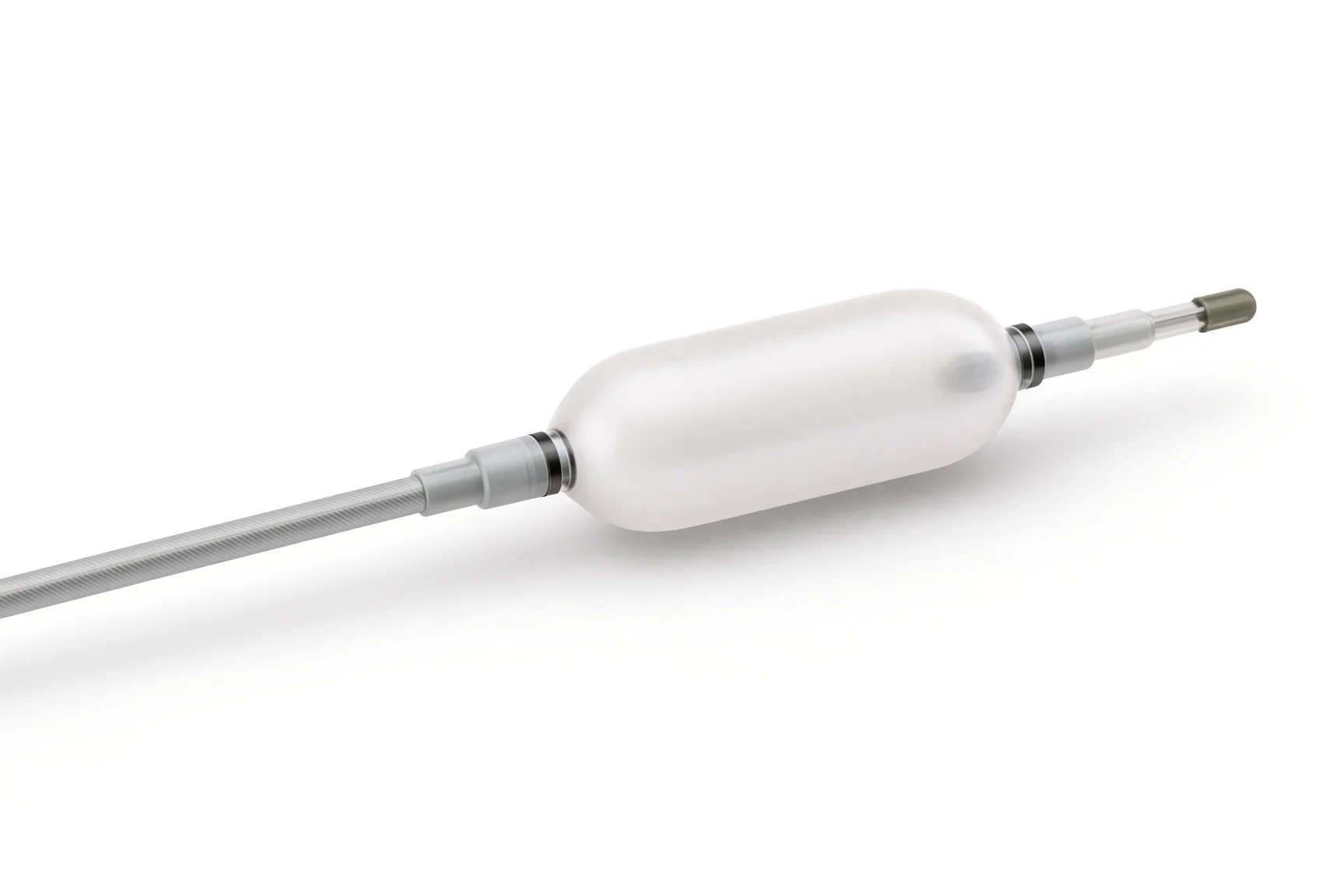
High-pressure balloons.
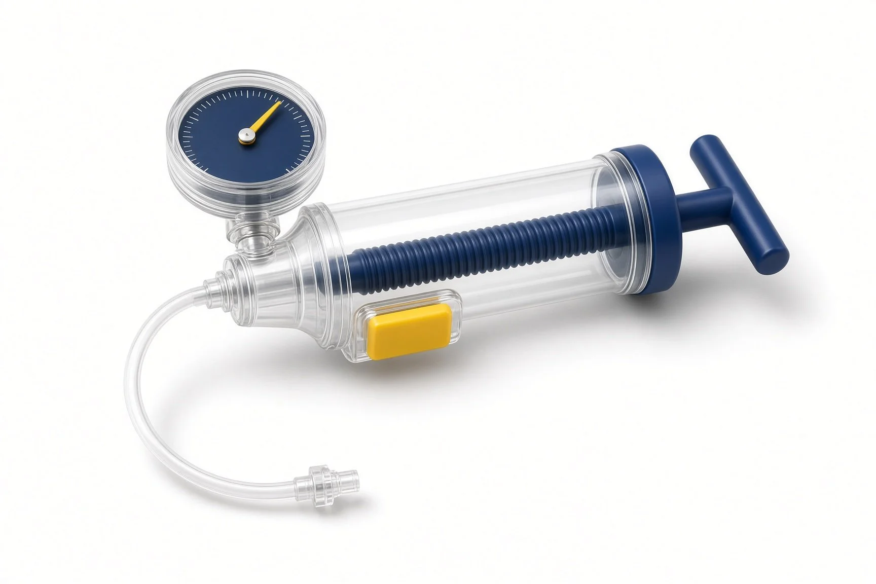
Inflation device.
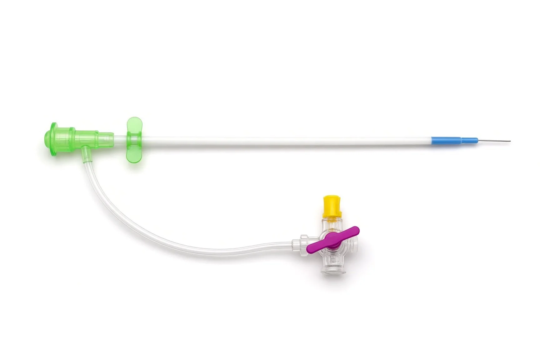
Short sheath.
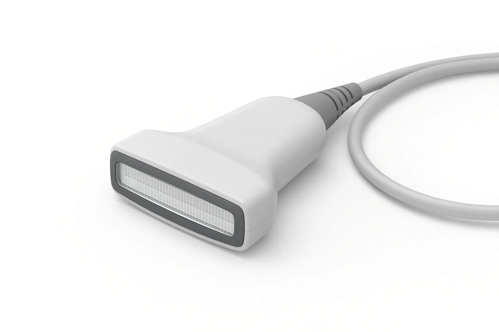
Ultrasound probe.
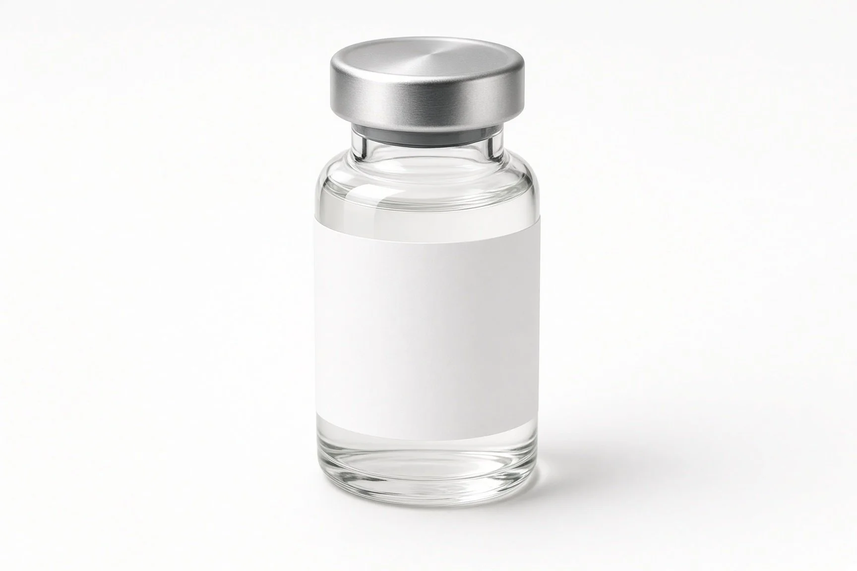
Contrast.
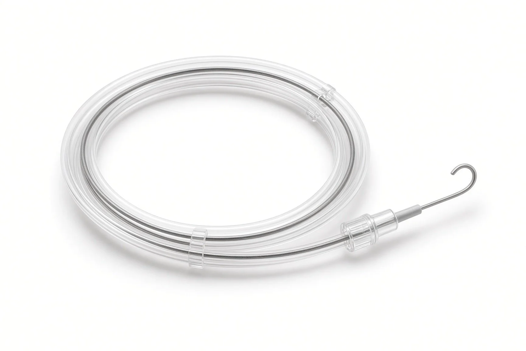
Guidewires.
Procedural steps (Barts example)
Getting ready
Access
Opening the narrowing
Finishing
Afterwards
First time in the cath lab?
Check the potassium. A high potassium level in someone on dialysis can cause dangerous heart rhythms; know the result before starting.
Protect the arm. No cuff, drip or tourniquet on the fistula arm, before, during or after.
Feel the thrill. Learn what a good buzz feels like before and after; it is the simplest check that the fistula works.
Expect high pressures. Fistula balloons often go to high pressures; know the inflation device and the balloon's rated pressure.
Protect the fistula arm. No cuff, tourniquet or cannula on it; put a sign on the arm board if needed.
Press, don't block. Hold enough pressure to stop the bleeding while you can still feel the thrill above the site.
FAQ
Will it hurt?
The local anaesthetic stings briefly. Balloon inflation can ache for a few seconds.
Can I have dialysis the same day?
Often yes, depending on the puncture site and your unit's advice.
Will it need doing again?
Narrowings often come back over months. Regular checks at dialysis help spot this early, and repeat treatment is common.
What should I do if the buzz stops?
Contact your dialysis unit straight away. A clotted fistula can often be saved if it is treated quickly.
Related Cath Lab 101 guides
Peripheral arterial intervention (leg arteries) (coming soon)
Venous stenting (iliac vein and SVC obstruction) (coming soon)
Sources
All checked on 12 November 2026. Each page's own date is shown where it gives one.
UK Kidney Association (formerly the Renal Association). Clinical practice guideline: vascular access for haemodialysis. ukkidney.org
Lok CE, et al. KDOQI clinical practice guideline for vascular access: 2019 update. Am J Kidney Dis 2020.
StatPearls (NCBI Bookshelf), search: arteriovenous fistula. ncbi.nlm.nih.gov/books
Disclaimer
Education only. This page is for general education for healthcare students, cath lab staff and curious readers. It isn't medical advice and doesn't replace advice from your own doctor, heart team or local unit protocols. Equipment and medicines are described generically. No product or manufacturer is recommended. If you think someone is having a heart emergency, call 999.
Disclosure: I work in the medical device industry. This page is independent education and isn't sponsored or reviewed by any company.
All diagrams on this page are original illustrations created for rodnieoro.com.